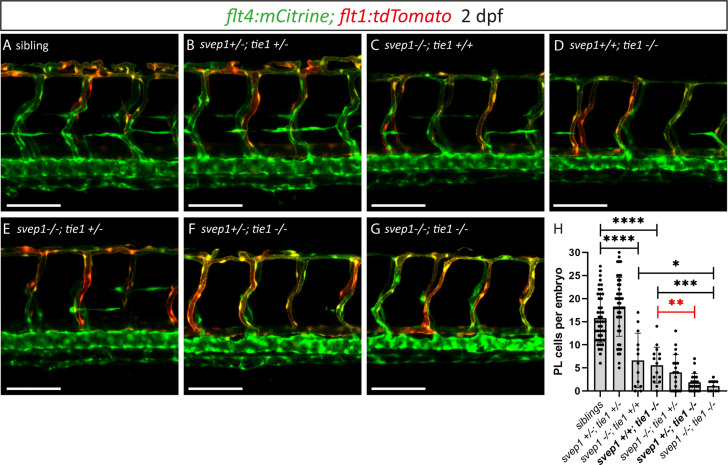
Figure 7. Heterozygous loss of svep1 exacerbates the PL phenotype in tie1 mutants, indicating genetic interaction between svep1 and tie1.
(A–G) Confocal images of blood and lymphatic vasculature in the trunk of 2-dpf-old embryos derived from svep1; tie1 double heterozygous fish, showing severely reduced PL numbers in svep1; tie1 double mutants and significant decrease of PL cell numbers in svep1+/−; tie1−/− compared to svep1+/+; tie1−/− (**p = 0.0012). (H) Quantification of PL cell numbers at 2 dpf using Mann–Whitney test (siblings: n = 45; svep1+/−; tie1+/−: n = 45; svep1−/−; tie1+/+: n = 13; svep1+/+; tie1−/−: n = 15; svep1−/−; tie1+/−: n = 20; svep1+/−; tie1−/−: n = 21; svep1−/−; tie1−/−: n = 11). Scale bar = 100 µm. Values are presented as means ± standard deviation (SD), ****p < 0.0001, ***p = 0.007, *p = 0.0163, ns = not significant. dpf, days post-fertilization; PL, parachordal lymphangioblast.


