ABSTRACT
There has been an increase in the prevalence of Clostridioides difficile (C. diff) causing significant economic impact on the health care system. Although toxigenic C. diff carriage is recognized in infancy, there is limited data regarding its longitudinal trends, associated epidemiolocal risk factors and intestinal microbiome characteristics. The objectives of our longitudinal cohort study were to investigate temporal changes in the prevalence of toxigenic C.diff colonization in children up to 2 years, associated epidemiological and intestinal microbiome characteristics. Pregnant mothers were enrolled prenatally, and serial stool samples were collected from their children for 2 years. 2608 serial stool samples were collected from 817 children. 411/817 (50%) were males, and 738/817 (90%) were born full term. Toxigenic C.diff was detected in 7/569 (1%) of meconium samples, 116/624 (19%) of 2 m (month), 221/606 (37%) of 6 m, 227/574 (40%) of 12 m and 18/235 (8%) of 24 m samples. Infants receiving any breast milk at 6 m were less likely to be carriers at 2 m, 6 m and 12 m than those not receiving it. (p = 0.002 at 2 m, p < 0.0001 at 6 m, p = 0.022 at 12 m). There were no robust differences in the underlying alpha or beta diversity between those with and without toxigenic C. diff carriage at any timepoint, although small differences in the relative abundance of certain taxa were found. In this largest longitudinal cohort study to date, a high prevalence of toxigenic C. diff carrier state was noted. Toxigenic C. diff carrier state in children is most likely a transient component of the dynamic infant microbiome.
KEYWORDS: Epidemiology, Clostridiodes difficile, microbiome, infants, children
Introduction
Clostridioides difficile (C. diff) is a Gram-positive, anaerobic spore forming pathogen that causes a spectrum of manifestations from asymptomatic carrier state to diarrhea, fulminant colitis, toxic megacolon, sepsis, and death.1 Epidemiological studies have shown an increasing prevalence of toxigenic C. diff infection (CDI) in both children and adults causing significant morbidity and economic impact on the health care system.2–4 Infants represent a unique cohort as cross-sectional studies have shown a high C. diff carrier state in infancy around 25-70% that approaches the adult carrier rate of 3% at around 8 years of age; although many of these studies did not differentiate toxigenic strains.5–12 Thus infants can be a potential reservoir for community spread of C.diff, specifically if they have disease causing toxigenic strains.13,14 Several factors that can detrimentally impact the intestinal microbiome have been associated with C.diff carriage in infancy.14–21 These include maternal factors (use of prenatal/perinatal antibiotics, mode of delivery) and infant factors (earlier gestational age, mode of feeding, hospitalizations, use of antibiotics and proton pump inhibitors).14,22,23 Intestinal dysbiosis has been associated with CDI and asymptomatic carriage in older children and adults.21,24,25 However there is paucity of data on the chronological drifts of toxigenic C.diff carriage during infancy, associated microbiome characteristics and epidemiological risk factors. Although previous studies have shown conflicting associations of C.diff carriage in infancy with atopic sensitization, there is limited understanding of potential long term health consequences.14–18–26–28
The aims of this study was to investigate the longitudinal trends in the prevalence of toxigenic C.diff carrier state through infancy to 2 years of age and identify associated clinical, demographic and intestinal microbiome characteristics.
Results
Subject demographics and clinical characteristics
A total of 2608 serial stool samples were collected from 817 children until around the age of 24 m from April 2018 to August 2021. 411/817 (50%) infants were males, 499/817 (61%) were born vaginally, and 738/817 (90%) were full term (>37 weeks of gestation) (Table 1). The study group had a diverse racial and ethnic background (Table 1). 170/817 (21%) and 456/817 (56%) mothers had received prenatal and perinatal antibiotics respectively.
Table 1.
Distribution of clinical and demographic variables ‘positive group’ (toxigenic clostridioides difficile positive) versus negative group (non-toxigenic clostridioides difficile positive/clostridioides difficile negative) at 2, 6, 12 and 24 months of age.
| Toxigenic Clostridioides difficile at 2 months |
Toxigenic Clostridioides difficile at 6 months |
Toxigenic Clostridioides difficile at 12 months |
Toxigenic Clostridioides difficile at 24 months |
|||||||||
|---|---|---|---|---|---|---|---|---|---|---|---|---|
| Positive (n = 116) |
Negative (n = 508) |
p- value | Positive (n = 221) | Negative (n = 385) |
p- value | Positive (n = 227) | Negative (n = 347) |
p- value | Positive (n = 18) | Negative (n = 217) |
p- value | |
|
Gender Male Female |
65 (56%) 51 (44%) |
259 (51%) 247 (49%) | 0.345 | 114 (52%) 105 (48%) |
184 (48%) 197 (51%) | 0.375 | 111 (49%) 116 (51%) |
170 (49%) 174 (50%) |
0.984 | 9 (50%) 9 (50%) |
110 (51%) 105 (48%) |
0.924 |
|
Maternal ethnicity Hispanic Non-Hispanic |
23 (20%) 93 (80%) |
99 (20%) 408 (80%) | 0.941 | 44 (20%) 177 (80%) |
78 (20%) 305 (79%) | 0.742 | 40 (18%) 187 (82%) |
91 (26%) 255 (74%) |
0.015a | 3 (17%) 15 (83%) |
34 (16%) 183 (84%) |
0.911 |
|
Maternal race Caucasian Asian African American or Black American Indian Native Hawaiian More than one race Other |
86 (74%) 9 (8%) 9 (8%) 1 (1%) 0 1 (1%) 10 (9%) |
349 (69%) 74 (15%) 20 (4%) 6 (1%) 0 25 (5%) 33 (7%) |
0.049a | 161 (73%) 19 (9%) 11 (5%) 2 (1%) 1 (1%) 11 (5%) 16 (7%) |
272 (71%) 55 (14%) 11 (3%) 3 (1%) 0 17 (4%) 25 (7%) |
0.275 | 164 (72%) 25 (11%) 10 (4%) 2 (1%) 0 11 (5%) 14 (6%) |
252 (73%) 43 (12%) 9 (3%) 3 (1%) 1 (1%) 15 (4%) 23 (7%) |
0.883 | 13 (72%) 2 (11%) 1 (6%) 0 0 2 (11%) 0 |
160 (74%) 23 (11%) 9 (4%) 4 (2%) 0 10 (5%) 11 (5%) |
0.745 |
| Maternal age in years (mean, SD) | 32.6±4.76 | 33.2 ± 4.43 | 0.195 | 33.09 ± 4.84 | 33.48 ± 4.42 | 0.307 | 33.17 ± 4.45 | 33.19 ± 4.61 | 0.950 | 34.61 ± 3.74 | 33.53 ± 4.45 | 0.322 |
|
Maternal weight gain in pregnancyc Less than recommended Recommended More than recommended |
15 (13%) 35 (30%) 65 (56%) |
87 (17%) 171 (34%) 246 (48%) |
0.291 | 39 (18%) 56 (25%) 124 (56%) |
66 (17%) 136 (35%) 177 (46%) |
0.025a | 37 (16%) 59 (26%) 129 (57%) |
66 (19%) 125 (36%) 152 (44%) |
0.008a | 4 (22%) 3 (17%) 11 (61%) |
40 (18%) 67 (31%) 109 (50%) |
0.442 |
|
Full term gestational age Yes No |
109 (94%) 7 (6%) |
461 (91%) 45 (9%) |
0.315 | 196 (89%) 24 (11%) |
352 (91%) 29 (8%) |
0.169 | 206 (91%) 20 (9%) |
310 (89%) 34 (10%) |
0.680 | 15 (83%) 3 (17%) |
192 (89%) 24 (11%) |
0.478 |
| Gestational age in weeks (mean, SD) | 38.6±-1.3 | 38.4 ± 1.7 | 0.421 | 38.3 ± 1.7 | 38.5 ± 1.5 | 0.130 | 38.4 ± 1.7 | 38.4 ± 1.9 | 0.900 | 38.2 ± 1.6 | 38.2 ± 2.1 | 0.971 |
|
Mode of delivery Vaginal Caesarean |
66 (57%) 50 (43%) ‘ |
320 (63%) 188 (37%) |
0.222 | 137 (62%) 84 (38%) |
229 (60%) 154 (40%) |
0.594 | 141 (62%) 86 (38%) |
215 (62%) 13 (38%) |
0.995 | 11 (61%) 7 (39%) |
127 (59%) 90 (42%) |
0.830 |
|
Prenatal antibiotics Yes No |
21 (18%) 91 (78%) |
106 (21%) 394 (78%) |
0.563 | 41 (19%) 175 (79%) |
81 (21%) 294 (76%) |
0.448 | 52 (23%) 171 (75%) |
66 (19%) 276 (80%) |
0.250 | 2 (11%) 16 (89%) |
43 (20%) 174 (80%) |
0.367 |
|
Peripartum antibiotics Yes No |
73 (63%) 42 (36%) |
274 (54%) 229 (45%) |
0.079 | 119 (54%) 101 (46%) |
158 (41%) 219 (57%) |
0.341 | 123 (54%) 101 (45%) |
195 (56%) 148 (43%) |
0.649 | 12 (67%) 5 (28%) |
128 (59%) 87 (40%) |
0.369 |
|
Infant antibiotic use by 2 months Yes No |
3 (3%) 107 (92%) |
23 (5%) 466 (92%) |
0.358 | 7 (3%) 158 (72%) |
14 (4%) 280 (73%) |
0.798 | 6 (3%) 181 (80%) |
10 (3%) 237 (68%) |
0.645 | 2 (11%) 12 (67%) |
9 (4%) 161 (74%) |
0.172 |
|
Infant antibiotic use by 6 months Yes No |
21 (10%) 192 (87%) |
27 (7%) 344 (90%) |
0.274 | 15 (7%) 168 (74%) |
22 (6%) 249 (72%) |
0.976 | 2 (11%) 14 (78%) |
19 (9%) 163 (75%) |
0.797 | |||
|
Infant antibiotic use by 12 months Yes No |
42 (19%) 179 (79%) |
61 (18%) 28 (81%) |
0.738 | 5(28%) 9 (50%) |
44 (20%) 148 (68%) |
0.277 | ||||||
|
Infant antibiotic use by 24 months Yes No |
3 (17%) 15 (83%) |
8 (13%) 184 (85%) |
0.679 | |||||||||
| Number of breast milk feeds/week at 6 months (mean, SD) | 22.3 ± 22.7 (n = 82) | 27.8 ± 22.8 (n = 339) |
0.053a | 19.8 ± 20.7 (n = 154) |
30.9 ± 22.9 (n = 279) |
0.001a | 25.8 ± 23.8 (n = 169) |
29.6 ± 23.08 (n = 229) |
0.113 | 30.5 ± 25.01 (n = 16) |
28.4 ± 22.8 (n = 184) |
0.735 |
| Number of breast milk feeds/week at 12 months (mean, SD) | 11.9 ± 16.6 (n = 72) |
12.7 ± 17.2 (n = 325) |
0.725 | 8.77 ± 14.52 (n = 142) |
14.94 ± 17.87 (n = 260) |
<0.001a | 11.2 ± 16.1 (n = 169) |
13.3 ± 17.7 (n = 247) |
0.219 | 11.9 ± 21.1 (n = 17) |
11.8 ± 16.8 (n = 167) |
0.983 |
|
Any breast milk at 6 months Yes No |
48 (41%) 34 (29%) |
256 (50%) 84 (17%) |
0.002a | 94 (43%) 60 (27%) |
219 (57%) 61 (16%) |
<0.001a | 114 (50%) 56 (25%) |
177 (51%) 52 (15%) |
0.022a | 12 (67%) 4 (22%) |
140 (65%) 45 (21%) |
0.951 |
|
Any breast milk at 12 months Yes No |
30 (26%) 42 (36%) |
144 (28%) 184 (36%) |
0.728 | 44 (20%) 98 (44%) |
135 (35%) 128 (33%) |
<0.001a | 67 (30%) 102 (45%) |
113 (33%) 137 (40%) |
0.259 | 5 (28%) 12 (67%) |
69 (32%) 98 (45%) |
0.340 |
|
Daycare at 6 months Yes No |
35 (30%) 43 (37%) |
137 (27%) 187 (37%) |
0.813 | 74 (34%) 77 (35%) |
98 (26%) 174 (45%) | 0.025a | 77 (34%) 81 (36%) |
89 (26%) 139 (40%) |
0.117 | 8 (44%) 10 (56%) |
89 (41%) 90 (42%) |
0.867 |
|
Daycare at 12 months Yes No |
31 (27%) 34 (29%) |
121 (24%) 178 (35%) |
0.284 | 63 (29%) 65 (29%) |
89 (23%) 154 (40%) |
0.008a | 68 (30%) 89 (39%) |
95 (27%) 134 (39%) |
0.721 | 7 (39%) 10 (56%) |
87 (40%) 67 (31%) |
0.228 |
|
Toxigenic carriage at 2 months Yes No |
60 (27%) 109 (49%) |
27 (7%) 275 (71%) |
<0.001a | 45 (20%) 144 (63%) |
29 (8%) 227 (65%) |
<0.001a | 2 (11%) 13 (72%) |
32 (15%) 141 (65%) |
0.618 | |||
|
Toxigenic carriage at 6 months Yes No |
90 (40%) 98 (43%) |
79 (23%) 197 (57%) |
<0.001a | 4 (22%) 13 (72%) |
68 (31%) 121 (56%) |
0.302 | ||||||
|
Toxigenic carriage at 12 months Yes No |
6 (33%) 9 (50%) |
77 (36%) 114 (53%) |
0.980 | |||||||||
a American College of Obstetricians and Gynecologists.
b Missing/Unknown values are not reported in the table.
c Significant p values.
Epidemiology of toxigenic C. diff and clinical associations
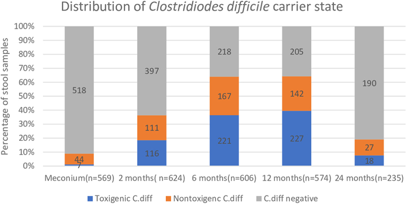
The prevalence of toxigenic C.diff at each timepoint was evaluated after longitudinal stool samples were collected. 7/569 (1%), 116/624 (19%), 221/606 (37%), 227/574 (40%) and 18/235 (8%) of meconium, 2 m, 6 m,12 m, 24 m were positive respectively (Figure 1). Longitudinal stool samples at 2 m, 6 m and 12 m were available in 181/817 (22%) infants and 27/181 (15%) were ‘always positive’. The inter-quartile ranges for CD 16s rRNA, tcdA, tcdB primer/probe sets were 20.32-31.2 (75% of reported detections with Ct/cycle threshold values under 31.20), 23.98-32.36 (75% of reported detections with Ct values under 32.36) and 24.24-32.76 (75% of reported detections with Ct values under 32.76) respectively. No infant was clinically diagnosed with CDI.
Figure 1.

Longitudinal distribution of toxigenic clostridioides difficile carrier state from birth to 2 years of age.
Clinical and demographic variables (as described in methods) were then studied for any significant association. Infants with non-Hispanic mothers were more likely to be positive when compared to those with Hispanic mothers at 12 m (42% in non-Hispanic group versus 31% in Hispanic group; p = 0.015, chi-square). Other demographic characteristics were similar between the two groups. Infants born to mothers with more than recommended weight gain during pregnancy (per American College of Obstetricians and Gynecologists guidelines) were more likely to be positive at 6 m and 12 m (p = 0.025 and 0.008 at 6 m and 12 m respectively).
Infants receiving any breast milk at 6 m were less likely to be positive at 2 m, 6 m and 12 m than those not receiving it (16% in breast milk group versus 29% in no breast milk group at 2 m, p = 0.002; 30% in breast milk group versus 50% in no breast milk group at 6 m, p < 0.001; 39% in breast milk group versus 52% in no breast milk group at 12 m, p = 0.022, chi-square). This was directly proportional to the number of times infants received breast milk per week (Table 1).
Infants attending daycare at 6 m were more likely to be positive (43% in daycare group versus 31% in no daycare group, p = 0.025, chi-square). There was no significant difference in carrier state with mode of delivery, prenatal or peripartum antibiotic use, child gender, gestational age, and infant antibiotic use.
Infants positive at 2 m were more likely to test positive at 6 m (69% in positive versus 28% in negative; p < 0.001) and 12 m (61% in positive versus 39% in negative, p < 0.001, chi-square). Infants positive at 6 m were more likely to be positive at 12 m (53% in positive versus 33% in negative, p < 0.001, chi-square).
Infants who were ‘always positive’ received more formula feeds per week when compared to ‘always negative’ at 12 m (mean = 13.0 and 7.02 in always positive and always negative respectively, p = 0.004, two sample t-test) (Table 2).
Table 2.
Distribution of clinical and demographic variables between ‘always positive’ toxigenic Clostridioides difficile (positive at 2 months, 6 months, 12 months) versus ‘always negative’ groups (negative at 2 months, 6 months and 12 months).
| “Always positive” for toxigenic Clostridioides difficile at 2,6 and 12 months (n = 27) |
“Always negative” for toxigenic Clostridioides difficile at 2,6 and 12 months (n = 154) |
p-value | |
|---|---|---|---|
|
Gender Male Female |
16 (59%) 11 (41%) |
83 (54%) 71 (46%) |
0.605 |
|
Maternal ethnicity Hispanic Non-Hispanic |
2 (7%) 25 (93%) |
25 (16%) 129 (84%) |
0.235 |
|
Maternal race Caucasian Asian African American American Indian Native Hawaiian More than one race Other |
21 (78%) 3 (11%) 1 (4%) 0 0 1 (4%) 1 (4%) |
116 (75%) 23 (15%) 2 (1%) 2 (1%) 0 8 (5%) 3 (2%) |
0.872 |
|
Maternal age in years (mean, SD) |
32.8 ± 5.2 | 33.7 ± 4.5 | 0.3197 |
|
Maternal weight gain in pregnancy (ACOG)b Less than recommended Recommended More than recommended |
2 (7%) 6 (22%) 18 (67%) |
25 (16%) 56 (36%) 71 (46%) |
0.101 |
|
Term gestational age Yes No |
25 (93%) 2 (7%) |
130 (84%) 16 (10%) |
0.632 |
|
Mode of delivery Vaginal Caesarean |
16 (59%) 11 (41%) |
86 (56%) 68 (44%) |
0.741 |
|
Prenatal antibiotics Yes No |
3 (11%) 23 (85%) |
25 (16%) 128 (83%) |
0.545 |
|
Peripartum antibiotics Yes No |
17 (63%) 10 (37%) |
97 (63%) 57 (37%) |
0.06 |
|
Infant antibiotic course by 2 months Yes No |
1 (4%) 26 (96%) |
9 (6%) 140 (91%) |
0.629 |
|
Infant antibiotic use by 6 months Yes No |
0 27 (100%) |
10 (7%) 141 (92%) |
0.168 |
|
Infant antibiotic use by 12 months Yes No |
6 (22%) 20 (74%) |
27 (18%) 125 (81%) |
0.519 |
| Infant antibiotic use at 24 months Yes No |
1 (4%) 21 (78%) |
12 (8%) 96 (62%) |
0.349 |
| Number of breast milk feeds/week at 6 months (mean, SD) | 25.8 ± 22 (n = 25) |
32.6 ± 23.8 (n = 123) |
0.188 |
| Number of breast milk feeds/week at 12 months (mean, SD) | 13.6 ± 15.5 (n = 23) |
19.38 ± 28.4 (n = 122) |
0.353 |
|
Any Breast milk at 6 months Yes No |
17 (63%) 8 (30%) |
100 (65%) 23 (15%) |
0.136 |
|
Any Breast milk at 12 months Yes No |
12 (45%) 11 (41%) |
66 (43%) 56 (36%) |
0.865 |
| Number of formula feeds/week at 6 months (mean, SD) | 8.9 ± 13.4 (n = 24) |
9.8 ± 14.6 (n = 123) |
0.789 |
| Number of formula feeds/week at 12 months (mean, SD) | 13.0 ± 15.5 (n = 23) |
7.02 ± 12.5 (n = 122) |
0.042a |
|
Daycare at 6 months Yes No |
11 (41%) 11 (41%) |
43 (28%) 75 (49%) |
0.441 |
|
Daycare at 12 months Yes No |
10 (37%) 10 (37%) |
40 (26%) 71 (46%) |
0.236 |
aSignificant p values.
bAmerican College of Obstetricians and Gynecologists.
cMissing/Unknown values are not reported in the table.
Microbiome associations with toxigenic C. diff carrier state
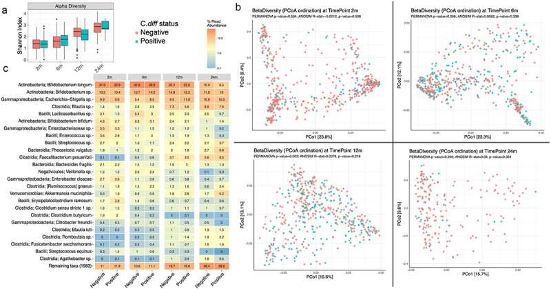
Microbiome data was available in 514/624 (82%), 509/606 (84%), 466/574 (81%) and 207/235 (88%) of 2 m, 6 m,12 m and 24 m samples respectively. The visual community diversity explorations and statistical diversity analyses showed no significant differences in the alpha diversity or beta diversity at 2 m, 6 m, and 24 m between positive and negative groups (Figure 2). Although statistically significant differences were detected in community diversities between groups at 12 m (ANOVA on Alpha diversity p = 0.001 and PERMANOVA on Beta diversity p = 0.003), further community explorations showed minimal factor contribution (ANOSIM effect size~0.8%, p >>0.5), location-based separation (PERMDISP F-value = 0.5, p >> 0.5) and no clear visual separation of the communities (Figure 2). Significant abundance changes were detected in a few taxa between the 2 groups (supplementary Figure S1). There was an overall decrease in Lacticaseibacillus species and an increase in Proteus mirabilis in the positive group at 12 m.
Figure 2.

Microbiome characteristics in ‘positive group’ (toxigenic clostridioides difficile positive) versus negative group (non-toxigenic clostridioides difficile positive/clostridioides difficile negative) at 2, 6, 12 and 24 months of age. A- distribution of alpha diversity (shannon index); B- Principal coordinate analysis plot for beta diversity (bray-curtis index), C-Heat map diagram of the relative abundance of taxa by carrier state.
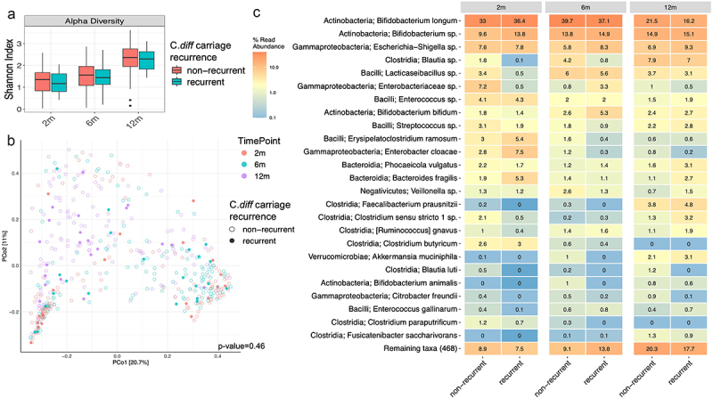
In the longitudinal cohort with samples at all time points (2 m, 6 m and 12 m) subjects were divided into two cohorts for microbiome analysis: ‘recurrent carriage group’ defined as positive at 2 or more timepoints and the rest were included in ‘non-recurrent carriage group’. There was no significant difference in the alpha diversity, beta diversity and relative abundance of most taxa between the two groups at all timepoints (Figure 3). There was a significant decrease in the relative abundance of some taxa (Bifidobacterium, Lacticaseibacillus, Lactobacillus, Veilonella, Streptococcus, certain Bacteriodes and Enterobacteriaceae species) and increase in some Firmicutes (Eisenbergiella species) in the recurrent carriage group (Supplementary Figure S2).
Figure 3.

Microbiome characteristics in ‘recurrent carriage group’ (defined as positive for toxigenic clostridioides difficile at 2 or more timepoints) versus ‘non-recurrent carriage group’ (rest of the subjects) at 2, 6, 12, 24 months. A- distribution of alpha diversity (shannon index); B- Principal coordinate analysis plot for beta diversity (bray-curtis index); C- heat map diagram of the relative abundance of taxa by carrier state.
There were no significant differences in the alpha diversity, beta diversity or relative abundance of most taxa between infants exclusively on breast milk versus formula at 6 m when adjusted for colonization status (Supplementary Figure S3). Bifidobacterium and Lacticaseibacillus species were most differentially abundant in negative infants on breast milk and least abundant in positive infants on formula. Firmicutes (Fusicatenibacter saccharivorans) and Gammaproteobacteria (Escherichia-Shigella species) were most differentially abundant in positive formula fed babies (Supplementary Figure S4)
Discussion
In the largest longitudinal study to date on the epidemiology of toxigenic C.diff carriage in children up to 2 years of age and its intestinal microbiome associations, a high prevalence rate was noted with a peak of 40% at 12 months. Infants receiving breast milk were less likely to be carriers and there were no major associated changes in microbiome diversity.
A recent metanalyses on the epidemiology of C.diff showed a pooled prevalence rate of toxigenic C.diff at 14% around 6-12 m.5 Even though the longitudinal trends for toxigenic carrier state in our study were consistent with the findings from previous studies, we had a much higher prevalence rate at 36% −40% at 6-12 m.5–7-9–19-29 These differences could be due to the increase in its prevalence with time, geographic variations in distribution and differences in study design (most of these studies were cross sectional with smaller sample size) and C.diff detection techniques used (validated detection of toxin gene by PCR utilized in our study is the most sensitive).30–32 The very low prevalence of C.diff in meconium samples and the steady increase during rest of infancy suggests that it is most likely environmentally acquired rather than maternally transmitted at birth; however maternal C.diff status at delivery was not obtained to confirm this hypothesis.14 Infants positive in earlier samples were more likely to again test positive. This implies that toxigenicity has a propensity for persistence through infancy once acquired. Thus infants may act as a significant reservoir for community spread of toxigenic C.diff given its high prevalence rate and tendency to persist after initial colonization.
Intestinal dysbiosis with an overall decrease in microbial diversity and richness has been associated with asymptomatic colonization and CDI in older children and adults.21,24,25 These changes include an increase in Firmicutes/Bacteriodes ratio, Proteobacteria (Enterobacteriaceae/Enterococcus), Lactobacillus and Veilonella species and a decrease in Lachnospiraceae, Ruminococcaceae, Clostridium IV and XIV clusters, Faecalibacterium prausnitzii and Bifidobacterium species.20,21,33 A limited number of cross-sectional studies investigating microbiome changes in colonization during infancy have described an increase in Firmicutes, Klebsiella species and decrease in Bifidobacterium species.19,23,29 It is not clearly understood if C. diff is a mediator of these changes or just a part of the dysbiotic state. To our knowledge no longitudinal studies to date have examined intestinal microbiome associations with toxigenic C.diff in infancy. We noted significant differences in the relative abundance of certain taxa similar to previous studies, however, without any robust changes in microbial diversity. A novel finding was a consistent decrease in Lacticaseibacillus species in carrier state. Contrary to previous adult studies, an increase in Enterobacteriaceae or Veilonella species was not noted with toxigenicity.24,34,35 Since there were no distinct microbial signatures causing robust changes in diversity with carriage, it is most likely a transient component in the dynamic infant microbiome as it transitions toward the relatively stable adult microbiome.
Our study noted that infants receiving breast milk during infancy were less likely to be positive. Breast milk has several antimicrobial factors including human milk oligosaccharides, lysozymes, secretory IgA and a unique microbiome (mostly commensal bacteria; Bifidibacterium and Lactobacillus) that possibly offer protection against colonization.36–38 Drall et al noted that exclusively breast-fed babies colonized with C.diff at 3 m had a different gut microbial profile resembling that of formula-fed babies and greater alpha-diversity than those not colonized.23 However in our study there was no significant difference in microbial diversity between infants exclusively on breast milk versus formula at 6 m when adjusted for carrier state. These contradicting findings could be due to differences in study design (subject age, diet and study methods as colonization status without toxigenicity was analyzed in Drall et al study). Our findings suggest that even though breast milk modulates the intestinal microbiome, the differences in carrier state with mode of feeding is less likely mediated through these changes.
A higher incidence of toxigenicity was noted in subjects with non-Hispanic mothers at 12 months. This aligns with findings from a recent case-control study that demonstrated higher risk of CDI in non-Hispanic children.6 The higher positive rate in infants of mothers with more than recommended weight gain during pregnancy could possibly be from maternal transmission postnatally as adult studies have shown association between obesity and CDI.39 The higher positive rate in infants attending day care again suggests that it is most likely environmentally acquired.40,41 Infant use of antibiotics and prolonged hospitalizations have been associated with carrier state and infection.14,16 Our study group consisted mostly of term infants with limited use of antibiotics and hence might not have captured a potential association.
Despite the high carrier state, there was no documented clinical CDI in our study group irrespective of feeding method and other clinical factors. The absence of toxigenic C.diff receptors is one of the postulated reasons; however this hypothesis is based on animal studies with small sample size.42,43 The overall features of the developing infant microbiome which differs from adult microbiome in structure and composition are another possible protective factor against CDI.44–46 Several studies have demonstrated differences in gut metabolomic profiles with CDI in adults characterized by a depletion of short chain fatty acid (SCFA) and secondary bile acids.47,48 The infant gut has a different SCFA metabolomic profile based on feeding (higher levels of acetate in infants receiving breast milk and prebiotic containing formula) and has a bile acid profile that is initially predominated by primary bile acids which may also be permissive to carriage.49–51 However, further studies are needed to elucidate comprehensive characteristics of infant gut microbiome and metabolome in carrier state and its role in protection against CDI. This may offer strategies against CDI in older children and adults. The high prevalence of asymptomatic carriage in infancy reiterates that testing for CDI in this population should be conducted with great caution, as per the IDSA guidelines, due to the possibility of detecting carriage rather than true infection.52 Children can develop humoral immune responses against C.diff toxin even in the absence of CDI; however the duration of protection from anti-toxin antibodies and its long term health implications need further investigation.53
The major strengths of our study included its large sample size, longitudinal study design and attention to toxigenicity that provided a better understanding of the dynamics of C.diff carrier state in infancy right from the neonatal period. The major limitation of our study was that it was a single center study and even though we had racially and ethnically diverse group the results of our study may be less generalizable to other regions of the world. Moreover, although this is the first longitudinal study to assess intestinal microbiome associations of toxigenic C. diff carriage in infancy, 16S rRNA gene sequencing was used. Utilization of shotgun metagenomic sequencing and metabolomic analysis may have provided further insight into characteristics of the microbiome at species level, in addition to non-bacterial microorganisms, as well as its functional properties.54 In addition our study consisted mostly of term infants and hence the prevalence rate may not be reflective of distributions in preterm infants with prolonged hospitalization and more frequent antibiotic exposure.55,56
Conclusions
This study gives novel insight into the longitudinal dynamics of toxigenic C. diff carriage in infancy and associated epidemiological, clinical and intestinal microbiome characteristics. Given its high prevalence rates, infants may act as potential community reservoir. Infants receiving breast milk were less likely to be carriers, most likely due to its anti-microbial properties rather than direct effects on the microbiome composition. Colonization in children is not associated with significant changes in microbial diversity and hence it is most likely a transient component of the dynamic infant microbiome.
Methods
Study design and sample collection
Mothers were enrolled prenatally with informed consent in a longitudinal, prospective cohort study “The First 1000 Days of Life and Beyond” within the Inova Health System. All experimental protocols were approved by the Inova Health System and WCG Institutional Review Board (Inova protocol #15-1804, WCG protocol #20120204). Serial stool samples were collected from infants starting in the first 2 days after birth (meconium) and at around 2 months(m), 6 m, 12 m, and 24 m of age. All samples except meconium were collected by caregivers at home and mailed back to the lab, using previously validated methods and stored at −80°C until analysis.57,58
Demographic information including infant gender, maternal race/ethnicity, pregnancy details including mode of delivery (vaginal delivery versus cesarean section), use of prenatal antibiotics (antibiotics given to mother from conception to 2 days before delivery) and peripartum antibiotics (antibiotics given to mother 2 days before and during delivery), gestational age at delivery, and maternal weight gain during pregnancy were collected through a questionnaire and review of electronic medical records. Details including infant feeding, daycare attendance, use of antibiotics were also collected from parents through a questionnaire.
DNA extraction and microbiome sequencing
DNA was extracted from stool aliquots using the DNeasy PowerSoil Pro kit (Qiagen, Valencia, CA) following manufacturer’s instructions. Barcoded PCR primers annealing to the V4 region of 16S ribosomal RNA gene were used for library generation (forward primer 5’-GTGYCAGCMGCC-GCGGTAA-3’, reverse primer: 5’-GGACTACN-VGGGTWTCTAAT-3’). Sequencing was performed on the Miseq platform (Illumina, CA, USA) with 2 × 250 base pair paired end reads. Positive controls (DNA sequences) and negative controls (DNA free water) were used.
qPCR for C. difficile detection
Primers and Taqman-based probes targeting C. diff specific 16S rRNA (CD16S rRNA), tcdA (Toxin A), and tcdB (Toxin B) genes were used (Invitrogen, MA, USA) on extracted DNA.31 Samples were run in duplicate for each assay and qPCR was performed in 384-well optical plates on ABI QuantStudio 6 Flex Real-Time PCR System (ThermoFisher, CA, USA). Target detection was considered positive with conclusive amplification curves in both replicates of a given primer/probe set; mean Ct values over 46 were considered negative (Ct > 50 was considered negative by the reference study and we lowered this cutoff to>46). Samples positive for CD16S rRNA but negative for both tcdA and tcdB were designated as non-toxigenic C. diff, samples positive for CD16S rRNA and either tcdA, tcdB, or both, were designated as positive for toxigenic C. diff and samples with no target detection for all three primer probe sets were designated as negative for C. diff.
Epidemiological and statistical analysis
The subjects were divided into following analytical categories: 1) toxigenic C.diff positive group hereafter referred to as ‘positive’ versus non-toxigenic C. diff positive/C. diff negative group hereafter referred to as ’negative’ and 2) ‘always positive’ for toxigenic C.diff (positive at time points 2 m, 6 m, 12 m) versus ‘always negative’ for toxigenic C.diff.
Correlation between clinical and demographic factors with the prevalence of toxigenic C.diff at each timepoint was evaluated using the chi-square test and two-sample t-test for categorical and continues variables respectively.
Microbiome analysis
Meconium samples were not included due to low C.diff and bacterial DNA contents. Amplicon sequence variants (ASV) were generated by denoising and merging reads with DADA2 v1.20.0 and subsequent taxonomic assignment with rdp and the SILVA v138.1 database.59,60 ASVs unable to be identified by SILVA were further queried against NCBI’s 16S ribosomal RNA database using BLAST+ v2.12.0.61 Sample read depth analysis and filtering were performed with R statistical language in R Studio (tidyverse, ampvis2, vegan, ggplot2).62–65 Samples with read abundance<15000 reads and ASVs with abundance<15 reads were excluded from downstream analysis. The 16S rRNA sequence data of qualified samples are available in NCBI Sequence Read Archive (SRA), PRJNA851805.
Community differences were explored visually and statistically between positive and negative groups with the help of R packages.66,67 Alpha diversity was explored using Shannon index and groups were compared statistically with ANOVA and Wilcoxon tests. Beta diversity was explored using Bray-Curtis dissimilarity matrix, distances visualized with PCoA and community contrasts explored statistically with PERMANOVA, ANOSIM and PERMDISP (where applicable). Differential abundance testing on species level taxonomy was performed using DESeq2 contrasting the positive with negative samples and p-values adjusted by the Benjamini-Hochberg method for multiple comparisons.68
Supplementary Material
Funding Statement
This work was supported by a National Institute of Child Health and Human Development of the National Institutes of Health K23 award (No. K23HD099240, Hourigan), the National Institute of Allergy and Infectious Diseases of the National Institutes of Health under the Intramural Research Program (Hourigan) and an Inova Health System Seed Grant (Mani, Hourigan). The content is solely the responsibility of the authors and does not necessarily represent the official views of the National Institutes of Health. Inova expresses its appreciation to the Fairfax County in VA, which has supported Inova’s research projects with annual funding from its Contributory Fund (Fund 10030, Maxwell).
Disclosure statement
No potential conflict of interest was reported by the author(s).
Contributors’ statement page
Dr. Mani conceptualized and designed the study, collected and interpreted data, drafted the initial manuscript, reviewed and revised the manuscript.
Ms. Levy conceptualized and designed the study, designed the data collection instruments, collected data and reviewed and revised the manuscript.
Mr. Fassnacht carried out the Clostridioides difficile PCR analysis and reviewed and revised the manuscript.
Dr. Hazrati carried out the initial epidemiological analyses and reviewed and revised the manuscript.
Dr. Angelova carried out microbiome analysis and reviewed and revised the manuscript.
Dr. Subramanian carried out microbiome analysis and reviewed and revised the manuscript
Ms. Richards collected data and reviewed and revised the manuscript.
Mr. Rice collected data and reviewed and revised the manuscript.
Dr. Niederhuber conceptualized and designed the study and reviewed and revised the manuscript.
Dr. Maxwell conceptualized and designed the study and reviewed and revised the manuscript.
Dr. Hourigan conceptualized and designed the study, collected and interpreted data, drafted the initial manuscript and critically revised the manuscript for important intellectual content.
All authors approved the final manuscript as submitted and agree to be accountable for all aspects of the work.
Data availability statement
The data that support the findings of this study are openly available in https://www.ncbi.nlm.nih.gov/bioproject/PRJNA851805 [ncbi.nlm.nih.gov]
Supplementary material
Supplemental data for this article can be accessed online at https://doi.org/10.1080/19490976.2023.2203969
References
- 1.Czepiel J, Dróżdż M, Pituch H, Kuijper EJ, Perucki W, Mielimonka A, Goldman S, Wultańska D, Garlicki A, Biesiada G.. Clostridium difficile infection: review. Eur J Clin Microbiol Infect Dis. 2019;38(7):1211–14. doi: 10.1007/s10096-019-03539-6. [DOI] [PMC free article] [PubMed] [Google Scholar]
- 2.De Roo AC, Regenbogen SE. Clostridium difficile infection: an epidemiology update. Clin Colon Rectal Surg. 2020;33(2):49–57. doi: 10.1055/s-0040-1701229. [DOI] [PMC free article] [PubMed] [Google Scholar]
- 3.Balsells E, Shi T, Leese C, Lyell I, Burrows J, Wiuff C, Campbell H, Kyaw MH, Nair H. Global burden of clostridium difficile infections: a systematic review and meta-analysis. J Glob Health. 2019;9(1):010407. doi: 10.7189/jogh.09.010407. [DOI] [PMC free article] [PubMed] [Google Scholar]
- 4.Depestel DD, Aronoff DM. Epidemiology of clostridium difficile infection. J Pharm Pract. 2013;26(5):464–475. doi: 10.1177/0897190013499521. [DOI] [PMC free article] [PubMed] [Google Scholar]
- 5.Tougas SR, Lodha N, Vandermeer B, Lorenzetti DL, Tarr PI, Tarr GA, Chui L, Vanderkooi OG, Freedman SB. Prevalence of detection of clostridioides difficile among asymptomatic children: a systematic review and meta-analysis. JAMA Pediatr. 2021;175(10):e212328. doi: 10.1001/jamapediatrics.2021.2328. [DOI] [PMC free article] [PubMed] [Google Scholar]
- 6.Miranda-Katz M, Parmar D, Dang R, Alabaster A, Greenhow TL. Epidemiology and risk factors for community associated clostridioides difficile in children. J Pediatr. 2020;221:99–106. doi: 10.1016/j.jpeds.2020.02.005. [DOI] [PubMed] [Google Scholar]
- 7.Stoesser N, Eyre DW, Quan TP, Godwin H, Pill G, Mbuvi E, Vaughan A, Griffiths D, Martin J, Fawley W, et al. Epidemiology of clostridium difficile in infants in oxfordshire, UK: risk sfactors for colonization and carriage, and genetic overlap with regional C. difficile infection strains. Plos One. 2017;12(8):e0182307. doi: 10.1371/journal.pone.0182307. [DOI] [PMC free article] [PubMed] [Google Scholar]
- 8.Rousseau C, Lemée L, Le Monnier A, Poilane I, Pons JL, Collignon A. Prevalence and diversity of clostridium difficile strains in infants. J Med Microbiol. 2011;60(Pt 8):1112–1118. doi: 10.1099/jmm.0.029736-0. [DOI] [PubMed] [Google Scholar]
- 9.Rousseau C, Poilane I, De Pontual L, Maherault AC, Le Monnier A, Collignon A. Clostridium difficile carriage in healthy infants in the community: a potential reservoir for pathogenic strains. Clin Infect Dis. 2012;55(9):1209–1215. doi: 10.1093/cid/cis637. [DOI] [PubMed] [Google Scholar]
- 10.Collignon A, Ticchi L, Depitre C, Gaudelus J, Delmée M, Corthier G. Heterogeneity of clostridium difficile isolates from infants. Eur J Pediatr. 1993;152(4):319–322. doi: 10.1007/BF01956743. [DOI] [PubMed] [Google Scholar]
- 11.Adlerberth I, Huang H, Lindberg E, Åberg N, Hesselmar B, Saalman R, Nord CE, Wold AE, Weintraub A. Toxin-producing clostridium difficile strains as long-term gut colonizers in healthy infants. J Clin Microbiol. 2014;52(1):173–179. doi: 10.1128/JCM.01701-13. [DOI] [PMC free article] [PubMed] [Google Scholar]
- 12.Cui QQ, Yang J, Sun SJ, Li ZR, Qiang CX, Niu YN, Li RX, Shi DY, Wei HL, Tian TT, et al. Carriage of clostridioides difficile in healthy infants in the community of Handan, China: a 1-year follow-up study. Anaerobe. 2021;67:102295. doi: 10.1016/j.anaerobe.2020.102295. [DOI] [PubMed] [Google Scholar]
- 13.Enoch DA, Butler MJ, Pai S, Aliyu SH, Karas JA. Clostridium difficile in children: colonisation and disease. J Infect. 2011;63(2):105–113. doi: 10.1016/j.jinf.2011.05.016. [DOI] [PubMed] [Google Scholar]
- 14.Jangi S, Lamont JT. Asymptomatic colonization by clostridium difficile in infants: implications for disease in later life. J Pediatr Gastroenterol Nutr. 2010;51(1):2–7. doi: 10.1097/MPG.0b013e3181d29767. [DOI] [PubMed] [Google Scholar]
- 15.Sammons JS, Toltzis P, Zaoutis TE. Clostridium difficile infection in children. JAMA Pediatr. 2013;167(6):567–573. doi: 10.1001/jamapediatrics.2013.441. [DOI] [PubMed] [Google Scholar]
- 16.Anjewierden S, Han Z, Foster CB, Pant C, Deshpande A. Risk factors for clostridium difficile infection in pediatric inpatients: a meta-analysis and systematic review. Infect Control Hosp Epidemiol. 2019;40(4):420–426. doi: 10.1017/ice.2019.23. [DOI] [PubMed] [Google Scholar]
- 17.Antharam VC, Li EC, Ishmael A, Sharma A, Mai V, KH R, Wang GP. Intestinal dysbiosis and depletion of butyrogenic bacteria in clostridium difficile infection and nosocomial diarrhea. J Clin Microbiol. 2013;51(9):2884–2892. doi: 10.1128/JCM.00845-13. [DOI] [PMC free article] [PubMed] [Google Scholar]
- 18.Vu K, Lou W, Tun HM, Konya TB, Morales-Lizcano N, Chari RS, Field CJ, Guttman DS, Mandal R, Wishart DS, et al. From birth to overweight and atopic disease: multiple and common pathways of the infant gut microbiome. Gastroenterol. 2021;160(1):128–144.e10. doi: 10.1053/j.gastro.2020.08.053. [DOI] [PubMed] [Google Scholar]
- 19.Rousseau C, Levenez F, Fouqueray C, Doré J, Collignon A, Lepage P. Clostridium difficile colonization in early infancy is accompanied by changes in intestinal microbiota composition. J Clin Microbiol. 2011;49(3):858–865. doi: 10.1128/JCM.01507-10. [DOI] [PMC free article] [PubMed] [Google Scholar]
- 20.Samarkos M, Mastrogianni E, Kampouropoulou O. The role of gut microbiota in clostridium difficile infection. Eur J Intern Med. 2018;50:28–32. doi: 10.1016/j.ejim.2018.02.006. [DOI] [PubMed] [Google Scholar]
- 21.Vasilescu IM, Chifiriuc MC, Pircalabioru GG, Filip R, Bolocan A, Lazăr V, Diţu LM, Bleotu C. Gut dysbiosis and clostridioides difficile infection in neonates and adults. Front Microbiol. 2021;12:651081. doi: 10.3389/fmicb.2021.651081. [DOI] [PMC free article] [PubMed] [Google Scholar]
- 22.Lees EA, Miyajima F, Pirmohamed M, Carrol ED. The role of clostridium difficile in the paediatric and neonatal gut - a narrative review. Eur J Clin Microbiol Infect Dis. 2016;35(7):1047–1057. doi: 10.1007/s10096-016-2639-3. [DOI] [PMC free article] [PubMed] [Google Scholar]
- 23.Drall KM, Tun HM, Morales-Lizcano NP, Konya TB, Guttman DS, Field CJ, Mandal R, Wishart DS, Becker AB, Azad MB, et al. Clostridioides difficile colonization is differentially associated with gut microbiome profiles by infant feeding modality at 3–4 months of age. Front Immunol. 2019;10:2866. doi: 10.3389/fimmu.2019.02866. [DOI] [PMC free article] [PubMed] [Google Scholar]
- 24.Kachrimanidou M, Tsintarakis E. Insights into the role of human gut microbiota in clostridioides difficile infection. Microorganisms. 2020;8(2):200. doi: 10.3390/microorganisms8020200. [DOI] [PMC free article] [PubMed] [Google Scholar]
- 25.Chen LA, Hourigan SK, Grigoryan Z, Gao Z, Clemente JC, Rideout JR, Chirumamilla S, Rabidazeh S, Saeed S, Elson CO, et al. Decreased fecal bacterial diversity and altered microbiome in children colonized with clostridium difficile. J Pediatr Gastroenterol Nutr. 2019;68(4):502–508. doi: 10.1097/MPG.0000000000002210. [DOI] [PubMed] [Google Scholar]
- 26.Kim H, Sitarik AR, Woodcroft K, Johnson CC, Zoratti E. Birth mode, breastfeeding, pet exposure, and antibiotic use: associations with the gut microbiome and sensitization in children. Curr Allergy Asthma Rep. 2019;19(4):22. doi: 10.1007/s11882-019-0851-9. [DOI] [PMC free article] [PubMed] [Google Scholar]
- 27.van Nimwegen FA, Penders J, Stobberingh EE, van Nimwegen FA, Postma DS, Koppelman GH, Kerkhof M, Reijmerink NE, Dompeling E, van den Brandt PA, et al. Mode and place of delivery, gastrointestinal microbiota, and their influence on asthma and atopy. J Allergy Clin Immunol. 2011;128(5):948–55.e1. doi: 10.1016/j.jaci.2011.07.027. [DOI] [PubMed] [Google Scholar]
- 28.Penders J, Thijs C, van den Brandt PA, Kummeling I, Snijders B, Stelma F, Adams H, van Ree R, Stobberingh EE. Gut microbiota composition and development of atopic manifestations in infancy: the KOALA birth cohort study. Gut. 2007;56(5):661–667. doi: 10.1136/gut.2006.100164. [DOI] [PMC free article] [PubMed] [Google Scholar]
- 29.Nicholson MR, Strickland B, Guiberson ER, Shilts M, Edwards K, Skaar EP, Das S. Mo1604: microbiome and bile acid profiles predict clostridioides difficile colonization versus symptomatic disease in children. Gastroenterol. 2022;162(7):S-833-S-834. doi: 10.1016/S0016-5085(22)61965-6. [DOI] [Google Scholar]
- 30.Okanda T, Mitsutake H, Aso R, Sekizawa R, Takemura H, Matsumoto T, Nakamura S. Rapid detection assay of toxigenic clostridioides difficile through PathOC rightgene, a novel high-speed polymerase chain reaction device. Diagn Microbiol Infect Dis. 2021;99(2):115247. doi: 10.1016/j.diagmicrobio.2020.115247. [DOI] [PubMed] [Google Scholar]
- 31.Kubota H, Sakai T, Gawad A, Makino H, Akiyama T, Ishikawa E, Oishi K. Development of TaqMan-based quantitative PCR for sensitive and selective detection of toxigenic clostridium difficile in human stools. Plos One. 2014;9(10):e111684. doi: 10.1371/journal.pone.0111684. [DOI] [PMC free article] [PubMed] [Google Scholar]
- 32.Kubota H, Makino H, Gawad A, Kushiro A, Ishikawa E, Sakai T, Akiyama T, Matsuda K, Martin R, Knol J, et al. Longitudinal investigation of carriage rates, counts, and genotypes of toxigenic clostridium difficile in early infancy. Appl Environ Microbiol. 2016;82(19):5806–5814. doi: 10.1128/AEM.01540-16. [DOI] [PMC free article] [PubMed] [Google Scholar]
- 33.Martinez E, Taminiau B, Rodriguez C, Daube G. Gut microbiota composition associated with clostridioides difficile colonization and infection. Pathogens. 2022;11(7):781. doi: 10.3390/pathogens11070781. [DOI] [PMC free article] [PubMed] [Google Scholar]
- 34.Crobach MJT, Ducarmon QR, Terveer EM, Harmanus C, Sanders IM, Verduin KM, Kuijper EJ, Zwittink RD. The bacterial gut microbiota of adult patients infected, colonized or noncolonized by clostridioides difficile. Microorganisms. 2020;8(5):677. doi: 10.3390/microorganisms8050677. [DOI] [PMC free article] [PubMed] [Google Scholar]
- 35.Seekatz AM, Young VB. Clostridium difficile and the microbiota. J Clin Invest. 2014;124(10):4182–4189. doi: 10.1172/JCI72336. [DOI] [PMC free article] [PubMed] [Google Scholar]
- 36.Lyons KE, Ryan CA, Dempsey EM, Ross RP, Stanton C. Breast milk, a source of beneficial microbes and associated benefits for infant health. Nutrients. 2020;12(4):1039. doi: 10.3390/nu12041039. [DOI] [PMC free article] [PubMed] [Google Scholar]
- 37.Rousseaux A, Brosseau C, Le Gall S, Piloquet H, Barbarot S, Bodinier M. Human milk oligosaccharides: their effects on the host and their potential as therapeutic agents. Front Immunol. 2021;12:680911. doi: 10.3389/fimmu.2021.680911. [DOI] [PMC free article] [PubMed] [Google Scholar]
- 38.Lawrence RM, Lawrence RA. Breast milk and infection. Clin Perinatol. 2004;31(3):501–528. doi: 10.1016/j.clp.2004.03.019. [DOI] [PMC free article] [PubMed] [Google Scholar]
- 39.Leung J, Burke B, Ford D, Garvin G, Korn C, Sulis C, Bhadelia N. Possible association between obesity and clostridium difficile infection. Emerg Infect Dis. 2013;19(11):1791–1798. doi: 10.3201/eid1911.130618. [DOI] [PMC free article] [PubMed] [Google Scholar]
- 40.Kim K, DuPont HL, Pickering LK. Outbreaks of diarrhea associated with clostridium difficile and its toxin in day-care centers: evidence of person-to-person spread. J Pediatr. 1983;102(3):376–382. doi: 10.1016/s0022-3476(83)80652-0. [DOI] [PubMed] [Google Scholar]
- 41.Matsuki S, Ozaki E, Shozu M, Inoue M, Shimizu S, Yamaguchi N, Karasawa T, Yamagishi T, Nakamura S. Colonization by clostridium difficile of neonates in a hospital, and infants and children in three day-care facilities of kanazawa, Japan. Int Microbiol. 2005;8(1):43–48. [PubMed] [Google Scholar]
- 42.Eglow R, Pothoulakis C, Itzkowitz S, Israel EJ, O’Keane CJ, Gong DA, Gao NI, Xu YL, Walker WA, LaMont JT, et al. Diminished clostridium difficile toxin a sensitivity in newborn rabbit ileum is associated with decreased toxin a receptor. J Clin Invest. 1992;90(3):822–829. doi: 10.1172/JCI115957. [DOI] [PMC free article] [PubMed] [Google Scholar]
- 43.Keel MK, Songer JG. The distribution and density of clostridium difficile toxin receptors on the intestinal mucosa of neonatal pigs. Vet Pathol. 2007;44(6):814–822. doi: 10.1354/vp.44-6-814. [DOI] [PubMed] [Google Scholar]
- 44.Beller L, Deboutte W, Falony G, Vieira-Silva S, Tito RY, Valles-Colomer M, Rymenans L, Jansen D, Van Espen L, Papadaki MI, et al. Successional stages in infant gut microbiota maturation. MBio. 2021;12(6):e0185721. doi: 10.1128/mBio.01857-21. [DOI] [PMC free article] [PubMed] [Google Scholar]
- 45.Avershina E, Lundgård K, Sekelja M, Dotterud C, Storrø O, Øien T, Johnsen R, Rudi K. Transition from infant- to adult-like gut microbiota. Environ Microbiol. 2016;18(7):2226–2236. doi: 10.1111/1462-2920.13248. [DOI] [PubMed] [Google Scholar]
- 46.Fletcher JR, Erwin S, Lanzas C, Theriot CM, D’Orazio SEF. Shifts in the gut metabolome and clostridium difficile transcriptome throughout colonization and infection in a mouse model. mSphere. 2018;3(2). doi: 10.1128/mSphere.00089-18. [DOI] [PMC free article] [PubMed] [Google Scholar]
- 47.Guilloteau P, Martin L, Eeckhaut V, Ducatelle R, Zabielski R, Van Immerseel F. From the gut to the peripheral tissues: the multiple effects of butyrate. Nutr Res Rev. 2010;23(2):366–384. doi: 10.1017/S0954422410000247. [DOI] [PubMed] [Google Scholar]
- 48.Thanissery R, Winston JA, Theriot CM. Inhibition of spore germination, growth, and toxin activity of clinically relevant C. difficile strains by gut microbiota derived secondary bile acids. Anaerobe. 2017;45:86–100. doi: 10.1016/j.anaerobe.2017.03.004. [DOI] [PMC free article] [PubMed] [Google Scholar]
- 49.Dimitrakopoulou EI, Pouliakis A, Falaina V, Xanthos T, Zoumpoulakis P, Tsiaka T, Sokou R, Iliodromiti Z, Boutsikou T, Iacovidou N. The metagenomic and metabolomic profile of the infantile gut: can they be “predicted” by the feed type? Children (Basel). 2022;9(2):154. doi: 10.3390/children9020154. [DOI] [PMC free article] [PubMed] [Google Scholar]
- 50.Gupta P, Yakubov S, Tin K, Zea D, Garankina O, Ghitan M, Chapnick EK, Homel P, Lin YS, Koegel MM. Does alkaline colonic pH predispose to clostridium difficile infection? South Med J. 2016;109(2):91–96. doi: 10.14423/SMJ.0000000000000414. [DOI] [PubMed] [Google Scholar]
- 51.Tanaka M, Sanefuji M, Morokuma S, Yoden M, Momoda R, Sonomoto K, Ogawa M, Kato K, Nakayama J. The association between gut microbiota development and maturation of intestinal bile acid metabolism in the first 3 y of healthy Japanese infants. Gut Microbes. 2020;11(2):205–216. doi: 10.1080/19490976.2019.1650997. [DOI] [PMC free article] [PubMed] [Google Scholar]
- 52.Johnson S, Lavergne V, Skinner AM, Gonzales-Luna AJ, Garey KW, Kelly CP, Wilcox MH. Clinical practice guideline by the infectious diseases society of America (IDSA) and society for healthcare epidemiology of America (SHEA): 2021 focused update guidelines on management of clostridioides difficile infection in adults. Clin Infect Dis. 2021;73(5):e1029–1044. doi: 10.1093/cid/ciab549. [DOI] [PubMed] [Google Scholar]
- 53.Kociolek LK, Espinosa RO, Gerding DN, Hauser AR, Ozer EA, Budz M, Balaji A, Chen X, Tanz RR, Yalcinkaya N, et al. Natural clostridioides difficile toxin immunization in colonized infants. Clin Infect Dis. 2020;70(10):2095–2102. doi: 10.1093/cid/ciz582. [DOI] [PMC free article] [PubMed] [Google Scholar]
- 54.Fishbein SR, Robinson JI, Hink T, Reske KA, Newcomer EP, Burnham CAD, Henderson JP, Dubberke ER, Dantas G. Multi-omics investigation of clostridioides difficile-colonized patients reveals pathogen and commensal correlates of C. difficile pathogenesis. eLife. 2022;11:11. doi: 10.7554/eLife.72801. [DOI] [PMC free article] [PubMed] [Google Scholar]
- 55.Pant C, Deshpande A, Gilroy R, Olyaee M, Donskey CJ. Rising incidence of clostridium difficile related discharges among hospitalized children in the United States. Infect Control Hosp Epidemiol. 2016;37(1):104–106. doi: 10.1017/ice.2015.234. [DOI] [PubMed] [Google Scholar]
- 56.Ferraris L, Couturier J, Eckert C, Delannoy J, Barbut F, Butel MJ, Aires J. Carriage and colonization of C. difficile in preterm neonates: a longitudinal prospective study. Plos One. 2019;14(2):e0212568. doi: 10.1371/journal.pone.0212568. [DOI] [PMC free article] [PubMed] [Google Scholar]
- 57.McDonald D, Hyde E, Debelius JW, Morton JT, Gonzalez A, Ackermann G, Aksenov AA, Behsaz B, Brennan C, Chen Y. American gut: an open platform for citizen science microbiome research. mSystems. 2018;3(3). doi: 10.1128/mSystems.00031-18. [DOI] [PMC free article] [PubMed] [Google Scholar]
- 58.Nashed L, Mani J, Hazrati S, Stern DB, Subramanian P, Mattei L, Bittinger K, Hu W, Levy S, Maxwell GL, et al. Gut microbiota changes are detected in asymptomatic very young children with SARS-CoV-2 infection. Gut. 2022;71(11):2371–2373. doi: 10.1136/gutjnl-2021-326599. [DOI] [PMC free article] [PubMed] [Google Scholar]
- 59.Quast C, Pruesse E, Yilmaz P, Gerken J, Schweer T, Yarza P, Peplies J, Glöckner FO. The SILVA ribosomal RNA gene database project: improved data processing and web-based tools. Nucleic Acids Res. 2013;41(Database issue):D590–6. doi: 10.1093/nar/gks1219. [DOI] [PMC free article] [PubMed] [Google Scholar]
- 60.Callahan BJ, McMurdie PJ, Holmes SP. Exact sequence variants should replace operational taxonomic units in marker-gene data analysis. Isme J. 2017;11(12):2639–2643. doi: 10.1038/ismej.2017.119. [DOI] [PMC free article] [PubMed] [Google Scholar]
- 61.Camacho C, Coulouris G, Avagyan V, Ma N, Papadopoulos J, Bealer K, Madden TL. BLAST+: architecture and applications. BMC Bioinform. 2009;10(1):421. doi: 10.1186/1471-2105-10-421. [DOI] [PMC free article] [PubMed] [Google Scholar]
- 62.R: the R project for statistical computing; [Accessed 2022. Sep 28]. https://www.r-project.org/.
- 63.Rstudio | open source & professional software for data science teams - RStudio; [Accessed 2022. September 28]. https://www.rstudio.com/.
- 64.Skytte Andersen KS, Kirkegaard RH, Karst SM, Albertsen M. Ampvis2: an R package to analyse and visualise 16S rRNA amplicon data. bioRxiv. 2018. Apr 11. doi: 10.1101/299537. [DOI] [Google Scholar]
- 65.Community ecology package [R package vegan version 2.6-2]; 2022. April 17 [accessed 2022 Sep 28]. https://cran.r-project.org/web/packages/vegan/index.html.
- 66.Wickham H, Averick M, Bryan J, Chang W, McGowan L, François R, Grolemund G, Hayes A, Henry L, Hester J, et al. Welcome to the tidyverse. JOSS. 2019;4(43):1686. doi: 10.21105/joss.01686. [DOI] [Google Scholar]
- 67.Bauer DF. Constructing confidence sets using rank statistics. J Am Stat Assoc. 1972;67(339):687–690. doi: 10.1080/01621459.1972.10481279. [DOI] [Google Scholar]
- 68.Love MI, Huber W, Anders S. Moderated estimation of fold change and dispersion for RNA-seq data with DESeq2. Genome Biol. 2014;15(12):550. doi: 10.1186/s13059-014-0550-8. [DOI] [PMC free article] [PubMed] [Google Scholar]
Associated Data
This section collects any data citations, data availability statements, or supplementary materials included in this article.
Supplementary Materials
Data Availability Statement
The data that support the findings of this study are openly available in https://www.ncbi.nlm.nih.gov/bioproject/PRJNA851805 [ncbi.nlm.nih.gov]


