Abstract
Cannabis sativa is a global multi-billion-dollar cash crop with numerous industrial uses, including in medicine and recreation where its value is largely owed to the production of pharmacological and psychoactive metabolites known as cannabinoids. Often underappreciated in this role, the lipoxygenase (LOX)-derived green leaf volatiles (GLVs), also known as the scent of cut grass, are the hypothetical origin of hexanoic acid, the initial substrate for cannabinoid biosynthesis. The LOX pathway is best known as the primary source of plant oxylipins, molecules analogous to the eicosanoids from mammalian systems. These molecules are a group of chemically and functionally diverse fatty acid-derived signals that govern nearly all biological processes including plant defense and development. The interaction between oxylipin and cannabinoid biosynthetic pathways remains to be explored. Despite their unique importance in this crop, there has not been a comprehensive investigation focusing on the genes responsible for oxylipin biosynthesis in any Cannabis species. This study documents the first genome-wide catalogue of the Cannabis sativa oxylipin biosynthetic genes and identified 21 LOX, five allene oxide synthases (AOS), three allene oxide cyclases (AOC), one hydroperoxide lyase (HPL), and five 12-oxo-phytodienoic acid reductases (OPR). Gene collinearity analysis found chromosomal regions containing several isoforms maintained across Cannabis, Arabidopsis, and tomato. Promoter, expression, weighted co-expression genetic network, and functional enrichment analysis provide evidence of tissue- and cultivar-specific transcription and roles for distinct isoforms in oxylipin and cannabinoid biosynthesis. This knowledge facilitates future targeted approaches towards Cannabis crop improvement and for the manipulation of cannabinoid metabolism.
Introduction
For millennia, the diploid dioecious shrub Cannabis sativa, has been cultivated for its fiber, grain, and pharmacological properties [1]. Originating in Asia and now grown globally [2], C. sativa is a multibillion-dollar cash crop [3, 4] with many uses [5] including feed [6], textile [7], biofuel [8], medicine [9], and recreation [10, 11]. The latter two of which are due to the production of biologically active metabolites, known as cannabinoids, within the cannabis trichomes [12]. Cannabinoids are a large chemical family with 130 currently known distinct chemical species [13] grouped into 10 structural types [14]. Undeniably, the best understood are the cannabidiol (CBD) [15] and psychoactive Δ9-tetrahydrocannabinol (THC) [16]. Given their growing therapeutic uses and social acceptance, efforts are underway to produce chemotypes with explicit cannabinoid content [17, 18], platforms for heterologous production [19], and novel cannabinoid analogues [20].
The understanding of cannabinoid biosynthesis has made strides in recent years [12, 19, 21]. Briefly, cannabinoids are synthesized from the convergence of a fatty acid and terpene pathway. Hexanoic acid is activated into hexanoyl-CoA by acyl-activating enzyme 1 and subsequently elongated with three molecules of malonyl-CoA by TETRAKETIDE SYNTHASE (TSK, also known as OLIVETOL SYNTHASE). TKS functions in concert with OLIVETOLIC ACID CYCLASE to generate olivetolic acid. Geranyl pyrophosphate produced from the non-mevalonate-dependent isoprenoid pathway (MEP) is used by CANNABIGEROLIC ACID SYNTHASE to prenylate olivetolic acid into cannabigerolic acid (CBGA), the first bona fide cannabinoid. CBGA can then be shunted into one of at least three oxidocyclization subbranches to produce tetrahydrocannabinolic acid (THCA), cannabidiolic acid (CBDA), and cannabichromenic acid (CBCA) by THCA SYNTHASE, CBDA-SYNTHASE, and CBCA SYNTHASE, respectively. Later, THCA, CBDA, CBCA may undergo nonenzymatic decarboxylation into the better-known THC, CBD, or cannabichromene (CBC). Lastly, cannabinoids may also undergo spontaneous rearrangements that are responsible for the structural variation found within this chemical family [12, 13, 22].
Despite the recent advances in the understanding of the cannabinoid biosynthetic pathway, the origin of hexanoic acid (also known as caproic acid) remains unclear. It is often the rate-limiting step in heterogenous systems [20, 23] requiring hexanoic acid as a feedstock [19]. In planta, trichome-specific transcriptomic analysis observed co-expression of genes involved in oxylipin biosynthesis, namely LIPOXYGENASE (LOX) and HYDROPEROXIDE LYASE (HPL), with genes previously established in cannabinoid biosynthesis [24, 25]. This has prompted the hypothesis that the oxylipin pathway provides the substrate required for cannabinoid biosynthesis. Further, this putative oxylipin-cannabinoid interaction invites exploration to understand the role of the oxylipins in cannabinoid biosynthesis and Cannabis biology.
Oxylipins are a large group of oxidized fatty acid-derivatives possessing potent signaling activities that govern a multitude of physiological processes including, growth, development, and defense [26]. In plants, the majority of oxylipin biosynthesis occurs via the LOX pathway. It begins with the regio- and stereo-specific incorporation of molecular oxygen at either the 9th—or 13th -carbon of linoleic (C18:2) or linolenic (C18:3) acid by 9- or 13-LOX isoforms [27]. The resulting 9- or 13-oxylipins can be fluxed into at least one of seven sub-branches to produce chemically diverse groups of distinct chemical species, including alcohols, aldehydes, divinyl ethers, esters, epoxides, hydroxides, hydroperoxides, ketols, ketones, and triols. Though the biological role for the vast majority of plant oxylipins remains to be deciphered, investigations of selected members of 13-oxylipins, namely jasmonates and green leaf volatiles (GLVs) have spearheaded a framework towards understanding their physiological and ecological roles.
Jasmonates are best known for their roles in providing defense against insects and necrotrophic pathogens [28]. Jasmonates are cyclopentones produced through the ALLENE OXIDE SYNTHASE (AOS) subbranch [29, 30]. Following the oxygenation of C18:3 to 13[S]-hydroperoxyoctadecatrienoic acid (13-HPOT) by 13-LOX activity, AOS [29, 31, 32] converts 13-HPOT to 12,13(S)-epoxy-octadecatrienoic acid (12,13-EOT) and ALLENE OXIDE CYCLASE (AOC) [33] converts 12,13-EOT to (+)-cis-12-oxo-phytodienoic acid (12-OPDA), the first jasmonate in the pathway. LOX, AOS, and AOC are closely associated with each other and participate in substrate channeling during jasmonate biosynthesis [34]. Aside from serving as a substrate for downstream reactions, 12-OPDA possesses its own distinct activity [35–37]. 12-OPDA is reduced via 12-OXO-PHYTODIENOIC ACID REDUCTASE (OPR) [38, 39] to 9S,13S-OPDA to 3-oxo-2-(2′[Z]-pentenyl)-cyclopentane-1-octanoic acid (OPC-8:0) and then undergoes three rounds of β-oxidations to produce (+)-7-iso-jasmonic acid (JA) [40]. Hexadecatrienoic acid (C16:3) may also serve as a substrate for jasmonate biosynthesis, eliminating the need for a round of β-oxidation [41–43]. While a multitude of JA derivates have been identified [29], (+)-7-iso-jasmonoyl-L-isoleucine (JA-Ile) is the best studied, owed to its service as a ligand in receptor-mediated perception and subsequent signaling [44].
GLVs are best known as the scent of cut grass and are involved in defense signaling, plant-to-plant and plant-to-insect communication [45]. They are C6 volatile aldehydes, alcohols, and their acetyl esters produced through the HPL subbranch [46, 47]. Here, the 13-hydroperoxy fatty acids produced from 13-LOX activity from either C18:2 or C18:3 are cleaved, respectively, by 13-HPL [48–50] into the hexanal or (3Z)-hexenal and 12-oxo-(9Z)-dodecenoic acid. The latter of which is the progenitor of the traumatin sub-group, of which some members display signaling activity independent of GLVs [51]. Additionally, C16:3 may also serve as a fatty acid substrate for GLV biosynthesis, yielding (7Z)-10-oxo-decenoic acid in place of traumatin [52]. The GLV aldehydes are reduced to alcohols through reductase [53] and acetylated through acetyltransferase [54]. Finally, (3Z)-hexenal can also be isomerized through (3Z):(2E)-ENAL ISOMERASE [55, 56].
Thus, this study sought to elucidate the Cannabis oxylipin biosynthetic pathway as a source for cannabinoid substrate and molecular signals important in defense and development. Here, a census was conducted on the C. sativa genome to catalog the major oxylipin biosynthetic genes from the LOX, AOS, AOC, and OPR gene families. Respectively, their chromosomal locations, conserved protein domains, genetic structures, and phylogenetic relationships were determined. Collinearity analysis identified several genes in syntenic regions with Arabidopsis and tomato. The promoter analysis revealed evidence for tissue- and stimulus-specific transcriptional regulation, which stands in agreement with expression observed from a Cannabis transcriptome atlas and publicly-available transcriptomes. Finally, gene co-expressional network and functional enrichment analysis identified genetic modules that implicate oxylipin biosynthetic genes in distinct Cannabis physiological processes.
Materials and methods
Gene model identification, protein domain analysis, and subcellular localization prediction
The Cannabis sativa representative genome cs10 [57] was surveyed for LOX, AOS, HPL, AOC, and OPR gene models using the NCBI BLASTP algorithm against the Arabidopsis sequences as the input queries. The corresponding public transcriptome data was explored through the NCBI Genome Data Viewer [58] to obtain gene models and evidence of transcription and putative translation. Amino acid sequences were examined against the NCBI Conserved Domain Database for the presence of canonical domains using Batch CD-Search [59, 60]. Conserved peptide sequence motifs were determined using MEME 5.4.1 [61, 62] for up to 25 motifs and any number of repetitions. Subcellular localization prediction analysis was performed using DeepLoc 1.0 [63], LOCALIZER [64], Plant-mSubP [65], and TargetP 2.0 [66] through their web servers with default settings.
Multiple sequence alignment, phylogenetic analysis, and genetic structure visualization
Peptide sequences were aligned through the online version of MAFFT [67, 68] using the FFT-NS-I iterative refinement strategy, leaving gappy regions, and with other settings as default. Phylogenetic trees were generated through the MAFFT website using neighbor-joining, Jones-Taylor-Thornton substitution model, with estimated heterogeneity. Trees were tested with bootstrapping of 1000 resampling and drawn with FigTree 1.4.1. Gene structures were visualized with TBtools v1.098689 [69] using the NCBI Cannabis sativa Annotation Release 100.
Promoter analysis of cis-regulatory elements
Putative cis-acting regulatory elements were determined using the 1.5 kb upstream nucleotide sequences of gene models, analyzed with PlantCARE [70], and promoter diagrams were constructed with TBtools.
Comparative genomic analysis
Gene collinearity between C. sativa-Arabidopsis thaliana (reference genome TAIR10.1) and C. sativa-Solanum lycopersicum (representative genome SL3.0) was performed using MCScanX [71] feature of TBtools with an e-value 1e-10 and 5 BLAST hit cutoffs.
Transcriptomic profiling, regulatory network analysis
RNA-Seq SRP achieved data was retrieved from GEO Datasets, SRP234963 [72] and SRP168446 [73], and trimmed with fastp v0.23.1 [74] using default settings. Transcripts Per Million (TPM) were calculated using salmon [75] mapping-based mode against a decoy-aware transcriptome file. Corrections were made for fragment-level GC content and random hexamer primer biases. Transcriptome indices were computed from the cs10 reference transcriptome against its respective genome.
Gene co-expression networks were generated through an iterative weighted correlation network analysis (WGCNA) using iterativeWGCNA with default parameters [76, 77]. Relationships between highly interconnected gene modules were explored through an eigengene score correlation matrix visualized through Cytoscape [78].
Functional enrichment analysis was performed with g:Profiler [79] using orthologues identified through PLAZA 5.0 [80] and cross-referenced against the Kyoto Encyclopedia of Genes and Genomes [81–83] and Gene Ontology Database [84, 85].
Results
Cannabis oxylipin biosynthetic genes
To establish a foundation for Cannabis oxylipin biology, the C. sativa oxylipin biosynthetic genes were determined. The C. sativa representative genome was queried against selected Arabidopsis LOX, AOS, HPL, AOC, and OPR amino acid [19] sequences with BLASTP to detect candidate C. sativa orthologues. The analysis identified 21 LOX, six CYP74, three AOC, and five OPR gene models. Moreover, each was predicted to encode proteins that were functionally annotated within NCBI as members of these groups of oxylipin biosynthetic genes (Table 1).
Table 1. Genes encoding LOX, CYP74, AOC, and OPR isoforms of C. sativa and their predicted peptide subcellular localization.
| Annotation | NCBI Annotation | Locus | Chromosome | Location | Sense | mRNA | Protein | AA | DeepLOC | LOCALIZER | mSubP | TargetP | |
|---|---|---|---|---|---|---|---|---|---|---|---|---|---|
| CsLOX1 | probable linoleate 9S-lipoxygenase 5 | LOC115719608 | 2 | 48717953..48726423 | - | XM_030648706.1 | XP_030504566 | 860 | Cytoplasm | - | Cyto | - | |
| CsLOX2 | probable linoleate 9S-lipoxygenase 5 | LOC115718693 | 2 | 48399032..48404403 | - | XM_030647516.1 | XP_030503376 | 955 | Plastid | - | Plastid | - | |
| CsLOX3 | probable linoleate 9S-lipoxygenase 5 | LOC115724062 | 9 | 45035547..45040076 | - | XM_030653517.1 | XP_030509377 | 872 | Cytoplasm | - | Cyto | - | |
| CsLOX4 | probable linoleate 9S-lipoxygenase 5 | LOC115709296 | 3 | 1987993..1993483 | + | XM_030637370.1 | XP_030493230 | 859 | Cytoplasm | - | Cyto | - | |
| CsLOX5 | probable linoleate 9S-lipoxygenase 5 | LOC115720291 | 2 | 48437789..48443653 | - | XM_030649445.1 | XP_030505305 | 848 | Cytoplasm | - | Cyto | - | |
| CsLOX6 | probable linoleate 9S-lipoxygenase 5 Isoform X1 | LOC115721132 | 2 | 48616833..48624053 | - | XM_030650384.1 | XP_030506244 | 859 | Cytoplasm | - | Cyto | - | |
| CsLOX7 | probable linoleate 9S-lipoxygenase 5 | LOC115719336 | 2 | 48633009..48641095 | - | XM_030648324.1 | XP_030504184 | 871 | Cytoplasm | - | Cyto | - | |
| CsLOX8 | probable linoleate 9S-lipoxygenase 5 | LOC115722276 | 9 | 58835655..58849094 | + | XM_030651442.1 | XP_030507302 | 868 | Cytoplasm | - | Cyto | - | |
| CsLOX9 | linolate 9S-lipoxygenase 6 | LOC115721268 | 2 | 3929913..3935080 | + | XM_030650533.1 | XP_030506393 | 875 | Cytoplasm | - | Cyto | - | |
| CsLOX10 | probable linoleate 9S-lipoxygenase 5 | LOC115722275 | 9 | 58804514..58809886 | + | XM_030651441.1 | XP_030507301 | 868 | Cytoplasm | - | Cyto | - | |
| CsLOX11 | probable linoleate 9S-lipoxygenase 5 | LOC115723988 | 9 | 58775266..58790926 | + | XM_030653445.1 | XP_030509305 | 873 | Cytoplasm | - | Cyto | - | |
| CsLOX12 | linoleate 9S-lipoxygenase 1 | LOC115718785 | 2 | 181580..185609 | - | XM_030647611.1 | XP_030503471 | 838 | Mitochondrion | Mitochondrion | Endoplasm | mTP | |
| CsLOX13 | lipoxygenase 6, chloroplastic isoform X1 | LOC115712696 | 4 | 8039511..8043316 | + | XM_030641024.1 | XP_030496884 | 935 | Plastid | cTP | Plastid | cTP | |
| CsLOX14 | linoleate 13S-lipoxygenase 3–1, chloroplastic | LOC115707105 | 1 | 89971772..89976562 | - | XM_030634953.1 | XP_030490813 | 931 | Plastid | cTP | Plastid | - | |
| CsLOX15 | linoleate 13S-lipoxygenase 2–1, chloroplastic isoform X1 | LOC115719612 | 2 | 93914675..93922215 | + | XM_030648714.1 | XP_030504574 | 929 | Plastid | cTP | Cyto | - | |
| CsLOX16 | linoleate 13S-lipoxygenase 2–1, chloroplastic | LOC115719614 | 2 | 93928923..93935412 | + | XM_030648717.1 | XP_030504577 | 926 | Plastid | - | Cyto | - | |
| CsLOX17 | linoleate 13S-lipoxygenase 2–1, chloroplastic | LOC115720530 | 2 | 93947032..93955004 | + | XM_030649675.1 | XP_030505535 | 1293 | Plastid | - | Plastid | - | |
| CsLOX18 | linoleate 13S-lipoxygenase 2–1, chloroplastic | LOC115719613 | 2 | 93802540..93809737 | + | XM_030648716.1 | XP_030504576 | 929 | Plastid | cTP | Plastid | cTP | |
| CsLOX19 | linoleate 13S-lipoxygenase 2–1, chloroplastic | LOC115719615 | 2 | 93892217..93901839 | + | XM_030648718.1 | XP_030504578 | 922 | Plastid | cTP | Plastid | - | |
| CsLOX20 | linoleate 13S-lipoxygenase 2–1, chloroplastic | LOC115719616 | 2 | 93836391..93848870 | + | XM_030648719.1 | XP_030504579 | 917 | Plastid | - | Plastid | - | |
| CsLOX21 | linoleate 13S-lipoxygenase 2–1, chloroplastic | LOC115719617 | 2 | 93822415..93829745 | + | XM_030648721.1 | XP_030504581 | 909 | Plastid | cTP | Plastid | - | |
| CsAOS1 | allene oxide synthase 1, chloroplastic-like | LOC115723125 | 9 | 2271373..2273482 | + | XM_030652553.1 | XP_030508413 | 525 | Plastid | cTP | Plastid | cTP | |
| CsAOS2 | allene oxide synthase 3 | LOC115703182 | X | 6627388..6629208 | + | XM_030630703.1 | XP_030486563 | 482 | Peroxisome | - | Celmemb | - | |
| CsAOS3 | allene oxide synthase 3 | LOC115708244 | 1 | 22613465..22615178 | + | XM_030636464.1 | XP_030492324 | 483 | Plastid | - | Celmemb | - | |
| CsAOS4 | allene oxide synthase 3 | LOC115722144 | 9 | 55962020..55963631 | + | XM_030651275.1 | XP_030507135 | 503 | Plastid | - | Celmemb | - | |
| CsAOS5 | allene oxide synthase 3 | LOC115703186 | X | 6682344..6684188 | - | XM_030630707.1 | XP_030486567 | 503 | Plastid | - | Celmemb | - | |
| CsHPL | fatty acid hydroperoxide lyase, chloroplastic | LOC115698766 | 8 | 51951025..51958560 | + | XM_030625939.1 | XP_030481799 | 485 | Plastid | - | Plastid | - | |
| CsAOC1 | allene oxide cyclase, chloroplastic | LOC115701324 | 8 | 54784220..54785234 | + | XM_030629083.1 | XP_030484943 | 183 | Cytoplasm | - | Celmemb | - | |
| CsAOC2 | allene oxide cyclase, chloroplastic | LOC115700991 | 8 | 54774852..54776132 | - | XM_030628671.1 | XP_030484531 | 249 | Plastid | cTP | Plastid | cTP | |
| CsAOC3 | allene oxide cyclase, chloroplastic | LOC115699327 | 8 | 35236892..35238108 | + | XM_030626678.1 | XP_030482538 | 257 | Plastid | cTP | Plastid | cTP | |
| CsOPR1 | 12-oxophytodienoate reductase 3 isoform X1 | LOC115706391 | 1 | 27999501..28002834 | - | XM_030634031.1 | XP_030489891 | 452 | Peroxisome | cTP | Plastid | - | |
| CsOPR2 | LOW QUALITY PROTEIN: 12-oxophytodienoate reductase 3-like | LOC115706393 | 1 | 27991920..27994962 | - | XM_030634033.1 | XP_030489893 | 392 | Peroxisome | - | - | - | |
| CsOPR3 | 12-oxophytodienoate reductase 3-like | LOC115706394 | 1 | 27977420..27994954 | - | XM_030634034.1 | XP_030489894 | 392 | Peroxisome | - | Cyto | - | |
| CsOPR4 | 12-oxophytodienoate reductase 3-like | LOC115703964 | 1 | 27889405..27891439 | - | XM_030631189.1 | XP_030487049 | 392 | Peroxisome | - | Cyto | - | |
| CsOPR5 | putative 12-oxophytodienoate reductase 11 | LOC115722904 | 9 | 35363370..35365841 | - | XM_030652270.1 | XP_030508130 | 371 | Peroxisome | cTP | Cyto | cTP |
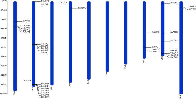
The genes were found asymmetrically distributed both across and within the 10 C. sativa chromosomes (Fig 1) with the largest concentration of genes found on Chromosome 1, 2, and 9. Over 60% of the LOX isoforms were found on Chromosome 2, and with the exception of two, were in large gene clusters. CsLOX1, 2, 5, 6, and 7 were located in a 327 kb region in the center of Chromosome 2, while CsLOX15, 16, 17, 18, 19, 20, and 21 were near the outer arm in a 152 kb region (S1 Fig). A smaller 74 kb region on Chromosome 9 contained the cluster of CsLOX8, 10, and 11 towards the end of a chromosomal arm. CsLOX9 and 12 were relatively close together on Chromosome 2, while Chromosomes 1, 3, and 4 contained single LOX isoforms, CsLOX14, 4, and 13, respectively.
Fig 1. Distribution of the LOX, AOS, HPL, AOC, and OPR gene families across the C. sativa genome.
Ruler depicts chromosome length.
Similar to the LOX genes, members of the CYP74, AOC, and OPR gene families were found in gene clusters at nearly the same proportions. On Chromosome 8, CsAOS2 and 5 were within 57 kb from each other (S2 Fig). The other CYP74 genes, CsAOS1, CsHPL, CsAOS1, and CsAOS4 were on Chromosomes 1, 8, and 9, respectively. AOC genes were found exclusively on Chromosome X with CsAOC1 and 2 only 10 kb apart (S2 Fig). Strikingly, with the exception of CsOPR5 located on Chromosome 9, the entire OPR gene family was confined to a 113 kb segment on Chromosome 1.
The C. sativa LOX gene family contains 21 members
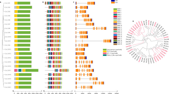
To verify that candidate LOX gene models encode functional isoforms, their peptide sequences were examined for the presence of the lipid-associated PLAT and catalytic LOX domains archetypical of LOX proteins [27]. Amino acids were examined through the NCBI Conserved Domain Database function and all 21 gene models encoded peptides that displayed both domains, providing support that C. sativa possesses 21, bona fide, LOX isoforms (Fig 2A). Unexpectedly, the analysis revealed that CsLOX17 also contained a RVT2 and a RNAse H-like domain which may indicate the insertion of a retrotransposon in its upstream region [86, 87].
Fig 2. Phylogenetic, genetic, motif, and domain analysis of the CsLOX gene family.
(A) Cladogram of peptide sequences and conserved domains. (B) Distribution of conserved peptide sequence motifs. Colors are described in legend; x-axis represents length of peptides in amino acids. (C) Diagram of genetic structure. Blue bars, orange bars, and gray lines represent untranslated regions, exons, and introns, respectively; x-axis represents length of gene in nucleotides. (D) Polar cladogram depicting evolutionary relationship with gene families of selected species. Node labels show confidence values from 1000 bootstrap replications.
Analysis of conserved motifs showed that the majority of the LOX isoforms contain similar composition and distribution of amino acid sequence patterns (Fig 2B). Nearly all isoforms displayed the same 13 motif arrangement, typical of LOX proteins, on the N-terminus, a highly conserved region which requires stringent composition for enzymatic activity. The exception was CsLOX12 which was absent of motif 13. Aside from CsLOX1 and 14, the arrangement of the first 5 motifs on the C-terminus was broadly consistent across all the proteins. However, several isoforms possessed regions where no motifs were detected. In LOX proteins, these regions typically contain highly divergent, transient peptide sequences that direct subcellular localization [88]. In support of this notion, their amino acid sequences were tested against four subcellular prediction algorithms, DeepLOC, LOCALIZER, mSUBP, and TargetP (Table 1). A consensus of plastid localization was reached for only CsLOX13 and CsLOX18, along with an agreement of the majority of the software localizing CsLOX2, CsLOX14, CsLOX15, CsLOX17, CsLOX18, CsLOX19, CsLOX20, and CsLOX21 to this organelle. CsLOX1 and CsLOX3-11 were predicted to be found in the cytoplasm. Interestingly, CsLOX12 was predicted to be associated with the mitochondria.
Motifs 17 and 20 located within the LOX domain showed the largest variability across the LOX isoforms. The former was absent from CsLOX3, 13, and 14, the latter was missing from CsLOX19 and 20, and both were missing from CsLOX12, 15, 16, 17, and 18, prompting the speculation of divergent catalytic activity among even closely related members [89].
The C. sativa LOX gene models ranged in size from around 4 to 16 kb and over 75% possessed eight introns (Fig 2C). A recurrent observation in the genetic structures of some LOX family members was the presence of a large first or second intron, e.g., CsLOX1, 6, 7, 8, 11, and 20 comprising of about 50 to 60% of the entire gene. No data is yet available for alternative splicing or transcript variants of these genes to provide insight into the role of these features.
Plant LOXs are typically grouped as 9- or 13-LOXs according to their major enzymatic product and isoforms with similar activity display higher sequence similarity and evolutionary relationships [27]. To designate the classification of C. sativa LOXs, their peptide sequences were compared to dicot and monocot plant species with well-characterized LOX protein families, namely, A. thaliana, C. sativa, S. lycopersicum, and Zea mays (Fig 2D). CsLOX1, 2, and 3 grouped with the Arabidopsis 9-LOXs, AtLOX1 and 5, respectively. Remarkably, eight Cannabis LOXs peptides, CsLOX4-11, formed a Cannabis specific clade nested within monocot and dicot 9-LOXs. CsLOX12 was difficult to place within the tree, a result which was likely related to the absence of the three variable peptide motifs in its catalytic domain (Fix. 2B). CsLOX13 and 14 clustered with clades containing dicot 13-LOXs. Interestingly, seven Cannabis LOX peptides, CsLOX15-21 grouped with the 13-LOX clade, and several of which may represent Cannabis-specific 13-LOXs. Thus, C. sativa possess eleven 9-LOXs (CsLOX1–11), nine 13-LOXs (CsLOX13–21), and one that remains to be empirically determined (CsLOX12).
The C. sativa CYP74 gene family contains six members
AOS and HPL belong to the atypical CYP74 clade of the P450 family involved in hydroperoxyl fatty acid rearrangement or dismutation [90]. Amino acid sequence analysis found a single CYP74 domain in all six Cannabis candidate CYP74s and four distinct patterns of conserved sequence motifs (Fig 3A and 3B). The pattern variations were most pronounced towards the N-termini where each possessed a distinctive starting motif. The N-terminus of CYP74s typically possess transient peptide signals involved in directing their subcellular localizations towards plastids, and while subsequent analysis prediction supported this notion in CsHPL and CsAOS1, a consensus among the four software prediction tools could not be reached for CsAOS2-5 (Table 1). For gene structure analysis, CsHPL was notable for the presence of two introns (Fig 3C), an unusual feature of CYP74 family members, one of which comprised the majority of its gene model sequence.
Fig 3. Phylogenetic, genetic, motif, and domain analysis of the C. sativa CYP74 gene family.
(A) Cladogram of peptide sequences and conserved domains. (B) Distribution of conserved peptide sequence motifs. Colors are described in legend; x-axis represents length of peptides in amino acids. (C) Diagram of genetic structure. Blue bars, orange bars, and gray lines represent untranslated regions, exons, and introns, respectively; x-axis represents length of gene in nucleotides. (D) Polar cladogram depicting evolutionary relationship with gene families of selected species. Node labels show confidence values from 1000 bootstrap replications.
While many CYP74 isoforms display multifunctionality [91], catalyzing varying proportions of AOS, epoxy alcohol synthase (EAS), divinyl ether synthase (DES), and HPL products, the CYP74A and CYP74B clade members show predominantly AOS and HPL activities, respectively. To understand the potential function of C. sativa CYP74 family members, a phylogenetic analysis assessed their relationship within the relatively well-characterized CYP74 subclades from diverse plant species (Fig 3D). CsAOS1 and CsHPL were grouped within the CYP74A and CYP74B clades, respectively. However, the placement of CsAOS2-4 proved challenging. Nonetheless, CsAOS2 and 3 and CsAOS4 and 5 grouped as pairs roughly within CYP74C.
Taken together, this analysis suggests that C. sativa contains at least two dedicated CYP74 enzymes that, given their phylogenies and subcellular localization, are specialized for 13-AOS and HPL activity, thus capable of providing substrate for JA or GLV biosynthesis. Cannabis also contains four CYP74 isoforms with unassigned activities.
The C. sativa AOC gene family contains three members
AOC provides steric hindrance for the stereospecific cyclization of allene oxide to 12-OPDA, the parent jasmonate species. All three CsAOC isoforms contain an AOC domain and similar amino acid sequence patterns on the last six motifs of their C-termini (Fig 4A and 4B). CsAOC2 and CsAOC3 displayed substantial variability in predicted motifs at their N-termini, likely corresponding to putative plastid transient peptide sequences (Table 1). CsAOC1 was predicted to localize outside of the plastids. The intron-exon distribution of this gene family differed mildly with CsAOC1 and 2 having three exons while CsAOC3 contained one (Fig 4C). To understand the evolutionary relationship of the C. sativa AOC gene family, their peptide sequences were analyzed phylogenetically with AOC members from other plant species [92]. CsAOC1 and 2 clustered close to each other within a dicot specific clade and CsAOC3 grouped into a separate dicot-specific clade that contained all Arabidopsis AOCs (Fig 4D).
Fig 4. Phylogenetic, genetic, motif, and domain analysis of the CsAOC gene family.
(A) Cladogram of peptide sequences and conserved domains. (B) Distribution of conserved peptide sequence motifs. Colors are described in legend; x-axis represents length of peptides in amino acids. (C) Diagram of genetic structure. Blue bars, orange bars, and gray lines represent untranslated regions, exons, and introns, respectively; x-axis represents length of gene in nucleotides. (D) Polar cladogram depicting evolutionary relationship with gene families of selected species. Node labels show confidence values from 1000 bootstrap replications.
The C. sativa OPR gene family contains five members
All five identified C. sativa OPR candidates were found to contain the conserved Old Yellow Enzyme-like domain necessary for their activity (Fig 5A) [93]. CsOPR1-4 had nearly identical patterns of amino acid sequence motifs and were largely similar to the motif arrangement of CsOPR5, however, the latter isoform lacked four conserved motifs (Fig 5B). In regard to their genetic structure, a substantial first intron was identified in CsOPR3, resulting in a gene model roughly three times larger than the other isoforms (Fig 5C). It is important to note that the current gene model of CsOPR3 appears chimeric with CsOPR2 in the current C. sativa representative genome (S2 Fig).
Fig 5. Phylogenetic, genetic, motif, and domain analysis of the CsOPR gene family.
(A) Cladogram of peptide sequences and conserved domains. (B) Distribution of conserved peptide sequence motifs. Colors are described in legend; x-axis represents length of peptides in amino acids. (C) Diagram of genetic structure. Blue bars, orange bars, and gray lines represent untranslated regions, exons, and introns, respectively; x-axis represents length of gene in nucleotides. (D) Polar cladogram depicting evolutionary relationship with gene families of selected species. Node labels show confidence values from 1000 bootstrap replications.
To investigate the evolutionary relationship between the C. sativa OPR gene family members, the amino acid sequences were compared to OPR family members from dicots and monocots. CsOPR1-4 formed a phylogenetic clade with the JA-producing AtOPR3. CsOPR5, however, was grouped with Type-II OPRs (Fig 5D). Taken together, C. sativa contains four Type-I OPR isoforms that are likely involved in classical JA biosynthesis.
Some LOX, CYP74, and AOC members are syntenic across Cannabis, Arabidopsis, and tomato
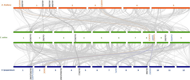
To infer the ancestry of the C. sativa oxylipin pathway, a genetic collinearity analysis was performed to compare physical co-localization of oxylipin biosynthetic genes in the genomes of C. sativa against two dicot genomes with well characterized oxylipin biosynthetic pathways, A. thaliana, and S. lycopersicum (tomato) (Fig 6). Remarkably, three LOX genes displayed collinearity across all three species. Of the 9-LOXs, CsLOX2 was in a syntenic region with AtLOX1 and SlLOX5 (also known as TOMLOXE). Two 13-LOXs display collinearity, CsLOX14 with both AtLOX3 and AtLOX4 and with SlLOX4/ LOXD, while CsLOX13 was collinear with AtLOX6 and SlLOX11. Two C. sativa CYP74 genes were in syntenic regions with tomato genes. CsAOS2 matched with SlDES (also known as LeDES) and CsHPL matched with both SlHPL and SlAOS3. Two CsAOC genes were collinear with either plant species although with dissimilarities. CsAOC2 was in a syntenic gene block with SlAOC and CsAOC3 matched with both AtAOC1 and AtAOC4.
Fig 6. Oxylipin biosynthetic gene collinearity between A. thaliana, C. sativa, and S. lycopersicum.
The grey lines represent collinear genetic blocks between the genomes of A. thaliana and C. sativa and between C. sativa and S. lycospersicum. Bars represent chromosomes and the black, blue, and orange lines represent the collinear pairs of LOX, CYP74, and AOC gene families, respectively.
Promoters of C. sativa oxylipin biosynthetic genes carry tissue-specific, conditional, and phytohormone-inducible cis-acting regulatory elements (CAREs)
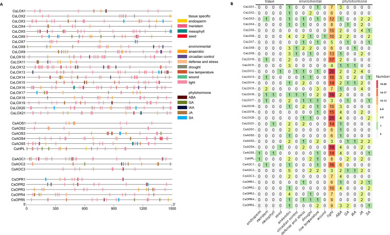
To elucidate the involvement of Cannabis oxylipin genes in diverse developmental and stress responses, an in silico promoter analysis was conducted to identify cis-regulatory elements involved in plant tissue-specific expression (endosperm, meristem, mesophyll, seed), adaptation to environmental conditions (anaerobic, circadian rhythm, drought, light, and low temperature), and phytohormone signaling, namely, abscisic acid (ABA), gibberellic acid (GA), indole-3-acetic acid (IAA), JA, and salicylic acid (SA).
All promoters varied considerably in the content, arrangement, and position of their CAREs (Fig 7 and S1 Table). Overall, the oxylipin biosynthetic gene promoters possessed CARE motifs with lengths of 8 to 32 nucleotides. For the LOX gene family, the promoter of CsLOX12 gene was found to contain the most regulatory elements with 32 and followed by CsLOX16 and 21, each with 28, while CsLOX2, 7, 11, and 17 displayed the least, with around 10 each. The Cannabis-specific 9-LOX group (CsLOX4-11) displayed the most motifs followed by the amplified GLV-producing 13-LOXs (CsLOX15-21). With the exception of CsLOX5, all contained several regulatory elements involved in conditional responses, though few contained elements involved in tissue expression. CAREs involved in light responses dominated most promoters with all genes having at least three such motifs. Interestingly, CsLOX12 light-responsive motifs constituted over 60% of all its identified motifs. Promoters associated with the presence of phytohormone inducible cis-regulatory elements varies considerably, spanning from 0 to 8 regulatory elements. With the exception of CsLOX2, 4, 7, 8, and 17, all had at least one CARE related to ABA responses. Incidentally, CsLOX2, 4, 7, 8, and 17 were also among the few members that showed motifs involved in GA responses, suggesting a role mediated by ABA-GA antagonism [94]. SA-responsive motifs were limited and predominately associated with the 9-LOXs.
Fig 7. Cis-acting regulatory element distribution across C. sativa oxylipin biosynthetic gene promoters.
(A) Physical distribution of motifs throughout a 1.5 kb region upstream of corresponding gene model. Gene symbols are presented on the left with grey line representing promoter regions with motif positions. Legend describes motifs associate with tissue-specific expression, environmental responses, and phytohormone inducibility. (B) Summary of cis-acting regulatory motif content. Heatmap depicts number of motifs identified in gene promoter region. The y-axis depicts C. sativa oxylipin biosynthetic genes and x-axis depicts associated condition for motif. Cell values are number of motifs identified and are colored according to legend.
Of the CYP74 gene family, the largest number of CAREs were identified on promoters of CsAOS4 and 5, followed by CsHPL (Fig 7). Relatively few were found on the putative JA-producing CsAOS1, namely those associated with anaerobic conditions, light, and ABA signaling responses. Unlike the pattern observed for LOXs, the disparity between light-related motifs and other CAREs was not as dramatic in the CYP74 family, with the exception of the CsAOS4 and 5 pair. Nearly all CYP74s had ABA-responsive motifs, with the exception of CsAOS3 and CsHPL which were also the only ones to possess GA-related motifs. CsAOS3, 4, and CsHPL each had two JA-related motifs, while CsAOS1 had none; this suggests a limited contribution of positive feedforward control of JA biosynthesis.
Analysis of the AOC gene family found only a single tissue-type related to CAREs across the promoters of all members: CsAOC1 for meristem expression. While non-light conditional-response motifs were the most numerous in CsAOC2 compared to the other two isomers, the opposite was seen for light-responsive motifs, where only three were found in CsAOC2. This was a sharp contrast with CsAOC1, which showed more than twice the number of these CAREs compared to its paralogs (Fig 7). No AOC was found to have SA-related, and only CsAOC2 had JA-related motifs, while all had ABA-related motifs.
Promoters of the OPR gene family contained similar patterns of CAREs compared with the other gene families, albeit with reduced CARE content. No tissue-related CARE was detected for any OPR member. However, all other members possessed motifs for anaerobic responses, with the exception of CsOPR1 (Fig 7). ABA motifs were found in CsOPR1, CsOPR2, and CsOPR5. CsOPR5 was also the only member to show GA motifs, and JA motifs were found in CsOPR1, 3, and 4. The notable difference in CARE patterns found within the JA-producing OPRs (CsOPR1-4), suggests that the 12-OPDA to JA balance is maintained in Cannabis via transcriptional control of distinct OPR members responses to the plant’s environment and during phytohormone signaling [95].
C. sativa oxylipin biosynthetic genes show tissue-, developmental-, and cultivar-specific expression
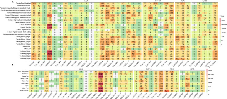
Promoter analysis suggested that expression of C. sativa oxylipin biosynthetic genes would display distinct patterns. To understand the transcriptional profile of oxylipin biosynthetic genes and to elucidate their role in Cannabis development and physiology, the gene expression of 23 tissues from a C. sativa transcriptome atlas [72] were examined (S2 Table). Overall, the major contributor associated with levels of expression appeared to be the identity of the specific gene family member, i.e., patterns of expression levels were generally consistent across the tissues examined (Fig 8A). Moreover, several members in all gene families had few if any detected transcripts across all tissues, suggesting a limited role for those isoforms under basal conditions or in the tissues profiled.
Fig 8.
Heatmaps showing expression of oxylipin biosynthetic genes from the C. sativa gene expression atlas (A) or female trichomes of diverse marijuana lines (B). Cells values are rounded TPM values and are colored according to the legend.
Of the 9-LOXs, CsLOX1, 2, 4, 7 and 8 showed pronounced levels of expression across most tissues. CsLOX4 is particularly notable as the highest expressed oxylipin biosynthetic gene analyzed, by one order of magnitude, in root and trichome tissues (Fig 8A), highlighting the probable importance of this isoform grouping within the Cannabis-specific 9-LOX clade (Fig 2). Curiously, CsLOX9 and 10 had minimal levels of expression in all but root tissues, where their levels were 20-fold higher compared to other tissues. With respect to the 13-LOXs, with the exception of CsLOX13 and 18, all had elevated expression in reproductive-related tissue. However, under these basal conditions, only CsLOX14 showed modest expression in roots. Remarkably, despite possessing the greatest number of identified CAREs (Fig 7B), nearly no expression was detected for CsLOX12 (Fig 8A) which implies a non-basal role for this gene.
In regards to the CYP74-related genes, the likely non-JA producing CsAOS2 displayed the most abundant expression levels, predominantly in early stages of flower and trichome development (Fig 8A, S3 Fig). The putative JA-producing, CsAOS1, also showed its greatest levels of expression in the female flowers, while minimally expressed in other tissues. While CsAOS5 showed modest levels of expression across tissues, transcripts of its closest paralogue CsAOS4 were only detected at low levels in few tissues. For the GLV- and cannabinoid-producing CsHPL, expression remained consistent across tissues, disregarding low levels in root tissues (Fig 8A). Interestingly, CsHPL expression remained relatively consistent across all stages of flower and trichome development (S3 Fig).
In the majority of tissues, CsAOC2 showed the most uniform levels of expression from this gene family (Fig 8), suggesting this member is the prominent isoform involved in JA biosynthesis under basal conditions. CsAOC1 and CsAOC3 showed moderate to modest levels of expression across most tissue types with several exceptions, prompting the idea that these members participate in inducible processes.
Only CsOPR2 and 5, displayed mentionable levels of expression. CsOPR2 transcript levels were an order of magnitude higher than the other Type II paralogs, suggesting this is the major JA-producing isoform under basal conditions (Fig 8). It is also notable that the majority of its expression was in tissues related to flower structures. The sole Type I OPR, CsOPR5 displayed even greater levels of expression in flower tissues, especially developing trichomes, raising the possibility for the involvement of CsOPR5 in reduction or detoxification of metabolites during trichome development.
To understand the variation in gene expression across Cannabis diversity, transcriptomes of trichomes from nine cultivars of mixed ancestry [73] were profiled for their expression of oxylipin biosynthetic genes (S3 Table). With the exception of expression of CsLOX15, of which transcript levels showed nearly a bimodal distribution across the cultivar tested, i.e., Black Berry Kush, Cherry Chem, Mamma Thai, and Terple possessed half of the expression compared to the five other lines (Fig 8B). Few differences were observed in the other oxylipin biosynthetic gene expression. In particular, decreased expression was seen in CsLOX14, CsLOX16, and CsOPR2 from Cherry Cham, Terple, and Black Berry Kush, respectively, relative to the other Cannabis lines. On the other hand, Mama Thai and Black Berry Kush showed elevated levels for transcripts of CsLOX4 and CsAOC1, respectively, and trichomes of White Cookies had increased expression of both CsAOS3 and CsAOC1. Interestingly, in sharp contrast to expression levels seen in the transcriptome atlas, transcripts of CsLOX15 dominated in the trichomes of these cultivars followed by those of CsLOX4. In agreement with the oxylipin-derived hexanoic acid hypothesis [96], the expression of CsHPL levels were also two- to 10-fold higher compared to the other CYP74s, likely owed to the selection of these cultivars for high cannabinoid production.
Cannabis oxylipin biosynthetic genes are found in gene networks associated with stress responses, growth, and development
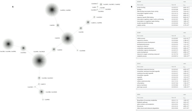
To obtain a more detailed picture of the regulation of the Cannabis oxylipin biosynthetic genes, an iterative weighted gene co-expression network analysis was performed, using multi-tissue expression data from the Cannabis transcriptome atlas [72]. This analysis revealed that 11 oxylipin biosynthesis genes were found clustering in seven multi-gene modules (Fig 9A), while the rest did not exhibit clustering with other genes, using the data available to us. The four largest modules contained between 1130–1852 genes each, in total containing a regulatory network up to 30% of the C. sativa gene models. One of these larger modules contained CsAOS5, while the other three modules contained multiple oxylipin biosynthetic genes: CsLOX3 and 10, CsOPR3 and 4, and CsLOX5, 9 and CsOPR1. The 15 smallest modules had under 200 genes each and together accounted for only 5% of the C. sativa transcriptome.
Fig 9. Weighted co-expression genetic network derived from the C. sativa transcriptome expression atlas.
(A) Clusters represent modules of highly connected genes containing at least one oxylipin biosynthetic gene. (B) Functional enrichment analysis of gene modules containing oxylipin biosynthetic genes: Gene Ontologies for Molecular Functions (MF), Biological Processes (BP), and Cellular Components (CC) and KEGG.
We also performed functional enrichment analysis of the oxylipin gene-associated co-expression network for Gene Ontology and KEGG identified terms related to molecular functions, biological process, and cellular component orthologs (Fig 9B). Among the top 10 terms for molecular functions, 60% were related to nucleic acid binding and transcription. Of the biological processes, 80% were related to development or physiological responses. For cellular components, 40% and 30% described organelle- and membrane-related terms, respectively. KEGG terms described secondary metabolism, particularly α-linolenic acid metabolism and branch-chain amino acid degradation. As expected, individual modules were enriched for differential functional terms (S4 Table). Taken together, these results suggest similar members of oxylipin biosynthesis pathways may be regulated in specialized manner, but in general, certain members may be part of transcriptome-wide regulatory networks. Further, focused expression, biochemical, and proteomic experiments will need to be performed to assess the importance of these patterns.
Discussion
Enthusiasm is growing to understand the physiological and ecological functions of plant oxylipins [90]. JA has long served [97] as the model oxylipin, and over the last fifty years, many aspects of its biosynthesis, metabolism, signaling, and activities have started to be defined [98–101]. However, this knowledge has been generated predominantly in the model plant species, Arabidopsis, and investigations focusing on oxylipin biology in crop species have been launched only in recent years. Here, we cataloged the major oxylipin biosynthetic genes families of the agro-economically important crop species, C. sativa, described their expression patterns, and delineated connections between their putative physiological functions.
LOX gene family
This study adds Cannabaceae to the collection of plant families with described LOX gene families, a group which so far includes Araceae: duckweed [102]; Actinidiaceae: kiwi [103]; Brassicaceae: Arabidopsis thaliana [104–106], radish [107], and turnip [108]; Caricaceae: papaya [106]; Cucurbitaceae: cucumber [109, 110], melon [111], and watermelon [112]; Fabaceae: Medicago truncatula, peanut [113], and soybean [114]; Malvaceae: cotton [115] Muscaceae: banana [116]; Poaceae: maize [117], rice [105], Setaria italica [118], sorghum [119]; Polygonaceae: buckwheat [120]; Rhamnaceae: jujube [121]; Rosaceae: apple [122], peach [123], pear [124]; Salicaceae: poplar [125]; Solanaceae: pepper [126] and tomato [127, 128]; Theaceae: Camellia sinensis [129]; and Vitaceae: grape [130].
Of the 21 C. sativa LOX isoforms identified (Fig 1), 11 are likely responsible for the production of 9-oxylipins, nine are producers of 13-oxylipins, and one member (CsLOX12) remains to be empirically determined. Of the hallmark 9-LOXs AtLOX1 and AtLOX5, two and one C. sativa LOX members grouped with each respectively, while eight formed their own clade. Within 9-LOX phylogenies, monocots typically display their own grouping [102, 116, 118], while the divergence of 9-LOXs in dicots is less common; yet still observed in Cucurbitaceae [111], Salicaceae [125], and Vitaceae [130]. Interestingly, seven members were identified in nearly one large tandem gene array (S1 Fig), which fell within the clade containing both monocot and dicot isoforms required for GLV biosynthesis [45, 131]. Similar substantial gene amplification of this LOX clade has been found to various degrees, in Cucurbitaceae [109–112], Salicaceae [125], and Vitaceae [130]. As AtLOX2 was not found to be syntenic across the Rosids [106], the gene duplications likely occurred independently in ancestors following the divergence of Rosales, Cucurbitales, Malpighiales, and Vitales.
Proteomic analysis [132] has verified that several lipoxygenases are found with spatial specificity in Cannabis flowers and components of their glandular trichomes. Of particular interest was the presence of isoforms from the tandem duplicated 13-LOXs, namely, CsLOX15, 16 and 17 within the trichome head. It is tempting to speculate that these gene duplications may serve to maintain the pool of hexanoic acid substrate available for cannabinoid biosynthesis. In Cannabis, tandem gene arrays have previously been implicated in regulating lipid biosynthesis, particularly the ratios of volatile terpenes that subsequently determines the scent of specific cultivars [133]. This amplification of the 13-LOX paralogues in a tandem gene array may be due to the origin of the representative C. sativa genome [57] from CBDRx, a high CBD producing line. The ubiquity of tandem gene arrays among other Cannabis species and cultivars remains to be examined, especially in regards to the agronomic characteristics that drive the selection of economically important cultivars. Interestingly, the 9-LOXs, CsLOX4 and 7 were also found within the flower and trichomes where they are likely to contribute to the 9-oxylipin volatiles of Cannabis [134].
CYP74 gene family
Despite its importance in oxylipin metabolism, few studies have examined CYP74 gene families from a genome-wide perspective [135–137]. In C. sativa, six CYP74 isoforms were identified Specifically CsAOS1 and CsHPL, were distinctly found within the JA- and GLV- producing CYP74 clades, respectively (Fig 3D). Both contained a transient peptide signal sequence predicting localization in plastids (Table 1), although the unique motifs found at their N-terminus suggest an association with different sub-plastid locations. This is similar to tomato where LeAOS was found with the inner chloroplast envelope while LeHPL was targeted to the outer plastid envelope [138]. Four isoforms were found with the CYP74C subfamily, a more irregular group of enzymes with members possessing varying levels of AOS, EAS, and/or HPL activity [139]. It is interesting to note that CsAOS2 was syntenic to the DES of tomato (Fig 6), however, a comprehensive survey and validation of Cannabis oxylipins remains to be performed to understand if this species indeed produces divinyl ethers. Ultimately, these isoforms will require biochemical characterization, as even a single amino acid change can result in a new function or protein activity [140, 141]. Four CYP74 peptides (CsAOS1,2,5 and CsHPL) have been detected in association with Cannabis flowers, and of these, CsHPL was the only isoform found within trichome heads and stalks [132].
AOC gene family
AOC activity yields the first JA and is the last enzymatic step of JA biosynthesis performed in the chloroplast [101]. Three members of the AOC gene family were identified in C. sativa, a number consistent with four of Arabidopsis [33], six of soybean [142], and two of maize [26]. Isoforms typically display organ- and tissue-specific expression and function as heterodimers [143]. Only CsAOC2 and CsAOC3 were predicted to localize to plastids (Table 1), supporting the function of these isoforms in JA-biosynthesis. Remarkably, CsAOC3 showed synteny with both Arabidopsis (AtAOC1 and AtAOC4) and tomato (SlAOC) orthologues. In Arabidopsis, heteromers containing AtAOC1 and AtAOC4 display the greatest enzymatic activity [144], suggesting an evolutionary advantage of this conserved genomic region for JA production.
OPR gene family
A larger variability in OPR gene content exists across plant species analyzed so far: three in Arabidopsis [38], five in pea [145], five in watermelon [146], eight in maize [147], 10 in cotton [148], 13 in rice [149], and 48 in wheat [150]. This study identified one Type I OPR and four Type II OPRs in the C. sativa genome. Both subgroups reduce α, β -unsaturated double bonds and are involved in reactive electrophilic species (RES) detoxification [151, 152]. While Type II OPRs are well-characterized for their peroxisomal role in reducing 12-OPDA to OPC8:0 during JA biosynthesis, Type I OPRs are less understood. They have been shown to reduce 4, 6-trinitrotoluene (TNT) [93] and were recently demonstrated to be involved in producing JA through reduction of an endogenous cyclopentenone JA-analog [42]. Tandem duplication has been implicated as playing a major role in the size of OPR gene families across plant species [153] and interestingly, while all Type II OPRs were found together in a gene cluster, no OPR displayed synteny across the species tested, suggesting OPR gene duplication was a relatively recent event in the C. sativa ancestor.
Oxylipins in Cannabis
To date, the interaction between the Cannabis oxylipin and cannabinoid pathways has largely been overlooked. Several investigations have sought to increase cannabinoid content using JA or methyl-JA treatments on Cannabis flowers [154], leaves [155], or cell cultures [156] and have been met with mixed results. Studies that analyze oxylipin levels in Cannabis tissues or products have focused on volatiles from above-ground tissues [157], seeds [158], or seed oil [159, 160] as components of their odor profiles. Recently, it was found that sex impacts the distinct oxylipin species that accumulate in the Cannabis flower. While male flowers have increased accumulation of the 9-LOX-derived, 9-oxo-octadecadienoic acid, female flowers accumulated higher levels of 12-OPDA [161].
Conclusion
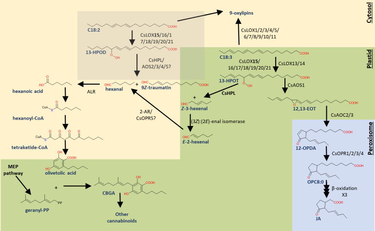
While the LOX pathway is the hypothetical origin of the hexanoic acid moiety used for cannabinoid biosynthesis [12, 19, 24, 25, 96], the precise mechanisms for its production are largely unknown. Here, we provide a comprehensive description of the C. sativa LOX pathway, including the major oxylipin biosynthetic gene families, to establish a working model (Fig 10) for further investigations.
Fig 10. Working model of C. sativa oxylipin biosynthetic enzyme isoforms and pathways involved in cannabinoid, GLV, and JA biosynthesis.
Abbreviations: [enzymes]: allene oxide cyclase (AOC), allene oxide synthase (AOS), 2-alkynal reductase (2-AR), hydroperoxide lyase (HPL), lipoxygenase (LOX), methylerythritol 4-phosphate pathway (MEP), 12-oxo-phytodienoic reductase (OPR); [metabolites]: (9Z, 11E, 13S, 15Z)-12,13-epoxy-9,11,15-octadecatrienoic acid (12,13-EOT), (9Z, 11E, 13S)-13-hydroperoxy-9,11-octadecadienoic acid (13-HPOD), (9Z, 11E, 13S, 15Z)-13-hydroperoxy-9,11,15-octadecatrienoic acid (13-HPOT), cannabigerolic acid (CBGA), linoleic acid (C18:2), α-linolenic acid (C18:3), jasmonic acid (JA), 12-oxo-phytodienoic acid (12-OPDA), 3-oxo-2-(2′[Z]-pentenyl)-cyclopentane-1-octanoic acid (OPC8:0). The gray box indicates uncertainty in subcellular localization.
Within this context, it will become important to understand the co-localization of the substrates with their corresponding enzymes. LOX activity depends on a 1,4-dipentadiene structure found in either C18:2 or C18:3 [27], both of which, in tomato, account for the most abundant fatty acids in its trichomes [162]. However, while HPL-cleavage of 13-hydroperoxydienoic acid would generate a statured C6-aldehyde, the GLV-producing 13-LOXs and HPL are likely localized to the C18:3-rich chloroplasts [163, 164]. Additionally, the GLV-producing AtLOX2 oxygenates C18:3 more efficiently than C18:2 [104], which together may explain why unsaturated GLVs prevail as the major C6 volatile in plants [165].
Enzymatic production of hexanoic acid from the C18:3 HPL-product, Z-3-hexenal is unexplored, but would reasonably require distinct enzymatic reactions. Chemical or enzymatic isomerization by (3Z):(2E)-enal isomerase would generate (2E)-hexenal [46, 55] which could be reduced to hexanal by 2-alkenal reductase [166] or by OPR [167]. The conversation of the fatty aldehyde to fatty acid likely requires an aldehyde dehydrogenase [168], the product of which would be readily diffused across cell membranes [169]. In an alternative scenario, 13-LOX may oxygenate C18:2 directly, however this would presumably require the cytosolic localization of both 13-LOX and HPL. A 13-LOX isoform in melon was shown to have preference for C18:2 and localized to non-chloroplast organelles [170]. This strategy would take advantage of the relatively high C18:2 content in the inflorescences of some Cannabis cultivars [171].
Understanding and applying knowledge of the Cannabis oxylipin biosynthetic pathway is expected to provide novel environmentally friendly approaches towards improving the crop and its desirable consumer traits. Similar technologies in maize and soybeans in development aim at increasing disease resistance [172], drought tolerance [173], and seed flavor [174]. Exploiting the diversity in Cannabis cultivars via oxylipin biosynthetic gene expression and variability in the predominant oxylipin biosynthetic pathway branch across cultivars and species (e.g., C. sativa vs C. indica) should also provide opportunities for manipulating cannabinoid content and other traits through marker-assisted selection or identification of superior alleles.
Supporting information
(TIF)
(TIF)
(TIF)
(XLSX)
(XLSX)
(XLSX)
(XLSX)
Acknowledgments
Thank you to Dr. Michael Kolomiets for helpful discussions and Dr. Michael Osier for the use of computational resources.
Data Availability
All relevant data are within the paper and its Supporting Information files.
Funding Statement
The authors would like to acknowledge the generous support provided by the Thomas H. Gosnell School of Life Sciences (GSoLS) and the College of Science (COS) at the Rochester Institute of Technology (RIT). The funders had no role in study design, data collection and analysis, decision to publish, or preparation of the manuscript.
References
- 1.Bonini SA, Premoli M, Tambaro S, Kumar A, Maccarinelli G, Memo M, et al. Cannabis sativa: A comprehensive ethnopharmacological review of a medicinal plant with a long history. J Ethnopharmacol. 2018;227:300–15. doi: 10.1016/j.jep.2018.09.004 [DOI] [PubMed] [Google Scholar]
- 2.Kovalchuk I, Pellino M, Rigault P, van Velzen R, Ebersbach J, Ashnest JR, et al. The Genomics of Cannabis and Its Close Relatives. Annu Rev Plant Biol. 2020;71:713–39. doi: 10.1146/annurev-arplant-081519-040203 [DOI] [PubMed] [Google Scholar]
- 3.de la Fuente A, Zamberlan F, Ferrán AS, Carrillo F, Tagliazucchi E, Pallavicini C. Relationship among subjective responses, flavor, and chemical composition across more than 800 commercial cannabis varieties. Journal of cannabis research. 2020;2(1):1–18. doi: 10.1186/s42238-020-00028-y [DOI] [PMC free article] [PubMed] [Google Scholar]
- 4.Torkamaneh D, Jones AMP. Cannabis, the multibillion dollar plant that no genebank wanted. Genome. 2021;99(999):1–5. doi: 10.1139/gen-2021-0016 [DOI] [PubMed] [Google Scholar]
- 5.Rehman M, Fahad S, Du G, Cheng X, Yang Y, Tang K, et al. Evaluation of hemp (Cannabis sativa L.) as an industrial crop: a review. Environ Sci Pollut Res Int. 2021;28(38):52832–43. doi: 10.1007/s11356-021-16264-5 [DOI] [PubMed] [Google Scholar]
- 6.Callaway JC. Hempseed as a nutritional resource: An overview. Euphytica. 2004;140(1–2):65–72. [Google Scholar]
- 7.Duque Schumacher AG, Pequito S, Pazour J. Industrial hemp fiber: A sustainable and economical alternative to cotton. Journal of Cleaner Production. 2020;268:122180. [Google Scholar]
- 8.Finnan J, Styles D. Hemp: A more sustainable annual energy crop for climate and energy policy. Energy Policy. 2013;58:152–62. [Google Scholar]
- 9.Citti C, Braghiroli D, Vandelli MA, Cannazza G. Pharmaceutical and biomedical analysis of cannabinoids: A critical review. J Pharm Biomed Anal. 2018;147:565–79. doi: 10.1016/j.jpba.2017.06.003 [DOI] [PubMed] [Google Scholar]
- 10.Keul A, Eisenhauer B. Making the high country: cannabis tourism in Colorado USA. Annals of Leisure Research. 2018;22(2):140–60. [Google Scholar]
- 11.Peng H, Shahidi F. Cannabis and Cannabis Edibles: A Review. J Agric Food Chem. 2021;69(6):1751–74. doi: 10.1021/acs.jafc.0c07472 [DOI] [PubMed] [Google Scholar]
- 12.Gulck T, Moller BL. Phytocannabinoids: Origins and Biosynthesis. Trends Plant Sci. 2020;25(10):985–1004. doi: 10.1016/j.tplants.2020.05.005 [DOI] [PubMed] [Google Scholar]
- 13.Lange BM, Zager JJ. Comprehensive inventory of cannabinoids in Cannabis sativa L.: Can we connect genotype and chemotype? Phytochemistry Reviews. 2021:1–41. [Google Scholar]
- 14.Flores-Sanchez IJ, Verpoorte R. Secondary metabolism in cannabis. Phytochemistry reviews. 2008;7(3):615–39. [Google Scholar]
- 15.Iseger TA, Bossong MG. A systematic review of the antipsychotic properties of cannabidiol in humans. Schizophr Res. 2015;162(1–3):153–61. doi: 10.1016/j.schres.2015.01.033 [DOI] [PubMed] [Google Scholar]
- 16.Mechoulam R, Parker LA. The endocannabinoid system and the brain. Annu Rev Psychol. 2013;64:21–47. doi: 10.1146/annurev-psych-113011-143739 [DOI] [PubMed] [Google Scholar]
- 17.Toth JA, Stack GM, Cala AR, Carlson CH, Wilk RL, Crawford JL, et al. Development and validation of genetic markers for sex and cannabinoid chemotype in Cannabis sativa L. Global Change Biology Bioenergy. 2020;12(3):213–22. [Google Scholar]
- 18.Johnson MS, Wallace JG. Genomic and Chemical Diversity of Commercially Available High-CBD Industrial Hemp Accessions. Front Genet. 2021;12(1160):682475. doi: 10.3389/fgene.2021.682475 [DOI] [PMC free article] [PubMed] [Google Scholar]
- 19.Blatt-Janmaat K, Qu Y. The Biochemistry of Phytocannabinoids and Metabolic Engineering of Their Production in Heterologous Systems. Int J Mol Sci. 2021;22(5):2454. doi: 10.3390/ijms22052454 [DOI] [PMC free article] [PubMed] [Google Scholar]
- 20.Luo X, Reiter MA, d’Espaux L, Wong J, Denby CM, Lechner A, et al. Complete biosynthesis of cannabinoids and their unnatural analogues in yeast. Nature. 2019;567(7746):123–6. doi: 10.1038/s41586-019-0978-9 [DOI] [PubMed] [Google Scholar]
- 21.Tahir MN, Shahbazi F, Rondeau-Gagne S, Trant JF. The biosynthesis of the cannabinoids. J Cannabis Res. 2021;3(1):7. doi: 10.1186/s42238-021-00062-4 [DOI] [PMC free article] [PubMed] [Google Scholar]
- 22.Hanus LO, Meyer SM, Munoz E, Taglialatela-Scafati O, Appendino G. Phytocannabinoids: a unified critical inventory. Nat Prod Rep. 2016;33(12):1357–92. doi: 10.1039/c6np00074f [DOI] [PubMed] [Google Scholar]
- 23.Tan Z, Clomburg JM, Gonzalez R. Synthetic Pathway for the Production of Olivetolic Acid in Escherichia coli. ACS Synth Biol. 2018;7(8):1886–96. doi: 10.1021/acssynbio.8b00075 [DOI] [PubMed] [Google Scholar]
- 24.Livingston SJ, Quilichini TD, Booth JK, Wong DCJ, Rensing KH, Laflamme-Yonkman J, et al. Cannabis glandular trichomes alter morphology and metabolite content during flower maturation. Plant J. 2020;101(1):37–56. doi: 10.1111/tpj.14516 [DOI] [PubMed] [Google Scholar]
- 25.Stout JM, Boubakir Z, Ambrose SJ, Purves RW, Page JE. The hexanoyl‐CoA precursor for cannabinoid biosynthesis is formed by an acyl‐activating enzyme in Cannabis sativa trichomes. The Plant Journal. 2012;71(3):353–65. doi: 10.1111/j.1365-313X.2012.04949.x [DOI] [PubMed] [Google Scholar]
- 26.Borrego EJ, Kolomiets MV. Synthesis and Functions of Jasmonates in Maize. Plants (Basel). 2016;5(4):41. doi: 10.3390/plants5040041 [DOI] [PMC free article] [PubMed] [Google Scholar]
- 27.Feussner I, Wasternack C. The lipoxygenase pathway. Annu Rev Plant Biol. 2002;53(1):275–97. doi: 10.1146/annurev.arplant.53.100301.135248 [DOI] [PubMed] [Google Scholar]
- 28.Yan Y, Christensen S, Isakeit T, Engelberth J, Meeley R, Hayward A, et al. Disruption of OPR7 and OPR8 reveals the versatile functions of jasmonic acid in maize development and defense. Plant Cell. 2012;24(4):1420–36. doi: 10.1105/tpc.111.094151 [DOI] [PMC free article] [PubMed] [Google Scholar]
- 29.Yan Y, Borrego E, Kolomiets MV. Jasmonate biosynthesis, perception and function in plant development and stress responses. Lipid metabolism InTech. 2013:393–442. [Google Scholar]
- 30.Wasternack C. Jasmonates: an update on biosynthesis, signal transduction and action in plant stress response, growth and development. Ann Bot. 2007;100(4):681–97. doi: 10.1093/aob/mcm079 [DOI] [PMC free article] [PubMed] [Google Scholar]
- 31.Laudert D, Pfannschmidt U, Lottspeich F, Hollander-Czytko H, Weiler EW. Cloning, molecular and functional characterization of Arabidopsis thaliana allene oxide synthase (CYP 74), the first enzyme of the octadecanoid pathway to jasmonates. Plant Mol Biol. 1996;31(2):323–35. doi: 10.1007/BF00021793 [DOI] [PubMed] [Google Scholar]
- 32.Park JH, Halitschke R, Kim HB, Baldwin IT, Feldmann KA, Feyereisen R. A knock‐out mutation in allene oxide synthase results in male sterility and defective wound signal transduction in Arabidopsis due to a block in jasmonic acid biosynthesis. The Plant Journal. 2002;31(1):1–12. doi: 10.1046/j.1365-313x.2002.01328.x [DOI] [PubMed] [Google Scholar]
- 33.Stenzel I, Hause B, Miersch O, Kurz T, Maucher H, Weichert H, et al. Jasmonate biosynthesis and the allene oxide cyclase family of Arabidopsis thaliana. Plant Mol Biol. 2003;51(6):895–911. doi: 10.1023/a:1023049319723 [DOI] [PubMed] [Google Scholar]
- 34.Pollmann S, Springer A, Rustgi S, von Wettstein D, Kang C, Reinbothe C, et al. Substrate channeling in oxylipin biosynthesis through a protein complex in the plastid envelope of Arabidopsis thaliana. J Exp Bot. 2019;70(5):1483–95. doi: 10.1093/jxb/erz015 [DOI] [PMC free article] [PubMed] [Google Scholar]
- 35.Park SW, Li W, Viehhauser A, He B, Kim S, Nilsson AK, et al. Cyclophilin 20–3 relays a 12-oxo-phytodienoic acid signal during stress responsive regulation of cellular redox homeostasis. Proc Natl Acad Sci U S A. 2013;110(23):9559–64. doi: 10.1073/pnas.1218872110 [DOI] [PMC free article] [PubMed] [Google Scholar]
- 36.Stintzi A, Weber H, Reymond P, Browse J, Farmer EE. Plant defense in the absence of jasmonic acid: the role of cyclopentenones. Proc Natl Acad Sci U S A. 2001;98(22):12837–42. doi: 10.1073/pnas.211311098 [DOI] [PMC free article] [PubMed] [Google Scholar]
- 37.Wang KD, Borrego EJ, Kenerley CM, Kolomiets MV. Oxylipins Other Than Jasmonic Acid Are Xylem-Resident Signals Regulating Systemic Resistance Induced by Trichoderma virens in Maize. Plant Cell. 2020;32(1):166–85. doi: 10.1105/tpc.19.00487 [DOI] [PMC free article] [PubMed] [Google Scholar]
- 38.Mussig C, Biesgen C, Lisso J, Uwer U, Weiler EW, Altmann T. A novel stress-inducible 12-oxophytodienoate reductase from Arabidopsis thaliana provides a potential link between Brassinosteroid-action and Jasmonic-acid synthesis. Journal of Plant Physiology. 2000;157(2):143–52. [Google Scholar]
- 39.Stintzi A, Browse J. The Arabidopsis male-sterile mutant, opr3, lacks the 12-oxophytodienoic acid reductase required for jasmonate synthesis. Proc Natl Acad Sci U S A. 2000;97(19):10625–30. doi: 10.1073/pnas.190264497 [DOI] [PMC free article] [PubMed] [Google Scholar]
- 40.Vick BA, Zimmerman DC. Biosynthesis of jasmonic Acid by several plant species. Plant Physiol. 1984;75(2):458–61. doi: 10.1104/pp.75.2.458 [DOI] [PMC free article] [PubMed] [Google Scholar]
- 41.Wasternack C, Hause B. A Bypass in Jasmonate Biosynthesis—the OPR3-independent Formation. Trends Plant Sci. 2018;23(4):276–9. doi: 10.1016/j.tplants.2018.02.011 [DOI] [PubMed] [Google Scholar]
- 42.Chini A, Monte I, Zamarreno AM, Hamberg M, Lassueur S, Reymond P, et al. An OPR3-independent pathway uses 4,5-didehydrojasmonate for jasmonate synthesis. Nat Chem Biol. 2018;14(2):171–8. doi: 10.1038/nchembio.2540 [DOI] [PubMed] [Google Scholar]
- 43.Weber H, Vick BA, Farmer EE. Dinor-oxo-phytodienoic acid: a new hexadecanoid signal in the jasmonate family. Proc Natl Acad Sci U S A. 1997;94(19):10473–8. doi: 10.1073/pnas.94.19.10473 [DOI] [PMC free article] [PubMed] [Google Scholar]
- 44.Fonseca S, Chini A, Hamberg M, Adie B, Porzel A, Kramell R, et al. (+)-7-iso-Jasmonoyl-L-isoleucine is the endogenous bioactive jasmonate. Nature chemical biology. 2009;5(5):344–50. doi: 10.1038/nchembio.161 [DOI] [PubMed] [Google Scholar]
- 45.Christensen SA, Nemchenko A, Borrego E, Murray I, Sobhy IS, Bosak L, et al. The maize lipoxygenase, Zm LOX 10, mediates green leaf volatile, jasmonate and herbivore‐induced plant volatile production for defense against insect attack. The Plant Journal. 2013;74(1):59–73. doi: 10.1111/tpj.12101 [DOI] [PubMed] [Google Scholar]
- 46.Ameye M, Allmann S, Verwaeren J, Smagghe G, Haesaert G, Schuurink RC, et al. Green leaf volatile production by plants: a meta-analysis. New Phytol. 2018;220(3):666–83. doi: 10.1111/nph.14671 [DOI] [PubMed] [Google Scholar]
- 47.Sugimoto K, Iijima Y, Takabayashi J, Matsui K. Processing of Airborne Green Leaf Volatiles for Their Glycosylation in the Exposed Plants. Front Plant Sci. 2021;12(2514):721572. doi: 10.3389/fpls.2021.721572 [DOI] [PMC free article] [PubMed] [Google Scholar]
- 48.Bate NJ, Sivasankar S, Moxon C, Riley JM, Thompson JE, Rothstein SJ. Molecular characterization of an Arabidopsis gene encoding hydroperoxide lyase, a cytochrome P-450 that is wound inducible. Plant Physiol. 1998;117(4):1393–400. doi: 10.1104/pp.117.4.1393 [DOI] [PMC free article] [PubMed] [Google Scholar]
- 49.Matsui K, Wilkinson J, Hiatt B, Knauf V, Kajiwara T. Molecular cloning and expression of Arabidopsis fatty acid hydroperoxide lyase. Plant Cell Physiol. 1999;40(5):477–81. doi: 10.1093/oxfordjournals.pcp.a029567 [DOI] [PubMed] [Google Scholar]
- 50.Duan H, Huang MY, Palacio K, Schuler MA. Variations in CYP74B2 (hydroperoxide lyase) gene expression differentially affect hexenal signaling in the Columbia and Landsberg erecta ecotypes of Arabidopsis. Plant Physiol. 2005;139(3):1529–44. doi: 10.1104/pp.105.067249 [DOI] [PMC free article] [PubMed] [Google Scholar]
- 51.Kallenbach M, Gilardoni PA, Allmann S, Baldwin IT, Bonaventure G. C12 derivatives of the hydroperoxide lyase pathway are produced by product recycling through lipoxygenase‐2 in Nicotiana attenuata leaves. New Phytologist. 2011;191(4):1054–68. doi: 10.1111/j.1469-8137.2011.03767.x [DOI] [PubMed] [Google Scholar]
- 52.Nakashima A, von Reuss SH, Tasaka H, Nomura M, Mochizuki S, Iijima Y, et al. Traumatin- and dinortraumatin-containing galactolipids in Arabidopsis: their formation in tissue-disrupted leaves as counterparts of green leaf volatiles. J Biol Chem. 2013;288(36):26078–88. doi: 10.1074/jbc.M113.487959 [DOI] [PMC free article] [PubMed] [Google Scholar]
- 53.Tanaka T, Ikeda A, Shiojiri K, Ozawa R, Shiki K, Nagai-Kunihiro N, et al. Identification of a Hexenal Reductase That Modulates the Composition of Green Leaf Volatiles. Plant Physiol. 2018;178(2):552–64. doi: 10.1104/pp.18.00632 [DOI] [PMC free article] [PubMed] [Google Scholar]
- 54.D’Auria JC, Pichersky E, Schaub A, Hansel A, Gershenzon J. Characterization of a BAHD acyltransferase responsible for producing the green leaf volatile (Z)-3-hexen-1-yl acetate in Arabidopsis thaliana. Plant J. 2007;49(2):194–207. doi: 10.1111/j.1365-313X.2006.02946.x [DOI] [PubMed] [Google Scholar]
- 55.Kunishima M, Yamauchi Y, Mizutani M, Kuse M, Takikawa H, Sugimoto Y. Identification of (Z)-3:(E)-2-Hexenal Isomerases Essential to the Production of the Leaf Aldehyde in Plants. J Biol Chem. 2016;291(27):14023–33. doi: 10.1074/jbc.M116.726687 [DOI] [PMC free article] [PubMed] [Google Scholar]
- 56.Spyropoulou EA, Dekker HL, Steemers L, van Maarseveen JH, de Koster CG, Haring MA, et al. Identification and Characterization of (3Z):(2E)-Hexenal Isomerases from Cucumber. Front Plant Sci. 2017;8:1342. doi: 10.3389/fpls.2017.01342 [DOI] [PMC free article] [PubMed] [Google Scholar]
- 57.Grassa CJ, Weiblen GD, Wenger JP, Dabney C, Poplawski SG, Timothy Motley S, et al. A new Cannabis genome assembly associates elevated cannabidiol (CBD) with hemp introgressed into marijuana. New Phytol. 2021;230(4):1665–79. doi: 10.1111/nph.17243 [DOI] [PMC free article] [PubMed] [Google Scholar]
- 58.Rangwala SH, Kuznetsov A, Ananiev V, Asztalos A, Borodin E, Evgeniev V, et al. Accessing NCBI data using the NCBI Sequence Viewer and Genome Data Viewer (GDV). Genome Res. 2021;31(1):159–69. doi: 10.1101/gr.266932.120 [DOI] [PMC free article] [PubMed] [Google Scholar]
- 59.Lu S, Wang J, Chitsaz F, Derbyshire MK, Geer RC, Gonzales NR, et al. CDD/SPARCLE: the conserved domain database in 2020. Nucleic Acids Res. 2020;48(D1):D265–D8. doi: 10.1093/nar/gkz991 [DOI] [PMC free article] [PubMed] [Google Scholar]
- 60.Marchler-Bauer A, Lu S, Anderson JB, Chitsaz F, Derbyshire MK, DeWeese-Scott C, et al. CDD: a Conserved Domain Database for the functional annotation of proteins. Nucleic Acids Res. 2011;39(Database issue):D225–9. doi: 10.1093/nar/gkq1189 [DOI] [PMC free article] [PubMed] [Google Scholar]
- 61.Bailey TL, Elkan C. Fitting a mixture model by expectation maximization to discover motifs in bipolymers. 1994. [PubMed] [Google Scholar]
- 62.Bailey TL, Johnson J, Grant CE, Noble WS. The MEME Suite. Nucleic Acids Res. 2015;43(W1):W39–49. doi: 10.1093/nar/gkv416 [DOI] [PMC free article] [PubMed] [Google Scholar]
- 63.Almagro Armenteros JJ, Sønderby CK, Sønderby SK, Nielsen H, Winther O. DeepLoc: prediction of protein subcellular localization using deep learning. Bioinformatics. 2017;33(21):3387–95. doi: 10.1093/bioinformatics/btx431 [DOI] [PubMed] [Google Scholar]
- 64.Sperschneider J, Catanzariti AM, DeBoer K, Petre B, Gardiner DM, Singh KB, et al. LOCALIZER: subcellular localization prediction of both plant and effector proteins in the plant cell. Sci Rep. 2017;7(1):44598. doi: 10.1038/srep44598 [DOI] [PMC free article] [PubMed] [Google Scholar]
- 65.Sahu SS, Loaiza CD, Kaundal R. Plant-mSubP: a computational framework for the prediction of single-and multi-target protein subcellular localization using integrated machine-learning approaches. AoB Plants. 2020;12(3):plz068. doi: 10.1093/aobpla/plz068 [DOI] [PMC free article] [PubMed] [Google Scholar]
- 66.Almagro Armenteros JJ, Salvatore M, Emanuelsson O, Winther O, von Heijne G, Elofsson A, et al. Detecting sequence signals in targeting peptides using deep learning. Life Sci Alliance. 2019;2(5). doi: 10.26508/lsa.201900429 [DOI] [PMC free article] [PubMed] [Google Scholar]
- 67.Katoh K, Rozewicki J, Yamada KD. MAFFT online service: multiple sequence alignment, interactive sequence choice and visualization. Brief Bioinform. 2019;20(4):1160–6. doi: 10.1093/bib/bbx108 [DOI] [PMC free article] [PubMed] [Google Scholar]
- 68.Kuraku S, Zmasek CM, Nishimura O, Katoh K. aLeaves facilitates on-demand exploration of metazoan gene family trees on MAFFT sequence alignment server with enhanced interactivity. Nucleic Acids Res. 2013;41(Web Server issue):W22–8. doi: 10.1093/nar/gkt389 [DOI] [PMC free article] [PubMed] [Google Scholar]
- 69.Chen C, Chen H, Zhang Y, Thomas HR, Frank MH, He Y, et al. TBtools: An Integrative Toolkit Developed for Interactive Analyses of Big Biological Data. Mol Plant. 2020;13(8):1194–202. doi: 10.1016/j.molp.2020.06.009 [DOI] [PubMed] [Google Scholar]
- 70.Lescot M, Dehais P, Thijs G, Marchal K, Moreau Y, Van de Peer Y, et al. PlantCARE, a database of plant cis-acting regulatory elements and a portal to tools for in silico analysis of promoter sequences. Nucleic Acids Res. 2002;30(1):325–7. doi: 10.1093/nar/30.1.325 [DOI] [PMC free article] [PubMed] [Google Scholar]
- 71.Wang Y, Tang H, Debarry JD, Tan X, Li J, Wang X, et al. MCScanX: a toolkit for detection and evolutionary analysis of gene synteny and collinearity. Nucleic Acids Res. 2012;40(7):e49. doi: 10.1093/nar/gkr1293 [DOI] [PMC free article] [PubMed] [Google Scholar]
- 72.Braich S, Baillie RC, Jewell LS, Spangenberg GC, Cogan NOI. Generation of a Comprehensive Transcriptome Atlas and Transcriptome Dynamics in Medicinal Cannabis. Sci Rep. 2019;9(1):16583. doi: 10.1038/s41598-019-53023-6 [DOI] [PMC free article] [PubMed] [Google Scholar]
- 73.Zager JJ, Lange I, Srividya N, Smith A, Lange BM. Gene Networks Underlying Cannabinoid and Terpenoid Accumulation in Cannabis. Plant Physiol. 2019;180(4):1877–97. doi: 10.1104/pp.18.01506 [DOI] [PMC free article] [PubMed] [Google Scholar]
- 74.Chen S, Zhou Y, Chen Y, Gu J. fastp: an ultra-fast all-in-one FASTQ preprocessor. Bioinformatics. 2018;34(17):i884–i90. doi: 10.1093/bioinformatics/bty560 [DOI] [PMC free article] [PubMed] [Google Scholar]
- 75.Patro R, Duggal G, Love MI, Irizarry RA, Kingsford C. Salmon provides fast and bias-aware quantification of transcript expression. Nat Methods. 2017;14(4):417–9. doi: 10.1038/nmeth.4197 [DOI] [PMC free article] [PubMed] [Google Scholar]
- 76.Langfelder P, Horvath S. WGCNA: an R package for weighted correlation network analysis. BMC Bioinformatics. 2008;9(1):559. [DOI] [PMC free article] [PubMed] [Google Scholar]
- 77.Greenfest-Allen E, Cartailler J-P, Magnuson MA, Stoeckert CJ. iterativeWGCNA: iterative refinement to improve module detection from WGCNA co-expression networks. bioRxiv. 2017:234062. [Google Scholar]
- 78.Shannon P, Markiel A, Ozier O, Baliga NS, Wang JT, Ramage D, et al. Cytoscape: a software environment for integrated models of biomolecular interaction networks. Genome Res. 2003;13(11):2498–504. doi: 10.1101/gr.1239303 [DOI] [PMC free article] [PubMed] [Google Scholar]
- 79.Raudvere U, Kolberg L, Kuzmin I, Arak T, Adler P, Peterson H, et al. g:Profiler: a web server for functional enrichment analysis and conversions of gene lists (2019 update). Nucleic Acids Res. 2019;47(W1):W191–W8. doi: 10.1093/nar/gkz369 [DOI] [PMC free article] [PubMed] [Google Scholar]
- 80.Van Bel M, Silvestri F, Weitz EM, Kreft L, Botzki A, Coppens F, et al. PLAZA 5.0: extending the scope and power of comparative and functional genomics in plants. Nucleic Acids Res. 2022;50(D1):D1468–D74. doi: 10.1093/nar/gkab1024 [DOI] [PMC free article] [PubMed] [Google Scholar]
- 81.Kanehisa M. Toward understanding the origin and evolution of cellular organisms. Protein Sci. 2019;28(11):1947–51. doi: 10.1002/pro.3715 [DOI] [PMC free article] [PubMed] [Google Scholar]
- 82.Kanehisa M, Furumichi M, Sato Y, Ishiguro-Watanabe M, Tanabe M. KEGG: integrating viruses and cellular organisms. Nucleic Acids Res. 2021;49(D1):D545–D51. doi: 10.1093/nar/gkaa970 [DOI] [PMC free article] [PubMed] [Google Scholar]
- 83.Kanehisa M, Goto S. KEGG: kyoto encyclopedia of genes and genomes. Nucleic Acids Res. 2000;28(1):27–30. doi: 10.1093/nar/28.1.27 [DOI] [PMC free article] [PubMed] [Google Scholar]
- 84.Ashburner M, Ball CA, Blake JA, Botstein D, Butler H, Cherry JM, et al. Gene ontology: tool for the unification of biology. The Gene Ontology Consortium. Nat Genet. 2000;25(1):25–9. doi: 10.1038/75556 [DOI] [PMC free article] [PubMed] [Google Scholar]
- 85.Gene Ontology C. The Gene Ontology resource: enriching a GOld mine. Nucleic Acids Res. 2021;49(D1):D325–D34. doi: 10.1093/nar/gkaa1113 [DOI] [PMC free article] [PubMed] [Google Scholar]
- 86.Majorek KA, Dunin-Horkawicz S, Steczkiewicz K, Muszewska A, Nowotny M, Ginalski K, et al. The RNase H-like superfamily: new members, comparative structural analysis and evolutionary classification. Nucleic Acids Res. 2014;42(7):4160–79. doi: 10.1093/nar/gkt1414 [DOI] [PMC free article] [PubMed] [Google Scholar]
- 87.Xiong Y, Eickbush TH. Origin and evolution of retroelements based upon their reverse transcriptase sequences. EMBO J. 1990;9(10):3353–62. doi: 10.1002/j.1460-2075.1990.tb07536.x [DOI] [PMC free article] [PubMed] [Google Scholar]
- 88.Farmaki T, Sanmartin M, Jimenez P, Paneque M, Sanz C, Vancanneyt G, et al. Differential distribution of the lipoxygenase pathway enzymes within potato chloroplasts. J Exp Bot. 2007;58(3):555–68. doi: 10.1093/jxb/erl230 [DOI] [PubMed] [Google Scholar]
- 89.Mikulska-Ruminska K, Shrivastava I, Krieger J, Zhang S, Li H, Bayir H, et al. Characterization of Differential Dynamics, Specificity, and Allostery of Lipoxygenase Family Members. J Chem Inf Model. 2019;59(5):2496–508. doi: 10.1021/acs.jcim.9b00006 [DOI] [PMC free article] [PubMed] [Google Scholar]
- 90.Wasternack C, Feussner I. The Oxylipin Pathways: Biochemistry and Function. Annu Rev Plant Biol. 2018;69:363–86. [DOI] [PubMed] [Google Scholar]
- 91.Toporkova YY, Smirnova EO, Iljina TM, Mukhtarova LS, Gorina SS, Grechkin AN. The CYP74B and CYP74D divinyl ether synthases possess a side hydroperoxide lyase and epoxyalcohol synthase activities that are enhanced by the site-directed mutagenesis. Phytochemistry. 2020;179:112512. doi: 10.1016/j.phytochem.2020.112512 [DOI] [PubMed] [Google Scholar]
- 92.Li JW, Guang YL, Yang YX, Zhou Y. Identification and Expression Analysis of Two allene oxide cyclase (AOC) Genes in Watermelon. Agriculture-Basel. 2019;9(10):225. [Google Scholar]
- 93.Beynon ER, Symons ZC, Jackson RG, Lorenz A, Rylott EL, Bruce NC. The role of oxophytodienoate reductases in the detoxification of the explosive 2,4,6-trinitrotoluene by Arabidopsis. Plant Physiol. 2009;151(1):253–61. doi: 10.1104/pp.109.141598 [DOI] [PMC free article] [PubMed] [Google Scholar]
- 94.Liu X, Hou X. Antagonistic Regulation of ABA and GA in Metabolism and Signaling Pathways. Front Plant Sci. 2018;9:251. doi: 10.3389/fpls.2018.00251 [DOI] [PMC free article] [PubMed] [Google Scholar]
- 95.Park S, Liu W. 12-oxo-phytodienoic acid: a fuse and/or switch of plant growth and defense responses? Frontiers in Plant Science. 2021:1687. doi: 10.3389/fpls.2021.724079 [DOI] [PMC free article] [PubMed] [Google Scholar]
- 96.van Bakel H, Stout JM, Cote AG, Tallon CM, Sharpe AG, Hughes TR, et al. The draft genome and transcriptome of Cannabis sativa. Genome Biol. 2011;12(10):R102. doi: 10.1186/gb-2011-12-10-r102 [DOI] [PMC free article] [PubMed] [Google Scholar]
- 97.Aldridge DC, Galt S, Giles D, Turner WB. Metabolites of Lasiodiplodia theobromae. Journal of the Chemical Society C: Organic. 1971(9):1623-&. [Google Scholar]
- 98.Huang H, Liu B, Liu L, Song S. Jasmonate action in plant growth and development. J Exp Bot. 2017;68(6):1349–59. doi: 10.1093/jxb/erw495 [DOI] [PubMed] [Google Scholar]
- 99.Howe GA, Major IT, Koo AJ. Modularity in Jasmonate Signaling for Multistress Resilience. Annu Rev Plant Biol. 2018;69:387–415. doi: 10.1146/annurev-arplant-042817-040047 [DOI] [PubMed] [Google Scholar]
- 100.Heitz T, Smirnova E, Marquis V, Poirier L. Metabolic Control within the Jasmonate Biochemical Pathway. Plant Cell Physiol. 2019;60(12):2621–8. doi: 10.1093/pcp/pcz172 [DOI] [PubMed] [Google Scholar]
- 101.Wasternack C, Hause B. Jasmonates: biosynthesis, perception, signal transduction and action in plant stress response, growth and development. An update to the 2007 review in Annals of Botany. Ann Bot. 2013;111(6):1021–58. doi: 10.1093/aob/mct067 [DOI] [PMC free article] [PubMed] [Google Scholar]
- 102.Upadhyay RK, Edelman M, Mattoo AK. Identification, Phylogeny, and Comparative Expression of the Lipoxygenase Gene Family of the Aquatic Duckweed, Spirodela polyrhiza, during Growth and in Response to Methyl Jasmonate and Salt. Int J Mol Sci. 2020;21(24):9527. doi: 10.3390/ijms21249527 [DOI] [PMC free article] [PubMed] [Google Scholar]
- 103.Zhang B, Chen K, Bowen J, Allan A, Espley R, Karunairetnam S, et al. Differential expression within the LOX gene family in ripening kiwifruit. J Exp Bot. 2006;57(14):3825–36. doi: 10.1093/jxb/erl151 [DOI] [PubMed] [Google Scholar]
- 104.Bannenberg G, Martinez M, Hamberg M, Castresana C. Diversity of the enzymatic activity in the lipoxygenase gene family of Arabidopsis thaliana. Lipids. 2009;44(2):85–95. doi: 10.1007/s11745-008-3245-7 [DOI] [PubMed] [Google Scholar]
- 105.Umate P. Genome-wide analysis of lipoxygenase gene family in Arabidopsis and rice. Plant Signal Behav. 2011;6(3):335–8. doi: 10.4161/psb.6.3.13546 [DOI] [PMC free article] [PubMed] [Google Scholar]
- 106.Chen Z, Chen D, Chu W, Zhu D, Yan H, Xiang Y. Retention and Molecular Evolution of Lipoxygenase Genes in Modern Rosid Plants. Front Genet. 2016;7:176. doi: 10.3389/fgene.2016.00176 [DOI] [PMC free article] [PubMed] [Google Scholar]
- 107.Wang J, Hu T, Wang W, Hu H, Wei Q, Wei X, et al. Bioinformatics Analysis of the Lipoxygenase Gene Family in Radish (Raphanus sativus) and Functional Characterization in Response to Abiotic and Biotic Stresses. Int J Mol Sci. 2019;20(23):6095. doi: 10.3390/ijms20236095 [DOI] [PMC free article] [PubMed] [Google Scholar]
- 108.Yan C, Jia K, Zhang J, Xiao Z, Sha X, Gao J, et al. Genome-wide identification and expression pattern analysis of lipoxygenase gene family in turnip (Brassica rapa L. subsp. rapa). PeerJ. 2022;10:e13746. doi: 10.7717/peerj.13746 [DOI] [PMC free article] [PubMed] [Google Scholar]
- 109.Yang XY, Jiang WJ, Yu HJ. The expression profiling of the lipoxygenase (LOX) family genes during fruit development, abiotic stress and hormonal treatments in cucumber (Cucumis sativus L.). Int J Mol Sci. 2012;13(2):2481–500. doi: 10.3390/ijms13022481 [DOI] [PMC free article] [PubMed] [Google Scholar]
- 110.Liu S, Liu X, Jiang L. Genome-wide identification, phylogeny and expression analysis of the lipoxygenase gene family in cucumber. Genet Mol Res. 2011;10(4):2613–36. doi: 10.4238/2011.October.25.9 [DOI] [PubMed] [Google Scholar]
- 111.Zhang C, Jin YZ, Liu JY, Tang YF, Cao SX, Qi HY. The phylogeny and expression profiles of the lipoxygenase (LOX) family genes in the melon (Cucumis melo L.) genome. Scientia Horticulturae. 2014;170:94–102. [Google Scholar]
- 112.Liu JP, Zhou Y, Li JW, Wang F, Yang YX. Comprehensive Genomic Characterization and Expression Analysis of the Lipoxygenase Gene Family in Watermelon under Hormonal Treatments. Agriculture-Basel. 2020;10(10):429. [Google Scholar]
- 113.Mou Y, Sun Q, Yuan C, Zhao X, Wang J, Yan C, et al. Identification of the LOX Gene Family in Peanut and Functional Characterization of AhLOX29 in Drought Tolerance. Front Plant Sci. 2022;13:832785. doi: 10.3389/fpls.2022.832785 [DOI] [PMC free article] [PubMed] [Google Scholar]
- 114.Shin JH, Van K, Kim DH, Kim KD, Jang YE, Choi BS, et al. The lipoxygenase gene family: a genomic fossil of shared polyploidy between Glycine max and Medicago truncatula. BMC Plant Biol. 2008;8(1):133. [DOI] [PMC free article] [PubMed] [Google Scholar]
- 115.Shaban M, Ahmed MM, Sun H, Ullah A, Zhu L. Genome-wide identification of lipoxygenase gene family in cotton and functional characterization in response to abiotic stresses. BMC Genomics. 2018;19(1):599. doi: 10.1186/s12864-018-4985-2 [DOI] [PMC free article] [PubMed] [Google Scholar]
- 116.Liu F, Li H, Wu J, Wang B, Tian N, Liu J, et al. Genome-wide identification and expression pattern analysis of lipoxygenase gene family in banana. Sci Rep. 2021;11(1):9948. doi: 10.1038/s41598-021-89211-6 [DOI] [PMC free article] [PubMed] [Google Scholar]
- 117.Ogunola OF, Hawkins LK, Mylroie E, Kolomiets MV, Borrego E, Tang JD, et al. Characterization of the maize lipoxygenase gene family in relation to aflatoxin accumulation resistance. PLoS One. 2017;12(7):e0181265. doi: 10.1371/journal.pone.0181265 [DOI] [PMC free article] [PubMed] [Google Scholar]
- 118.Zhang Q, Zhao Y, Zhang J, Li X, Ma F, Duan M, et al. The Responses of the Lipoxygenase Gene Family to Salt and Drought Stress in Foxtail Millet (Setaria italica). Life (Basel). 2021;11(11):1169. doi: 10.3390/life11111169 [DOI] [PMC free article] [PubMed] [Google Scholar]
- 119.Shrestha K, Pant S, Huang Y. Genome-wide identification and classification of Lipoxygenase gene family and their roles in sorghum-aphid interaction. Plant Mol Biol. 2021;105(4–5):527–41. doi: 10.1007/s11103-020-01107-7 [DOI] [PubMed] [Google Scholar]
- 120.Ji QQ, Zhang TY, Zhang D, Lv SM, Tan AJ. Genome-wide identification and expression analysis of lipoxygenase genes in Tartary buckwheat. Biotechnology & Biotechnological Equipment. 2020;34(1):273–86. [Google Scholar]
- 121.Li JD, Chen LC, Chen P, Li QC, Yang QQ, Zhang Y, et al. Genome-wide identification and expression of the lipoxygenase gene family in jujube (Ziziphus jujuba) in response to phytoplasma infection. Journal of Plant Biochemistry and Biotechnology. 2022;31(1):139–53. [Google Scholar]
- 122.Vogt J, Schiller D, Ulrich D, Schwab W, Dunemann F. Identification of lipoxygenase (LOX) genes putatively involved in fruit flavour formation in apple (Malus x domestica). Tree Genetics & Genomes. 2013;9(6):1493–511. [Google Scholar]
- 123.Guo SL, Song ZZ, Ma RJ, Yang Y, Yu ML. Genome-wide identification and expression analysis of the lipoxygenase gene family during peach fruit ripening under different postharvest treatments. Acta Physiologiae Plantarum. 2017;39(5):111. [Google Scholar]
- 124.Li M, Li L, Dunwell JM, Qiao X, Liu X, Zhang S. Characterization of the lipoxygenase (LOX) gene family in the Chinese white pear (Pyrus bretschneideri) and comparison with other members of the Rosaceae. BMC Genomics. 2014;15(1):444. doi: 10.1186/1471-2164-15-444 [DOI] [PMC free article] [PubMed] [Google Scholar]
- 125.Chen Z, Chen X, Yan H, Li W, Li Y, Cai R, et al. The Lipoxygenase Gene Family in Poplar: Identification, Classification, and Expression in Response to MeJA Treatment. PLoS One. 2015;10(4):e0125526. doi: 10.1371/journal.pone.0125526 [DOI] [PMC free article] [PubMed] [Google Scholar]
- 126.Sarde SJ, Kumar A, Remme RN, Dicke M. Genome-wide identification, classification and expression of lipoxygenase gene family in pepper. Plant Mol Biol. 2018;98(4–5):375–87. doi: 10.1007/s11103-018-0785-y [DOI] [PMC free article] [PubMed] [Google Scholar]
- 127.Upadhyay RK, Handa AK, Mattoo AK. Transcript Abundance Patterns of 9- and 13-Lipoxygenase Subfamily Gene Members in Response to Abiotic Stresses (Heat, Cold, Drought or Salt) in Tomato (Solanum lycopersicum L.) Highlights Member-Specific Dynamics Relevant to Each Stress. Genes (Basel). 2019;10(9):683. doi: 10.3390/genes10090683 [DOI] [PMC free article] [PubMed] [Google Scholar]
- 128.Upadhyay RK, Mattoo AK. Genome-wide identification of tomato (Solanum lycopersicum L.) lipoxygenases coupled with expression profiles during plant development and in response to methyl-jasmonate and wounding. J Plant Physiol. 2018;231:318–28. doi: 10.1016/j.jplph.2018.10.001 [DOI] [PubMed] [Google Scholar]
- 129.Zhu J, Wang X, Guo L, Xu Q, Zhao S, Li F, et al. Characterization and Alternative Splicing Profiles of the Lipoxygenase Gene Family in Tea Plant (Camellia sinensis). Plant Cell Physiol. 2018;59(9):1765–81. doi: 10.1093/pcp/pcy091 [DOI] [PMC free article] [PubMed] [Google Scholar]
- 130.Podolyan A, White J, Jordan B, Winefield C. Identification of the lipoxygenase gene family from Vitis vinifera and biochemical characterisation of two 13-lipoxygenases expressed in grape berries of Sauvignon Blanc. Functional Plant Biology. 2010;37(8):767–84. [Google Scholar]
- 131.Mochizuki S, Sugimoto K, Koeduka T, Matsui K. Arabidopsis lipoxygenase 2 is essential for formation of green leaf volatiles and five‐carbon volatiles. FEBS letters. 2016;590(7):1017–27. doi: 10.1002/1873-3468.12133 [DOI] [PubMed] [Google Scholar]
- 132.Conneely LJ, Mauleon R, Mieog J, Barkla BJ, Kretzschmar T. Characterization of the Cannabis sativa glandular trichome proteome. PLoS One. 2021;16(4):e0242633. doi: 10.1371/journal.pone.0242633 [DOI] [PMC free article] [PubMed] [Google Scholar]
- 133.Watts S, McElroy M, Migicovsky Z, Maassen H, van Velzen R, Myles S. Cannabis labelling is associated with genetic variation in terpene synthase genes. Nat Plants. 2021;7(10):1330–4. doi: 10.1038/s41477-021-01003-y [DOI] [PMC free article] [PubMed] [Google Scholar]
- 134.Rice S, Koziel JA. Characterizing the Smell of Marijuana by Odor Impact of Volatile Compounds: An Application of Simultaneous Chemical and Sensory Analysis. PLoS One. 2015;10(12):e0144160. doi: 10.1371/journal.pone.0144160 [DOI] [PMC free article] [PubMed] [Google Scholar]
- 135.Zhou Y, Guang YL, Li JW, Wang F, Ahammed GJ, Yang YX. The CYP74 Gene Family in Watermelon: Genome-Wide Identification and Expression Profiling Under Hormonal Stress and Root-Knot Nematode Infection. Agronomy-Basel. 2019;9(12):872. [Google Scholar]
- 136.De Domenico S, Taurino M, Gallo A, Poltronieri P, Pastor V, Flors V, et al. Oxylipin dynamics in Medicago truncatula in response to salt and wounding stresses. Physiol Plant. 2019;165(2):198–208. doi: 10.1111/ppl.12810 [DOI] [PubMed] [Google Scholar]
- 137.Xiong R, He T, Wang Y, Liu S, Gao Y, Yan H, et al. Genome and transcriptome analysis to understand the role diversification of cytochrome P450 gene under excess nitrogen treatment. BMC Plant Biol. 2021;21(1):447. doi: 10.1186/s12870-021-03224-x [DOI] [PMC free article] [PubMed] [Google Scholar]
- 138.Froehlich JE, Itoh A, Howe GA. Tomato allene oxide synthase and fatty acid hydroperoxide lyase, two cytochrome P450s involved in oxylipin metabolism, are targeted to different membranes of chloroplast envelope. Plant Physiol. 2001;125(1):306–17. doi: 10.1104/pp.125.1.306 [DOI] [PMC free article] [PubMed] [Google Scholar]
- 139.Toporkova YY, Askarova EK, Gorina SS, Ogorodnikova AV, Mukhtarova LS, Grechkin AN. Epoxyalcohol synthase activity of the CYP74B enzymes of higher plants. Biochim Biophys Acta Mol Cell Biol Lipids. 2020;1865(9):158743. doi: 10.1016/j.bbalip.2020.158743 [DOI] [PubMed] [Google Scholar]
- 140.Toporkova YY, Gogolev YV, Mukhtarova LS, Grechkin AN. Determinants governing the CYP74 catalysis: conversion of allene oxide synthase into hydroperoxide lyase by site-directed mutagenesis. FEBS Lett. 2008;582(23–24):3423–8. doi: 10.1016/j.febslet.2008.09.005 [DOI] [PubMed] [Google Scholar]
- 141.Lee DS, Nioche P, Hamberg M, Raman CS. Structural insights into the evolutionary paths of oxylipin biosynthetic enzymes. Nature. 2008;455(7211):363–8. doi: 10.1038/nature07307 [DOI] [PubMed] [Google Scholar]
- 142.Wu Q, Wu J, Sun H, Zhang D, Yu D. Sequence and expression divergence of the AOC gene family in soybean: insights into functional diversity for stress responses. Biotechnol Lett. 2011;33(7):1351–9. doi: 10.1007/s10529-011-0585-9 [DOI] [PubMed] [Google Scholar]
- 143.Stenzel I, Otto M, Delker C, Kirmse N, Schmidt D, Miersch O, et al. ALLENE OXIDE CYCLASE (AOC) gene family members of Arabidopsis thaliana: tissue- and organ-specific promoter activities and in vivo heteromerization. J Exp Bot. 2012;63(17):6125–38. doi: 10.1093/jxb/ers261 [DOI] [PMC free article] [PubMed] [Google Scholar]
- 144.Otto M, Naumann C, Brandt W, Wasternack C, Hause B. Activity Regulation by Heteromerization of Arabidopsis Allene Oxide Cyclase Family Members. Plants (Basel). 2016;5(1):3. doi: 10.3390/plants5010003 [DOI] [PMC free article] [PubMed] [Google Scholar]
- 145.Matsui H, Nakamura G, Ishiga Y, Toshima H, Inagaki Y, Toyoda K, et al. Structure and expression of 12-oxophytodienoate reductase (subgroup I) genes in pea, and characterization of the oxidoreductase activities of their recombinant products. Mol Genet Genomics. 2004;271(1):1–10. doi: 10.1007/s00438-003-0948-6 [DOI] [PubMed] [Google Scholar]
- 146.Guang Y, Luo S, Ahammed GJ, Xiao X, Li J, Zhou Y, et al. The OPR gene family in watermelon: Genome-wide identification and expression profiling under hormone treatments and root-knot nematode infection. Plant Biol (Stuttg). 2021;23 Suppl 1:80–8. [DOI] [PubMed] [Google Scholar]
- 147.Zhang J, Simmons C, Yalpani N, Crane V, Wilkinson H, Kolomiets M. Genomic analysis of the 12-oxo-phytodienoic acid reductase gene family of Zea mays. Plant Mol Biol. 2005;59(2):323–43. doi: 10.1007/s11103-005-8883-z [DOI] [PubMed] [Google Scholar]
- 148.Liu S, Sun R, Zhang X, Feng Z, Wei F, Zhao L, et al. Genome-Wide Analysis of OPR Family Genes in Cotton Identified a Role for GhOPR9 in Verticillium dahliae Resistance. Genes (Basel). 2020;11(10):1134. doi: 10.3390/genes11101134 [DOI] [PMC free article] [PubMed] [Google Scholar]
- 149.Li W, Zhou F, Liu B, Feng D, He Y, Qi K, et al. Comparative characterization, expression pattern and function analysis of the 12-oxo-phytodienoic acid reductase gene family in rice. Plant Cell Rep. 2011;30(6):981–95. doi: 10.1007/s00299-011-1002-5 [DOI] [PubMed] [Google Scholar]
- 150.Mou Y, Liu Y, Tian S, Guo Q, Wang C, Wen S. Genome-Wide Identification and Characterization of the OPR Gene Family in Wheat (Triticum aestivum L.). Int J Mol Sci. 2019;20(8):1914. doi: 10.3390/ijms20081914 [DOI] [PMC free article] [PubMed] [Google Scholar]
- 151.Sobajima H, Takeda M, Sugimori M, Kobashi N, Kiribuchi K, Cho EM, et al. Cloning and characterization of a jasmonic acid-responsive gene encoding 12-oxophytodienoic acid reductase in suspension-cultured rice cells. Planta. 2003;216(4):692–8. doi: 10.1007/s00425-002-0909-z [DOI] [PubMed] [Google Scholar]
- 152.Farmer EE, Davoine C. Reactive electrophile species. Curr Opin Plant Biol. 2007;10(4):380–6. doi: 10.1016/j.pbi.2007.04.019 [DOI] [PubMed] [Google Scholar]
- 153.Li W, Liu B, Yu L, Feng D, Wang H, Wang J. Phylogenetic analysis, structural evolution and functional divergence of the 12-oxo-phytodienoate acid reductase gene family in plants. BMC Evol Biol. 2009;9(1):90. doi: 10.1186/1471-2148-9-90 [DOI] [PMC free article] [PubMed] [Google Scholar]
- 154.Apicella PV, Sands LB, Ma Y, Berkowitz GA. Delineating genetic regulation of cannabinoid biosynthesis during female flower development in Cannabis sativa. Plant Direct. 2022;6(6):e412. doi: 10.1002/pld3.412 [DOI] [PMC free article] [PubMed] [Google Scholar]
- 155.Garrido J, Rico S, Corral C, Sánchez C, Vidal N, Martínez-Quesada JJ, et al. Exogenous application of stress-related signaling molecules affect growth and cannabinoid accumulation in medical cannabis (Cannabis sativa L.). Frontiers in Plant Science. 2022;13:5219. doi: 10.3389/fpls.2022.1082554 [DOI] [PMC free article] [PubMed] [Google Scholar]
- 156.Flores-Sanchez IJ, Peč J, Fei J, Choi YH, Dušek J, Verpoorte R. Elicitation studies in cell suspension cultures of Cannabis sativa L. Journal of biotechnology. 2009;143(2):157–68. doi: 10.1016/j.jbiotec.2009.05.006 [DOI] [PubMed] [Google Scholar]
- 157.Kumeroa F, Komahan S, Sofkova-Bobcheva S, Clavijo McCormick A. Characterization of the Volatile Profiles of Six Industrial Hemp (Cannabis sativa L.) Cultivars. Agronomy. 2022;12(11):2651. [Google Scholar]
- 158.Song W, Yin H, Zhong Y, Wang D, Xu W, Deng Y. Regional differentiation based on volatile compounds via HS-SPME/GC–MS and chemical compositions comparison of hemp (Cannabis sativa L.) seeds. Food Research International. 2022;162:112151. doi: 10.1016/j.foodres.2022.112151 [DOI] [PubMed] [Google Scholar]
- 159.Pavlovic R, Nenna G, Calvi L, Panseri S, Borgonovo G, Giupponi L, et al. Quality traits of “cannabidiol oils”: cannabinoids content, terpene fingerprint and oxidation stability of European commercially available preparations. Molecules. 2018;23(5):1230. doi: 10.3390/molecules23051230 [DOI] [PMC free article] [PubMed] [Google Scholar]
- 160.Tura M, Ansorena D, Astiasarán I, Mandrioli M, Toschi TG. Evaluation of hemp seed oils stability under accelerated storage test. Antioxidants. 2022;11(3):490. doi: 10.3390/antiox11030490 [DOI] [PMC free article] [PubMed] [Google Scholar]
- 161.Welling MT, Deseo MA, Bacic A, Doblin MS. Untargeted metabolomic analyses reveal chemical complexity of dioecious cannabis flowers. Australian Journal of Chemistry. 2021;74(6):463–79. [Google Scholar]
- 162.Balcke GU, Bennewitz S, Bergau N, Athmer B, Henning A, Majovsky P, et al. Multi-Omics of Tomato Glandular Trichomes Reveals Distinct Features of Central Carbon Metabolism Supporting High Productivity of Specialized Metabolites. Plant Cell. 2017;29(5):960–83. doi: 10.1105/tpc.17.00060 [DOI] [PMC free article] [PubMed] [Google Scholar]
- 163.Crombie WM. Fatty Acids in Chloroplasts and Leaves. Journal of Experimental Botany. 1958;9(26):254–61. [Google Scholar]
- 164.Poincelot RP. Lipid and Fatty Acid composition of chloroplast envelope membranes from species with differing net photosynthesis. Plant Physiol. 1976;58(4):595–8. doi: 10.1104/pp.58.4.595 [DOI] [PMC free article] [PubMed] [Google Scholar]
- 165.Engelberth J, Engelberth M. Variability in the Capacity to Produce Damage-Induced Aldehyde Green Leaf Volatiles among Different Plant Species Provides Novel Insights into Biosynthetic Diversity. Plants (Basel). 2020;9(2):213. doi: 10.3390/plants9020213 [DOI] [PMC free article] [PubMed] [Google Scholar]
- 166.Mano J, Belles-Boix E, Babiychuk E, Inze D, Torii Y, Hiraoka E, et al. Protection against photooxidative injury of tobacco leaves by 2-alkenal reductase. Detoxication of lipid peroxide-derived reactive carbonyls. Plant Physiol. 2005;139(4):1773–83. doi: 10.1104/pp.105.070391 [DOI] [PMC free article] [PubMed] [Google Scholar]
- 167.Maynard D, Kumar V, Sproß J, Dietz K-J. 12-oxophytodienoic acid reductase 3 (OPR3) functions as NADPH-dependent α, β-ketoalkene reductase in detoxification and monodehydroascorbate reductase in redox homeostasis. Plant and Cell Physiology. 2020;61(3):584–95. [DOI] [PubMed] [Google Scholar]
- 168.Tola AJ, Jaballi A, Germain H, Missihoun TD. Recent Development on Plant Aldehyde Dehydrogenase Enzymes and Their Functions in Plant Development and Stress Signaling. Genes (Basel). 2020;12(1):51. doi: 10.3390/genes12010051 [DOI] [PMC free article] [PubMed] [Google Scholar]
- 169.Li S, Hu PC, Malmstadt N. Imaging molecular transport across lipid bilayers. Biophys J. 2011;101(3):700–8. doi: 10.1016/j.bpj.2011.06.044 [DOI] [PMC free article] [PubMed] [Google Scholar]
- 170.Zhang C, Cao S, Jin Y, Ju L, Chen Q, Xing Q, et al. Melon13-lipoxygenase CmLOX18 may be involved in C6 volatiles biosynthesis in fruit. Scientific Reports. 2017;7(1):2816. doi: 10.1038/s41598-017-02559-6 [DOI] [PMC free article] [PubMed] [Google Scholar]
- 171.Piovesana S, Aita SE, Cannazza G, Capriotti AL, Cavaliere C, Cerrato A, et al. In-depth cannabis fatty acid profiling by ultra-high performance liquid chromatography coupled to high resolution mass spectrometry. Talanta. 2021;228:122249. doi: 10.1016/j.talanta.2021.122249 [DOI] [PubMed] [Google Scholar]
- 172.Pathi KM, Rink P, Budhagatapalli N, Betz R, Saado I, Hiekel S, et al. Engineering Smut Resistance in Maize by Site-Directed Mutagenesis of LIPOXYGENASE 3. Front Plant Sci. 2020;11:543895. doi: 10.3389/fpls.2020.543895 [DOI] [PMC free article] [PubMed] [Google Scholar]
- 173.Abbaraju HKR, Gupta R, Appenzeller LM, Fallis LP, Hazebroek J, Zhu G, et al. A vegetative storage protein improves drought tolerance in maize. Plant Biotechnol J. 2022;20(2):374–89. doi: 10.1111/pbi.13720 [DOI] [PMC free article] [PubMed] [Google Scholar]
- 174.Wang J, Kuang HQ, Zhang ZH, Yang YQ, Yan L, Zhang MC, et al. Generation of seed lipoxygenase-free soybean using CRISPR-Cas9. Crop Journal. 2020;8(3):432–9. [Google Scholar]
Associated Data
This section collects any data citations, data availability statements, or supplementary materials included in this article.
Supplementary Materials
(TIF)
(TIF)
(TIF)
(XLSX)
(XLSX)
(XLSX)
(XLSX)
Data Availability Statement
All relevant data are within the paper and its Supporting Information files.


