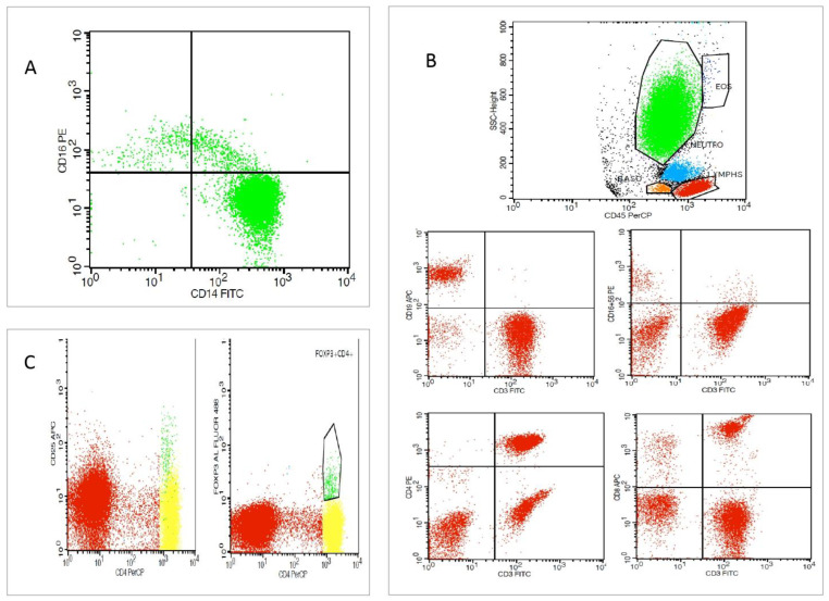
Figure 2.
Flow cytometric analysis of a type 2 CRS patient. (A) Representative dot plots depicting monocyte subsets (green clolour) with regard to surface expression of CD14 and CD16 in CD14++CD16−, CD14++C16+, and CD14+CD16+ subpopulations. (B) Representative dot plots depicting lymphocyte gating (red colour) with B-lymphocytes, T-lymphocytes, and natural killer (NK) cells defined as CD16+CD56+ cells, CD4+ T cells, and CD8+ T cells. (C). Representative dot plots depicting T regulatory cells (Tregs) defined as CD4+ FoxP3+ CD25high positive cells (green colour).

