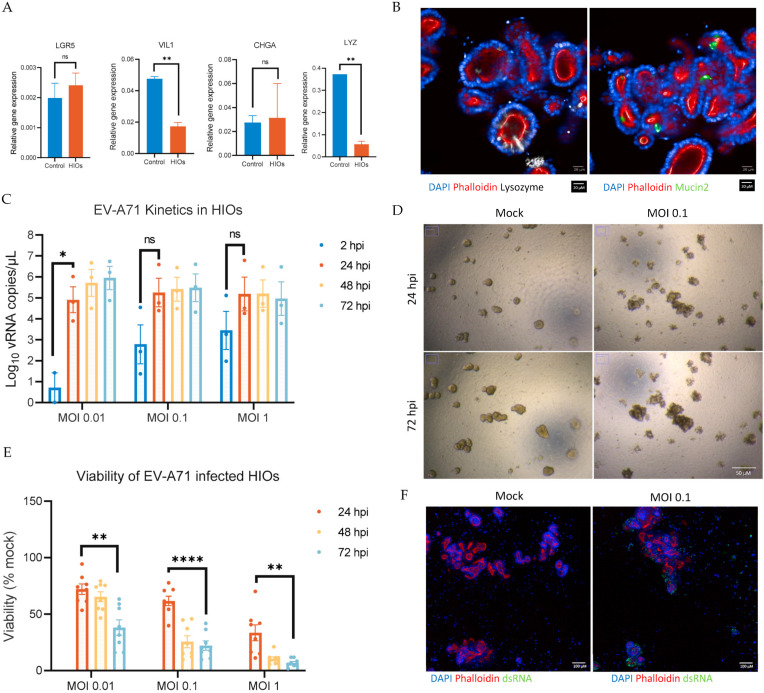
Figure 1.
Characterization of the differentiation of human induced pluripotent stem-cells-derived small intestine organoids (HIOs) and Enterovirus A71 (EV-A71) replication kinetics in the derived HIOs and viability post infection. (A) Relative expression of total RNA of adult small intestine as a control and HIOs. The expression level of each gene was calculated relative to EEF1A1 gene expression as housekeeping gene. (B) Images represent differentiated HIOs: DAPI (blue) for nucleus, phalloidin (red) for F-actin, lysozyme (white) for Paneth cells, and mucin 2 (green) for goblet cells. Scale bar, 20 µM. (C) HIOs were infected with EV-A71 virus at multiplicity of infection (MOI) 0.01, 0.1, and 1. At the indicated time points, total culture was collected by combining the triplicates to measure the levels of viral RNA yield by RT-qPCR. (D) Brightfield images of mock HIOs and MOI 0.1 infected HIOs at 24 h post-infection (hpi) and 72 hpi show a difference in morphology indicating cytopathic effect (CPE) due to infection. All images were acquired using the same objective. Scale bar, 50 µM. (E) To measure viability of HIOs, Cell Titer-Glo 3D was used at the indicated time points post-infection. Mock-infected HIOs were used to normalize the viability as a percentage. (F) Images represent mock- and EV-A71-infected HIOs (0.1 MOI) at 24 hpi: DAPI (blue) for nucleus, phalloidin (red) for F-actin, and dsRNA (green). Scale bar, 100 µm. Data are the mean ± SEM of three independent experiments, each carried out in triplicate; * indicates p < 0.05, ** indicates p < 0.01, and **** indicates p < 0.0001; ns indicates not significant.

