Abstract
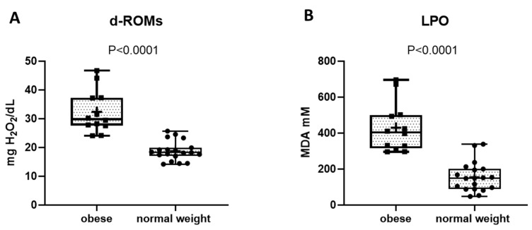
Oxidative stress, a key mediator of cardiovascular disease, metabolic alterations, and cancer, is independently associated with menopause and obesity. Yet, among postmenopausal women, the correlation between obesity and oxidative stress is poorly examined. Thus, in this study, we compared oxidative stress states in postmenopausal women with or without obesity. Body composition was assessed via DXA, while lipid peroxidation and total hydroperoxides were measured in patient’s serum samples via thiobarbituric-acid-reactive substances (TBARS) and derivate-reactive oxygen metabolites (d-ROMs) assays, respectively. Accordingly, 31 postmenopausal women were enrolled: 12 with obesity and 19 of normal weight (mean (SD) age 71.0 (5.7) years). Doubled levels of serum markers of oxidative stress were observed in women with obesity in women with obesity compared to those of normal weight (H2O2: 32.35 (7.3) vs. 18.80 (3.4) mg H2O2/dL; malondialdehyde (MDA): 429.6 (138.1) vs. 155.9 (82.4) mM in women with or without obesity, respectively; p < 0.0001 for both). Correlation analysis showed that both markers of oxidative stress increased with an increasing body mass index (BMI), visceral fat mass, and trunk fat percentage, but not with fasting glucose levels. In conclusion, obesity and visceral fat are associated with a greater increase in oxidative stress in postmenopausal women, possibly increasing cardiometabolic and cancer risks.
Keywords: obesity, oxidative stress, menopause, visceral fat, lipid peroxidation, oxidative damage
1. Introduction
Menopause is a condition precipitated by the permanent cessation of ovarian follicular function, leading to a decline in estrogen levels combined with an enhanced risk of cardiovascular disease (CVD), including arterial hypertension, coronary artery disease (CAD), and cerebrovascular disease [1]. Menopause is also associated with weight gain and worsening body composition with a shift from gynoid to android fat distribution [2]. Although the responsible mechanisms have not been completely clarified, changes in energy homeostasis due to aging; estrogen deficiency; sleep disturbances; changes in adipokines; gut hormones; and gut microbiota seem to play a role in weight gain and the worsening of body composition during menopause [3]. It has been estimated that approximately 70% of women of perimenopausal age are overweight or obese, both of which are associated with excess morbidity and mortality [4]. Obesity may have detrimental effects on a postmenopausal woman’s health, which are related to an increased risk of cancer [5], particularly breast cancer [6,7], and an increased risk of CVD [8]. Obesity is also associated with increased oxidative stress [9], a key mediator of CVD. In fact, the generation of reactive oxygen/nitrogen species (ROS/RNS) such as superoxide (O2−), hydrogen peroxide (H2O2), and peroxynitrite (ONOO−) is promoted by an imbalance between antioxidant and oxidant factors [10,11]. These factors have the potential to induce the oxidative modification of major cellular macromolecules (i.e., proteins, DNA, and lipids), thereby altering subcellular organelles and causing endothelial dysfunctions that promote atherosclerosis, which are fundamental events in the development of CVD [12]. Polyunsaturated fatty acids are particularly vulnerable to ROS, to which they yield primary products of lipid peroxidation, i.e., lipid hydroperoxides, and reactive aldehydes such as malondialdehyde (MDA), i.e., secondary products of lipid peroxidation [13]. In turn, MDA derivatives such as malondialdehyde acetaldehyde adducts may promote intra- or inter-molecular protein/DNA crosslinking, thus altering the biochemical properties of several proteins, including enzymes, carriers, and cytoskeletal, mitochondrial, and antioxidant proteins that are involved in aging and chronic diseases [14]. In patients with type 2 diabetes (T2D), elevated levels of MDA, a marker of lipid peroxidation, are associated with an increased risk of cardiovascular disease and complications of diabetes [15], thus contributing to the pathogenesis of atherosclerosis. An increase in lipid peroxidation levels has also been reported in the visceral adipose tissue (VAT) of postmenopausal women suffering from gynecological cancer [16].
Previous studies have demonstrated that oxidative stress is increased in obese patients, particularly in visceral obesity [9]. It has also been shown that postmenopausal women have greater levels of reactive oxygen species compared with premenopausal women [17,18] and men [19], indicating oxidative stress has been developed. It is likely that weight gain and the worsening of body composition play a role in increasing oxidative stress after menopause. However, very few data exist on the relationship between obesity and oxidative stress in postmenopausal women; specifically, there are no studies on this topic investigating the relationship between oxidative stress and adiposity, including with respect to VAT measured with a technique that provides high precision and accuracy such as dual-energy X-ray absorptiometry (DXA) [20]. The aim of this study was to fill this gap by investigating the association between oxidative stress and the key parameters related to fat accumulation. We show a positive correlation between BMI, VAT mass, and trunk fat percentage with oxidative stress markers, for which there is an almost two-fold increase in those markers among obese individuals compared to age-matched normal weight women. These findings underline the importance of keeping oxidative stress under control and managing fat-related parameters to reduce chronic cardiometabolic complications at such a critical age.
2. Materials and Methods
2.1. Study Subjects
We studied a subset of postmenopausal women who were participating in a previous cross-sectional study. Details of this subset of participants have been described previously [21]. Briefly, postmenopausal women (aged <65 years) undergoing hip arthroplasty for osteoarthritis were consecutively screened for participation in this study. Obesity was confirmed by the treating physician. Obesity was diagnosed when patients had a BMI ≥ 30 kg/m2, which is in accordance with the World Health Organization’s diagnostic criteria [22]. Malignancy was an exclusion criterion. Additionally, individuals treated with medications affecting bone such as estrogen, raloxifene, tamoxifen, bisphosphonates, teriparatide, denosumab, thiazolidinediones, glucocorticoids, anabolic steroids, and phenytoin, and those with hypercalcemia or hypocalcemia, hepatic or renal disorder, hypercortisolism, or currently consuming alcohol or taking anti-oxidant supplements, were excluded. This study was approved by the Ethics Committee of the Campus Bio-Medico University of Rome, and all participants provided written informed consent.
2.2. Body Composition/Anthropometric Measurements
Body scans were conducted with a Lunar ProdigyTM (GE Healthcare, Madison, WI, USA) DXA scanner. Body composition parameters were analyzed using the software enCORETM (version 17, 2016), and VAT was measured with the CoreScanTM (GE Healthcare, USA). Quality control and calibration of the model were performed each day following the protocol provided by the manufacturer. All participants were examined after completing a fasting period of at least 8 h. Blood samples were collected 24 h before surgery.
For this analysis, the variables of interest derived from the DXA dataset were trunk fat mass (gr), trunk total mass (gr), and defined anatomical regions (android and gynoid) and visceral adipose tissue (VAT mass (g) and VAT volume, (cm3)). BMI was calculated as weight/height2 (kg/m2).
2.3. Serum Lipid Peroxidation (LPO) Assay
Serum LPO was analyzed by the thiobarbituric-acid-reactive substance (TBARS) assay (OxiSelect™ TBARS Assay kit, Cell Biolabs, Inc., San Diego, CA, USA) as previously reported [23]. Briefly, serum samples were immediately stored at −80 °C after blood sampling; then, 20 µL was incubated in sodium dodecylsulphate (SDS) lysis solution to denature the proteins. Then, thiobarbituric acid (TBA) was added, and the samples were incubated for 50 min at 95 °C. The tubes, before being centrifuged at 735× g for 15 min., were kept at room temperature. The supernatants were recovered, and optical density was measured by a multilabel plate reader at 490 nm (Victor XTM, Perkin Elmer, Inc., Waltham, MA, USA).
2.4. Serum Levels of Total Hydroperoxide (TH)
Derivate-reactive oxygen metabolites (d-ROMs) Kit (Diacron Srl, Grosseto, Italy), was employed to measured TH levels, as reported in [24]. This method estimates the total amount of hydroperoxide present in a 10 μL serum sample by using a spectrophotometric procedure. Optical density was measured by a multilabel plate reader at 505 nm (Victor XTM, Perkin Elmer, USA), and the results were expressed in Carratelli units (UC) as conventional arbitrary units. The value of 1 Carratelli unit corresponds to a concentration of 0.08 mg/dL of hydrogen peroxide. A hydrogen peroxide calibration curve was developed using titrated H2O2 solutions.
2.5. Statistical Analysis
Data were analyzed using GraphPad Prism 9.0 (GraphPad Software, San Diego, CA, USA). Patients’ characteristics were described using means and standard deviation or counts and percentages. Group data are presented in boxplots with median and interquartile ranges; whiskers represent maximum and minimum values. We assessed data for normality. The unpaired t-test or the Chi square test were used to assess differences in the primary endpoints (d-ROMs) and in other variables between groups. Pearson’s correlation coefficients were used to assess the relationship between variables. Missing data were not imputed.
3. Results
3.1. Subject Characteristics
Of the 92 postmenopausal women originally enrolled in our previous study [21], 19 were excluded because they had diabetes, 11 because they were taking anti-oxidant nutritional supplements, and 31 because they did not have samples available for the oxidative stress analysis. A total of 31 postmenopausal women (12 with obesity and 19 of normal weight) were analyzed. As shown in Table 1, the clinical data of the study subjects highlighted that there were no differences in age and menopausal age between women with or without obesity. As expected, the BMI was significantly higher in the group with obesity compared to the group of women of normal weight. Fasting glucose levels were also higher in the subjects with obesity compared to those of normal weight (Table 1), although none of them were affected by diabetes as reported in their medical records. There were no differences in serum creatinine, urea, sodium, and potassium levels and the percentage of smokers between the groups (Table 1). The medications used by subjects with obesity included thyroid hormone replacement therapy (n = 1), statins (n = 1), and irbesartan/hydrochlorothiazide combination therapy (n = 1), whereas none of the normal weight subjects took any medications.
Table 1.
Demographic and clinical features of the study subjects.
| Parameters | Obesity (n = 12) |
Normal Weight (n = 19) |
p Value |
|---|---|---|---|
| Age (years) | 71.0 (5.7) | 67.7 (9.6) | 0.2579 |
| BMI (kg/m2) | 32.4 (2.2) | 23.1 (2.3) | <0.0001 |
| Menopausal age (years) | 49.7 (3.9) | 49.5 (8.3) | 0.9615 |
| Fasting glucose (mg/dL) | 105.4 (11.1) | 96.3 (11.8) | 0.0503 |
| Creatinine (mg/dL) | 0.76 (0.10) | 0.71 (0.12) | 0.3662 |
| Urea (mg/dL) | 39.3 (5.2) | 39.7 (7.2) | 0.9074 |
| Sodium (mmol/L) | 141.0 (1.06) | 141.0 (2.14) | <0.9999 |
| Potassium (mEq/L) | 3.8 (0.2) | 3.9 (0.3) | 0.6706 |
| Smokers (%) | 3.00 | 12.50 | 0.0628 |
Data were analyzed using unpaired t-test and are presented as means (SD) or counts (percentage). Statistical significance was considered when p value was ≤0.05.
3.2. Body Composition
Six subjects with obesity and seven subjects of normal weight underwent a DXA scan for an assessment of body composition. All the parameters related to body composition are reported in Table 2. Trunk total mass was significantly higher in the subjects with obesity compared to those without obesity, and so was trunk fat mass. Consistently, the percentage of trunk fat was higher in the subjects with obesity compared to those without obesity. Analysis of fat distribution highlighted that the subjects with obesity had both a higher percentage of android fat and gynoid fat compared to the normal weight subjects (Table 2). Finally, the levels of VAT mass and VAT volume were significantly greater in the subjects with obesity compared to the subjects of normal weight (Table 2).
Table 2.
Body composition.
| Parameters | Obesity (n = 6) |
Normal Weight (n = 7) |
p Value |
|---|---|---|---|
| Trunk fat mass (gr) | 23,226 (4975) | 13,845 (2963) | 0.0012 |
| Trunk total mass (gr) | 42,895 (5309) | 32,029 (3623) | 0.0012 |
| Trunk fat percentage (%) | 53.83 (6.4) | 42.83 (4.6) | 0.0047 |
| Android fat percentage (%) | 56.57 (4.6) | 44.77 (5.1) | 0.0012 |
| Gynoid fat percentage (%) | 51.80 (4.4) | 44.90 (3.8) | 0.0140 |
| VAT mass (gr) | 1292 (479.3) | 589.2 (178.9) | 0.0159 |
| VAT volume (cm3) | 1397 (518.3) | 636.8 (193.7) | 0.0159 |
Data were analyzed using unpaired t-test and are presented as means (SD). VAT, visceral adipose tissue.
3.3. Oxidative Stress Markers
We measured serum d-ROMs and LPO levels to study the levels of oxidative stress between the two groups (Figure 1). H2O2 levels were significantly higher in the subjects with obesity compared to those of normal weight (Figure 1A, p < 0.0001). The same trend was observed for MDA levels, which reflects the presence of lipid peroxidation damage in patients with obesity (Figure 1B, p < 0.0001).
Figure 1.
Oxidative stress and oxidative lipid damage in serum from study groups. (A) H2O2 and (B) MDA levels were evaluated in obese and normal weight postmenopausal women. All data represent the means of at least 3 replicates ± standard deviation. The analysis of difference between groups was performed using the unpaired t-test.
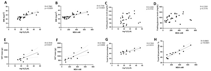
Correlation analysis including all study subjects showed that both H2O2 (R = 0.7962, p < 0.0001) and MDA levels (R = 0.7749, p < 0.0001) increased with an increasing BMI (Figure 2A,B), but we found no correlation with fasting glucose levels for either H2O2 (R = 0.2369, p = 0.2159) or MDA (R = 0.2361, p = 0.2176) (Figure 2C,D).
Figure 2.
Correlation analysis of oxidative stress markers and body composition parameters. H2O2 and MDA levels were correlated with BMI (A,B), fasting glucose levels (C,D), VAT mass (E,F), and trunk fat percentage (G,H) in obese and normal weight postmenopausal women. Analysis was performed using Pearson comparison test. R indicates Pearson correlation coefficient.
Finally, oxidative stress markers showed a positive correlation (Figure 2E–H) with VAT mass (d-ROMs R = 0.7016, p = 0.0237; LPO R = 0.7892, p = 0.0066) and percentage of trunk fat (d-ROMs R = 0.7843, p = 0.0015: LPO R = 0.8983, p < 0.0001).
4. Discussion
In this cross-sectional study, total hydroperoxides and malondealdehyde, markers of oxidative stress and lipid peroxidation, respectively, were evaluated in postmenopausal women both without and with obesity. Our results suggest that oxidative stress is significantly increased in postmenopausal women living with obesity compared to their normal weight counterparts. Moreover, markers of oxidative stress were significantly associated with BMI, VAT, and trunk fat but not with fasting plasma glucose. To the best of our knowledge, this is the first study that specifically investigates the relationship between oxidative stress and DXA-derived measures of adiposity, including VAT, in postmenopausal women with or without obesity.
In fact, obesity is characterized by an excessive increase in fat storage, which, in turn, promotes lipid peroxidation. Studies suggest that this condition promotes oxidative stress by increasing NADPH oxidase activity (NOX) and decreasing mRNA expression and the activity of major antioxidant enzymes such as superoxide dismutase (SOD) and catalase (CAT). Ultimately, this pro-oxidant environment leads to a chronic proinflammatory state related to comorbidities and poor clinical outcome [25]. Oxidative stress is recognized as an important contributor to the pathogenesis of metabolic alterations including metabolic syndrome, type 2 diabetes and its complications [26], cancer [27], and CVD [12]. High total hydroperoxide plasma levels constitute an independent predictor of CVD events and mortality [28,29] and predict CAD in women [19]. Oxidative stress also plays a role in the decline of muscle mass and function (i.e., sarcopenia) observed during the aging process [10] and has been postulated to enhance bone resorption, possibly contributing to osteoporosis [30] and osteosarcopenic obesity [31].
The menopausal transition is associated with increases in total body fat mass and VAT [32]. It has been reported that in the shift from a premenopausal to postmenopausal status, VAT increases from 5–8% to 15–20% of total body fat [33,34]. In this study, we show that oxidative stress increases with an increasing VAT. This finding suggests that the increase in oxidative stress observed in postmenopausal women [17,19] might be due, at least in part, to changes in body composition after menopause. Our findings are consistent with most of the few previous studies that investigated the relationship between oxidative stress and obesity in postmenopausal women. In a study conducted in Mexico involving more than 500 pre- and postmenopausal women, oxidative stress markers were higher in overweight/obese women compared to normal weight women regardless of their menopausal status [35]. Furthermore, the authors found a weak but significant correlation (r = 0.298; p < 0.0001) between plasma MDA and the waist-to-height ratio, an indicator of central obesity. A positive correlation between oxidative stress (as assessed by a whole-blood free oxygen radical test) and abdominal obesity as assessed by either waist circumference (r = 0.345; p = 0.02) or the waist-to-hip ratio (r = 0.540; p = 0.0001) was reported by Cagnacci and colleagues [36]. A very limited number of studies assessed the relationship between DXA-derived measurements of central adiposity and oxidative stress in postmenopausal women. Pansini and colleagues found that antioxidant status (r = 0.46; p < 0.001) and hydroperoxide levels (r = 0.26; p < 0.05) increased with an increasing trunk fat mass [37] in pre- and postmenopausal women with a wide range of BMI values. In a different analysis of the same population, the same group reported a positive correlation between trunk fat mass and hydroperoxides and a negative correlation between trunk fat mass and antioxidant capacity (standardized multiple regression coefficients for the association of trunk fat mass with hydroperoxides and residual antioxidant power were equal to 0.324 and −0.495, respectively, for which p < 0.05 for both) among postmenopausal women aged ≤ 65 years without obesity [38]. In a similar population, Crist and colleagues showed that the androidal/gynoidal fat mass ratio (the sum of waist and hip fat mass divided by thigh fat mass) was a significant determinant of oxidative stress (estimated either by oxidized low-density lipoprotein or 15-isoprostane F2α) [39]. However, in a large study of older (≥50 years) adults followed-up for up to 14 years, longitudinal changes in hydroperoxide levels were significantly associated with a BMI ≥ 35 kg/m2, but there was no significant association with central obesity (either assessed by waist circumference or waist-to-hip ratio) in women [40]. Unlike previous studies, we included the DXA-derived measurement of VAT, which is strongly associated with cardiometabolic risk and is superior to anthropometric surrogates of abdominal obesity such as waist circumference or waist-to-hip ratio and waist-to-height-ratios [41]. Furthermore, DXA-derived VAT is highly correlated with VAT measured by magnetic resonance imaging and is preferable to the measurement of trunk fat, which includes subcutaneous adipose tissue [42].
The reduction in estrogen levels after menopause impacts body composition [3] and appears to induce molecular changes that may lead to an increase in oxidative stress in adipose tissue mainly due to reductions in antioxidant capacity. Data from animal studies indicate that the reduction in estrogen levels following an oophorectomy decreases the expression of glutathione peroxidase 3 (Gpx3), a gene involved in the protection of cells from ROS, in white adipose tissue [43]. There is also preclinical evidence that estrogen treatment reduces oxidative stress by promoting the expression of macrophage heme oxygenase-1, NAD(P)H:quinone oxidoreductase 1, and glutamate-cysteine ligase [16,44]. It has also been postulated that a decrease in antioxidant defenses might be mediated by low levels of follicle-stimulating hormone (FSH) in postmenopausal women. Klisic and colleagues showed that postmenopausal women were more likely to be overweight/obese and found a positive correlation between FSH levels and the activity of the antioxidant enzyme glutathione peroxidase. However, when multivariable regression analysis was performed, the relationship between FSH and glutathione peroxidase disappeared, and abdominal obesity, which was assessed by waist circumference, was the only variable significantly associated with lower FSH levels, suggesting that abdominal obesity may be a major determinant and modulator of antioxidant capacity as assessed by glutathione peroxidase [45]. Altogether, these results and our findings of a strong association between abdominal adiposity/VAT and markers of oxidative stress suggest that the increase in VAT due to estrogen reduction and aging [46,47] is a powerful mediator of oxidative stress in postmenopausal women.
The lack of correlation between fasting plasma glucose and markers of oxidative stress is an unexpected finding that might be due to the relatively small range of glucose values measured glucose values. Furthermore, we did not measure plasma insulin concentrations and, therefore, were not able to calculate indices of insulin resistance, which have been shown to correlate with oxidative stress in several studies [48].
Our findings indicate the suitability of VAT accumulation as a therapeutic target to reduce oxidative stress (and, therefore, cardiometabolic risk) among postmenopausal women. Nutrition prevention strategies could help tackle VAT and VAT-accumulation-associated oxidative stress, as calorie restriction per se is associated with reductions in oxidative stress, due to an enhancement of mitochondrial biogenesis and turnover, antioxidant defenses, and decreased ROS production [49]. Several nutritional approaches have been proposed for postmenopausal women, which could help counteract the metabolic derangements associated with menopause [50].
The strengths of this study include its use of the d-ROMs test, i.e., a simple test that is already available as a point-of-care in vitro diagnostic and could, therefore, be easily implemented in clinical practice [29], and its use of DXA to determine VAT. Some limitations should be acknowledged. The number of participants was relatively low, and larger studies will be needed to confirm our results. It should be noted, however, that despite the small sample, the correlations between oxidative stress markers and adiposity parameters were very strong and significant. We did not include a control group of premenopausal women. Although this would have provided further insight into the relationship between oxidative stress and adiposity across different stages of a woman’s life, it has previously been shown that postmenopausal women have greater levels of oxidative stress compared with premenopausal women [17,18]. Therefore, we focused on the effect of obesity and fat accumulation, for which postmenopausal women of normal weight served as the control group. Additionally, we did not measure insulin levels or estrogen levels, as the focus of our study was the relationship between obesity, adiposity measures, and oxidative stress. Finally, this study’s cross-sectional design does not allow one to infer the direction of the associations found.
5. Conclusions
Menopause per se is associated with increased oxidative stress. We have shown that obesity and visceral fat are associated with a further increase in oxidative stress in postmenopausal women. These findings highlight the importance of keeping oxidative stress under control and preventing/managing excess adiposity to reduce chronic cardiometabolic complications at such a critical age. Clinicians who manage postmenopausal women should be aware of this abnormal association.
Author Contributions
Conceptualization, N.N., A.M.S., M.M. and R.S.; methodology, A.M.S., G.L. and A.P.; software, C.C.Q.; validation, A.M.S. and G.L.; formal analysis, G.L.; investigation, N.N. and A.M.S.; resources, N.N, A.M.S., M.M., C.C.Q., R.P. and V.D.; data curation, G.L. and C.C.; writing—original draft preparation, G.L. and C.C.; writing—review and editing, A.M.S., C.I., F.C., N.N. and M.M.; visualization, N.N. and A.M.S.; supervision, N.N. and A.M.S.; project administration, N.N. and G.L.; funding acquisition, N.N. and R.S. All authors have read and agreed to the published version of the manuscript.
Institutional Review Board Statement
The study was conducted in accordance with the Declaration of Helsinki and approved by the Ethics Committee of Campus Bio-Medico University of Rome (Protocol name: “Evaluation of bone strength and Wnt pathway in obese patients”, approved on 26 October 2015).
Informed Consent Statement
Informed consent was obtained from all subjects involved in the study.
Data Availability Statement
The data presented in this study are available on request from the corresponding author.
Conflicts of Interest
The authors declare no conflict of interest.
Funding Statement
This research was funded by an internal grant of Campus Bio-Medico University of Rome.
Footnotes
Disclaimer/Publisher’s Note: The statements, opinions and data contained in all publications are solely those of the individual author(s) and contributor(s) and not of MDPI and/or the editor(s). MDPI and/or the editor(s) disclaim responsibility for any injury to people or property resulting from any ideas, methods, instructions or products referred to in the content.
References
- 1.Harvey R.E., Coffman K.E., Miller V.M. Women-specific factors to consider in risk, diagnosis and treatment of cardiovascular disease. Womens Health. 2015;11:239–257. doi: 10.2217/WHE.14.64. [DOI] [PMC free article] [PubMed] [Google Scholar]
- 2.Farahmand M., Bahri Khomamid M., Rahmati M., Azizi F., Ramezani Tehrani F. Aging and changes in adiposity indices: The impact of menopause. J. Endocrinol. Investig. 2022;45:69–77. doi: 10.1007/s40618-021-01616-2. [DOI] [PubMed] [Google Scholar]
- 3.Fenton A. Weight, Shape, and Body Composition Changes at Menopause. J. Midlife Health. 2021;12:187–192. doi: 10.4103/jmh.jmh_123_21. [DOI] [PMC free article] [PubMed] [Google Scholar]
- 4.Knight M.G., Anekwe C., Washington K., Akam E.Y., Wang E., Stanford F.C. Weight regulation in menopause. Menopause. 2021;28:960–965. doi: 10.1097/GME.0000000000001792. [DOI] [PMC free article] [PubMed] [Google Scholar]
- 5.Anand P., Kunnumakkara A.B., Sundaram C., Harikumar K.B., Tharakan S.T., Lai O.S., Sung B., Aggarwal B.B. Cancer is a preventable disease that requires major lifestyle changes. Pharm. Res. 2008;25:2097–2116. doi: 10.1007/s11095-008-9661-9. [DOI] [PMC free article] [PubMed] [Google Scholar]
- 6.Reeves G.K., Pirie K., Beral V., Green J., Spencer E., Bull D., Million Women Study C. Cancer incidence and mortality in relation to body mass index in the Million Women Study: Cohort study. BMJ. 2007;335:1134. doi: 10.1136/bmj.39367.495995.AE. [DOI] [PMC free article] [PubMed] [Google Scholar]
- 7.Renehan A.G., Tyson M., Egger M., Heller R.F., Zwahlen M. Body-mass index and incidence of cancer: A systematic review and meta-analysis of prospective observational studies. Lancet. 2008;371:569–578. doi: 10.1016/S0140-6736(08)60269-X. [DOI] [PubMed] [Google Scholar]
- 8.Lloyd-Jones D.M., Leip E.P., Larson M.G., D’Agostino R.B., Beiser A., Wilson P.W., Wolf P.A., Levy D. Prediction of lifetime risk for cardiovascular disease by risk factor burden at 50 years of age. Circulation. 2006;113:791–798. doi: 10.1161/CIRCULATIONAHA.105.548206. [DOI] [PubMed] [Google Scholar]
- 9.Vincent H.K., Taylor A.G. Biomarkers and potential mechanisms of obesity-induced oxidant stress in humans. Int. J. Obes. 2006;30:400–418. doi: 10.1038/sj.ijo.0803177. [DOI] [PubMed] [Google Scholar]
- 10.Baumann C.W., Kwak D., Liu H.M., Thompson L.V. Age-induced oxidative stress: How does it influence skeletal muscle quantity and quality? J. Appl. Physiol. 1985. 2016;121:1047–1052. doi: 10.1152/japplphysiol.00321.2016. [DOI] [PMC free article] [PubMed] [Google Scholar]
- 11.Jackson M.J. Redox regulation of muscle adaptations to contractile activity and aging. J. Appl. Physiol. 1985. 2015;119:163–171. doi: 10.1152/japplphysiol.00760.2014. [DOI] [PMC free article] [PubMed] [Google Scholar]
- 12.Dubois-Deruy E., Peugnet V., Turkieh A., Pinet F. Oxidative Stress in Cardiovascular Diseases. Antioxidants. 2020;9:864. doi: 10.3390/antiox9090864. [DOI] [PMC free article] [PubMed] [Google Scholar]
- 13.Ayala A., Munoz M.F., Arguelles S. Lipid peroxidation: Production, metabolism, and signaling mechanisms of malondialdehyde and 4-hydroxy-2-nonenal. Oxid. Med. Cell. Longev. 2014;2014:360438. doi: 10.1155/2014/360438. [DOI] [PMC free article] [PubMed] [Google Scholar]
- 14.Zarkovic N., Cipak A., Jaganjac M., Borovic S., Zarkovic K. Pathophysiological relevance of aldehydic protein modifications. J. Proteom. 2013;92:239–247. doi: 10.1016/j.jprot.2013.02.004. [DOI] [PubMed] [Google Scholar]
- 15.Shabalala S.C., Johnson R., Basson A.K., Ziqubu K., Hlengwa N., Mthembu S.X.H., Mabhida S.E., Mazibuko-Mbeje S.E., Hanser S., Cirilli I., et al. Detrimental Effects of Lipid Peroxidation in Type 2 Diabetes: Exploring the Neutralizing Influence of Antioxidants. Antioxidants. 2022;11:2071. doi: 10.3390/antiox11102071. [DOI] [PMC free article] [PubMed] [Google Scholar]
- 16.Narumi M., Takahashi K., Yamatani H., Seino M., Yamanouchi K., Ohta T., Takahashi T., Kurachi H., Nagase S. Oxidative Stress in the Visceral Fat Is Elevated in Postmenopausal Women with Gynecologic Cancer. J. Womens Health. 2018;27:99–106. doi: 10.1089/jwh.2016.6301. [DOI] [PubMed] [Google Scholar]
- 17.Sanchez-Rodriguez M.A., Zacarias-Flores M., Arronte-Rosales A., Correa-Munoz E., Mendoza-Nunez V.M. Menopause as risk factor for oxidative stress. Menopause. 2012;19:361–367. doi: 10.1097/gme.0b013e318229977d. [DOI] [PubMed] [Google Scholar]
- 18.Bourgonje A.R., Abdulle A.E., Al-Rawas A.M., Al-Maqbali M., Al-Saleh M., Enriquez M.B., Al-Siyabi S., Al-Hashmi K., Al-Lawati I., Bulthuis M.L.C., et al. Systemic Oxidative Stress Is Increased in Postmenopausal Women and Independently Associates with Homocysteine Levels. Int. J. Mol. Sci. 2020;21:314. doi: 10.3390/ijms21010314. [DOI] [PMC free article] [PubMed] [Google Scholar]
- 19.Vassalle C., Sciarrino R., Bianchi S., Battaglia D., Mercuri A., Maffei S. Sex-related differences in association of oxidative stress status with coronary artery disease. Fertil. Steril. 2012;97:414–419. doi: 10.1016/j.fertnstert.2011.11.045. [DOI] [PubMed] [Google Scholar]
- 20.Ponti F., Santoro A., Mercatelli D., Gasperini C., Conte M., Martucci M., Sangiorgi L., Franceschi C., Bazzocchi A. Aging and Imaging Assessment of Body Composition: From Fat to Facts. Front. Endocrinol. 2019;10:861. doi: 10.3389/fendo.2019.00861. [DOI] [PMC free article] [PubMed] [Google Scholar]
- 21.Piccoli A., Cannata F., Strollo R., Pedone C., Leanza G., Russo F., Greto V., Isgro C., Quattrocchi C.C., Massaroni C., et al. Sclerostin Regulation, Microarchitecture, and Advanced Glycation End-Products in the Bone of Elderly Women With Type 2 Diabetes. J. Bone Miner. Res. 2020;35:2415–2422. doi: 10.1002/jbmr.4153. [DOI] [PMC free article] [PubMed] [Google Scholar]
- 22.World Health Organization WHO Consultation on Obesity (1999: Geneva, Switzerland) & World Health Organization. Obesity: Preventing and Managing the Global Epidemic: Report of a WHO Consultation. World Health Organization. 2000. [(accessed on 2 February 2023)]. Available online: https://apps.who.int/iris/handle/10665/42330. [PubMed]
- 23.Martino N.A., Marzano G., Mangiacotti M., Miedico O., Sardanelli A.M., Gnoni A., Lacalandra G.M., Chiaravalle A.E., Ciani E., Bogliolo L., et al. Exposure to cadmium during in vitro maturation at environmental nanomolar levels impairs oocyte fertilization through oxidative damage: A large animal model study. Reprod. Toxicol. 2017;69:132–145. doi: 10.1016/j.reprotox.2017.02.005. [DOI] [PubMed] [Google Scholar]
- 24.Buonocore G., Perrone S., Longini M., Terzuoli L., Bracci R. Total hydroperoxide and advanced oxidation protein products in preterm hypoxic babies. Pediatr. Res. 2000;47:221–224. doi: 10.1203/00006450-200002000-00012. [DOI] [PubMed] [Google Scholar]
- 25.Evans J.L., Goldfine I.D., Maddux B.A., Grodsky G.M. Oxidative stress and stress-activated signaling pathways: A unifying hypothesis of type 2 diabetes. Endocr. Rev. 2002;23:599–622. doi: 10.1210/er.2001-0039. [DOI] [PubMed] [Google Scholar]
- 26.Halim M., Halim A. The effects of inflammation, aging and oxidative stress on the pathogenesis of diabetes mellitus (type 2 diabetes) Diabetes Metab. Syndr. 2019;13:1165–1172. doi: 10.1016/j.dsx.2019.01.040. [DOI] [PubMed] [Google Scholar]
- 27.Calaf G.M., Urzua U., Termini L., Aguayo F. Oxidative stress in female cancers. Oncotarget. 2018;9:23824–23842. doi: 10.18632/oncotarget.25323. [DOI] [PMC free article] [PubMed] [Google Scholar]
- 28.Hitomi Y., Masaki N., Ishinoda Y., Ido Y., Iwashita M., Yumita Y., Kagami K., Yasuda R., Ikegami Y., Toya T., et al. Effectiveness of the d-ROMs oxidative stress test to predict long-term cardiovascular mortality. Int. J. Cardiol. 2022;354:43–47. doi: 10.1016/j.ijcard.2022.03.001. [DOI] [PubMed] [Google Scholar]
- 29.Pigazzani F., Gorni D., Dyar K.A., Pedrelli M., Kennedy G., Costantino G., Bruno A., Mackenzie I., MacDonald T.M., Tietge U.J.F., et al. The Prognostic Value of Derivatives-Reactive Oxygen Metabolites (d-ROMs) for Cardiovascular Disease Events and Mortality: A Review. Antioxidants. 2022;11:1541. doi: 10.3390/antiox11081541. [DOI] [PMC free article] [PubMed] [Google Scholar]
- 30.Cervellati C., Bonaccorsi G., Cremonini E., Romani A., Fila E., Castaldini M.C., Ferrazzini S., Giganti M., Massari L. Oxidative stress and bone resorption interplay as a possible trigger for postmenopausal osteoporosis. BioMed Res. Int. 2014;2014:569563. doi: 10.1155/2014/569563. [DOI] [PMC free article] [PubMed] [Google Scholar]
- 31.Di Filippo L., De Lorenzo R., Giustina A., Rovere-Querini P., Conte C. Vitamin D in Osteosarcopenic Obesity. Nutrients. 2022;14:1816. doi: 10.3390/nu14091816. [DOI] [PMC free article] [PubMed] [Google Scholar]
- 32.Abdulnour J., Doucet E., Brochu M., Lavoie J.M., Strychar I., Rabasa-Lhoret R., Prud’homme D. The effect of the menopausal transition on body composition and cardiometabolic risk factors: A Montreal-Ottawa New Emerging Team group study. Menopause. 2012;19:760–767. doi: 10.1097/gme.0b013e318240f6f3. [DOI] [PubMed] [Google Scholar]
- 33.Karvonen-Gutierrez C., Kim C. Association of Mid-Life Changes in Body Size, Body Composition and Obesity Status with the Menopausal Transition. Healthcare. 2016;4:42. doi: 10.3390/healthcare4030042. [DOI] [PMC free article] [PubMed] [Google Scholar]
- 34.Gambacciani M., Ciaponi M., Cappagli B., Benussi C., De Simone L., Genazzani A.R. Climacteric modifications in body weight and fat tissue distribution. Climacteric. 1999;2:37–44. doi: 10.3109/13697139909025561. [DOI] [PubMed] [Google Scholar]
- 35.Rodriguez-San Nicolas A., MA S.A.-R., Zacarias-Flores M., Correa-Munoz E., Mendoza-Nunez V.M. Relationship between central obesity and oxidative stress in premenopausal versus postmenopausal women. Nutr. Hosp. 2020;37:267–274. doi: 10.20960/nh.02552. [DOI] [PubMed] [Google Scholar]
- 36.Cagnacci A., Cannoletta M., Palma F., Bellafronte M., Romani C., Palmieri B. Relation between oxidative stress and climacteric symptoms in early postmenopausal women. Climacteric. 2015;18:631–636. doi: 10.3109/13697137.2014.999659. [DOI] [PubMed] [Google Scholar]
- 37.Pansini F., Cervellati C., Guariento A., Stacchini M.A., Castaldini C., Bernardi A., Pascale G., Bonaccorsi G., Patella A., Bagni B., et al. Oxidative stress, body fat composition, and endocrine status in pre- and postmenopausal women. Menopause. 2008;15:112–118. doi: 10.1097/gme.0b013e318068b285. [DOI] [PubMed] [Google Scholar]
- 38.Cervellati C., Bonaccorsi G., Cremonini E., Romani A., Fila E., Castaldini C., Ferrazzini S., Massari L., Squerzanti M., Sticozzi C., et al. Accumulation of central fat correlates with an adverse oxidative balance in non-obese postmenopausal women. Gynecol. Endocrinol. 2013;29:1063–1066. doi: 10.3109/09513590.2013.831829. [DOI] [PubMed] [Google Scholar]
- 39.Crist B.L., Alekel D.L., Ritland L.M., Hanson L.N., Genschel U., Reddy M.B. Association of oxidative stress, iron, and centralized fat mass in healthy postmenopausal women. J. Womens Health. 2009;18:795–801. doi: 10.1089/jwh.2008.0988. [DOI] [PMC free article] [PubMed] [Google Scholar]
- 40.Anusruti A., Jansen E., Gao X., Xuan Y., Brenner H., Schottker B. Longitudinal Associations of Body Mass Index, Waist Circumference, and Waist-to-Hip Ratio with Biomarkers of Oxidative Stress in Older Adults: Results of a Large Cohort Study. Obes. Facts. 2020;13:66–76. doi: 10.1159/000504711. [DOI] [PMC free article] [PubMed] [Google Scholar]
- 41.Konieczna J., Abete I., Galmes A.M., Babio N., Colom A., Zulet M.A., Estruch R., Vidal J., Toledo E., Diaz-Lopez A., et al. Body adiposity indicators and cardiometabolic risk: Cross-sectional analysis in participants from the PREDIMED-Plus trial. Clin. Nutr. 2019;38:1883–1891. doi: 10.1016/j.clnu.2018.07.005. [DOI] [PubMed] [Google Scholar]
- 42.Bea J.W., Chen Z., Blew R.M., Nicholas J.S., Follis S., Bland V.L., Cheng T.D., Ochs-Balcom H.M., Wactawski-Wende J., Banack H.R., et al. MRI Based Validation of Abdominal Adipose Tissue Measurements From DXA in Postmenopausal Women. J. Clin. Densitom. 2022;25:189–197. doi: 10.1016/j.jocd.2021.07.010. [DOI] [PMC free article] [PubMed] [Google Scholar]
- 43.Lundholm L., Putnik M., Otsuki M., Andersson S., Ohlsson C., Gustafsson J.A., Dahlman-Wright K. Effects of estrogen on gene expression profiles in mouse hypothalamus and white adipose tissue: Target genes include glutathione peroxidase 3 and cell death-inducing DNA fragmentation factor, alpha-subunit-like effector A. J. Endocrinol. 2008;196:547–557. doi: 10.1677/JOE-07-0277. [DOI] [PubMed] [Google Scholar]
- 44.Sul O.J., Hyun H.J., Rajasekaran M., Suh J.H., Choi H.S. Estrogen enhances browning in adipose tissue by M2 macrophage polarization via heme oxygenase-1. J. Cell Physiol. 2021;236:1875–1888. doi: 10.1002/jcp.29971. [DOI] [PubMed] [Google Scholar]
- 45.Klisic A., Kotur-Stevuljevic J., Kavaric N., Martinovic M., Matic M. The association between follicle stimulating hormone and glutathione peroxidase activity is dependent on abdominal obesity in postmenopausal women. Eat. Weight Disord. 2018;23:133–141. doi: 10.1007/s40519-016-0325-1. [DOI] [PubMed] [Google Scholar]
- 46.Bjune J.I., Stromland P.P., Jersin R.A., Mellgren G., Dankel S.N. Metabolic and Epigenetic Regulation by Estrogen in Adipocytes. Front. Endocrinol. 2022;13:828780. doi: 10.3389/fendo.2022.828780. [DOI] [PMC free article] [PubMed] [Google Scholar]
- 47.Ko S.H., Jung Y. Energy Metabolism Changes and Dysregulated Lipid Metabolism in Postmenopausal Women. Nutrients. 2021;13:4556. doi: 10.3390/nu13124556. [DOI] [PMC free article] [PubMed] [Google Scholar]
- 48.Yaribeygi H., Farrokhi F.R., Butler A.E., Sahebkar A. Insulin resistance: Review of the underlying molecular mechanisms. J. Cell Physiol. 2019;234:8152–8161. doi: 10.1002/jcp.27603. [DOI] [PubMed] [Google Scholar]
- 49.Amorim J.A., Coppotelli G., Rolo A.P., Palmeira C.M., Ross J.M., Sinclair D.A. Mitochondrial and metabolic dysfunction in ageing and age-related diseases. Nat. Rev. Endocrinol. 2022;18:243–258. doi: 10.1038/s41574-021-00626-7. [DOI] [PMC free article] [PubMed] [Google Scholar]
- 50.Silva T.R., Oppermann K., Reis F.M., Spritzer P.M. Nutrition in Menopausal Women: A Narrative Review. Nutrients. 2021;13:2149. doi: 10.3390/nu13072149. [DOI] [PMC free article] [PubMed] [Google Scholar]
Associated Data
This section collects any data citations, data availability statements, or supplementary materials included in this article.
Data Availability Statement
The data presented in this study are available on request from the corresponding author.


