

Abstract
Introduction and importance
Clavicular tumors are rare, consisting of <1 % of all skeletal tumors. In this series, we described our experience of treating medial clavicular tumors.
Case presentation
We treated three patients with medial clavicle tumors at a national tertiary referral hospital in Jakarta, Indonesia. The patients were treated with wide excision following bony reconstruction from fibular bone and one patient was treated by marginal excision. Each patient was treated by surgery and one patient underwent reconstruction using non-vascularized fibular graft and composite using bone cement.
Clinical discussion
All patients resulted in restoration of symmetry of the lower neck and upper chest and no post-surgical complication. Based on these cases above and the extension of tumor, we recommend medial clavicle tumor resection classification divided into three type to decide which type of surgical procedure that should be performed. In our report, all patients resulted in restoration of symmetry of the lower neck and upper chest and no post-surgical complication.
Conclusion
Clavicle resection in management of medial third clavicle tumor is technically demanding. We proposed three types of clavicular resection based on tumor extension. The surgical technique of medial end clavicle in this patient resulted in tumor free margin of medial clavicular, medial scapula, and lateral scapular incision. Reconstruction surgery following clavicle resection can be done in order to restore symmetry of the lower neck and upper chest, protect nearby neurovascular bundle, and rarely associated with significant shoulder function loss.
Keywords: Medial clavicular tumor, Clavicle resection
Highlights
-
•
Clavicular tumors are rare, consisting of <1 % of all skeletal tumors.
-
•
Clavicle resection in management of medial third clavicle tumor is technically demanding.
-
•
We recommended three types of clavicular resection based on tumor extension.
1. Introduction and importance
The development and structure of the clavicle are markedly different form that of other tubular bones in many respects [1]. Clavicular tumors are rare; most of them are primary malignant lesions [2]. Such malignancies may be related to the unique structure of the clavicle. However, due to the lack of sufficient number of cases, clinical characteristics of clavicle tumors and tumorous lesions remain unclear [1].
Primary clavicle tumors and tumorous lesions are mainly treated with surgery. Total claviculectomy is often required when a radical resection for malignant tumors is indicated. For Ewing sarcoma, surgery plus chemotherapy have been shown to provide better local control and survival compared to radiotherapy plus chemotherapy. Morbidity of the upper limb from a claviculectomy is obviously a concern. Studies have shown that most functions of the upper limb even after a total claviculectomy can be preserved without reconstruction of the clavicle. Therefore, complicated reconstruction of the clavicle, especially with vascularized fibular flaps, may not be necessary. However, the clavicles function as struts to prevent the upper limb and scapula from falling downward through the acromioclavicular joint and coracoclavicular ligament [1]. In this series, we described our experience in treating clavicular tumor using wide excision and bone reconstruction. This series is written in accordance with the SCARE 2020 guideline [3].
2. Presentation of cases
We included three patients diagnosed with medial clavicular tumor (Table 1). The first patient, a 40-year-old female, presented with right clavicular pain since one year before admission. Physical examination demonstrated hard mass measured 4 × 4 × 1.5 cm on sternoclavicular joint region (Fig. 1). Abduction of the right shoulder was limited due to pain. The patient was diagnosed with primary bone tumor of the right clavicle suspected benign tumor with differential diagnosis of osteochondroma. We performed marginal exicision to the patient.
Table 1.
Characteristics of the patients.
| Case No | Age | Sex | Diagnosis | Enneking | Metastasis | Chemotherapy | Procedures |
|---|---|---|---|---|---|---|---|
| 1 | 40 | F | Primary bone tumor of the right clavicle suspected benign dd/ osteochondroma | N/A | N/A | N/A | Marginal excision |
| 2 | 19 | M | Osteosarcoma of the left clavicle | IIB | N/A | Post neoadjuvant chemotherapy | Wide excision and reconstruction using non-vascularized fibular graft |
| 3 | 52 | M | Benign fibrous histiocytoma of left distal clavicle | N/A | N/A | N/A | Wide excision and acromioclavicular joint reconstruction |
Fig. 1.

Case 1, a 40-year-old female with primary bone tumor of the right clavicle suspected benign dd/ osteochondroma. (a) Clinical appearance of the tumor. (b) X-ray of the right clavicle. (c) MRI T2 axial of the right clavicle. (d) CT-scan of the MRI. (e) The tumor exposed. (f) Gross pathology of the tumor.
The second patient, a 19-year-old male, presented with left clavicular pain since ten months before admission. Initially, physical examination demonstrated palpable solid mass measured 4 × 4 × 2 cm on the medial part of the clavicle (Fig. 2). However, four months later, the mass grew enlarged extending to 8 × 8 × 2 cm despite chemotherapy. Pathological anatomy examination demonstrated osteosarcoma. The patient was diagnosed with osteosarcoma of the left clavicle, and we performed wide excision and reconstruction using non-vascularized fibular graft.
Fig. 2.

Case 2, a 19-year-old male with osteosarcoma of the left clavicle. (a) Clinical appearance of the tumor. (b) X-ray of the left clavicle. (c) MRI of the left clavicle. (d) CT-scan of the left clavicle. (e) Post-tumor removal. (f) Free fibular graft. (g) Postoperative X-ray of the left clavicle. (h, i) Gross pathology of the tumor. (j) Histopathological findings of the tumor.
The third patient, a 52-year-old male, presented with limited left shoulder motion since eight month before admission. He previously underwent acromioclavicular joint reconstruction. The patient was diagnosed with benign fibrous histiocytoma of the left distal clavicle, and we performed implant removal (Fig. 3).
Fig. 3.

Case 3, a 52-year-old male with benign fibrous histiocytoma of the left medial clavicle. (a) Clinical appearance of the tumor. (b) Chest X-ray of the patient. (c) Chest CT-scan of the patient. (d) MRI of the left clavicle. (e) The tumor exposed. (f, g) Wide excision and b one reconstruction. (g) Post-operative X-ray. (h) Gross pathology of the tumor. (i) Histopathological findings demonstrated mildly pleimorphic, vesicular, hyperchromatic, vesicular, rounded tumor cell, some parts were with nucleolus cell, eosinophilic cytoplasm.
3. Discussion
The clavicle is a rare site for bone tumors [4]. Moreover, its peculiarities have clinical significance for the orthopaedic oncologist. As a consequence, orthopaedic oncologists often have limited experience in the diagnosis and management of tumors and tumorous conditions of the clavicle. The clavicle shares its oncological characteristics with flat bones and not with other long bones [4]. That a majority of tumors of the clavicle are malignant is a fact reflected in many of the previous reports [4].
The most common types of clavicular malignant and benign tumors are plasma cell tumor and osteochondroma are, respectively. The main clinical symptoms include pain and local masses. Additionally, the presenting pain will be more notable and severe if the patient simultaneously suffers from pathological clavicular fractures [5]. At present, a partial or total resection of the clavicle is considered the most optimal treatment option [4,[6], [7], [8], [9]].
As in other areas of the body, the nature of the tumors determines the method of treatment. Due to the risk of injury to important neighboring neurovascular structures, needle aspiration biopsy is relatively infrequently performed for clavicular lesions, especially in areas where CT_guided needle biopsy has not been popularized. Therefore, in screening, it might be helpful to distinguish easily between benign and malignant lesions of the clavicle based on the clinical and epidemiological characteristics for identifying the candidates who need prompt further examination in superior medical institutions in central city [1].
Increased age is a significant risk factor for malignant primary clavicle tumors. There were few malignant lesions in patients under 10 years of age, but the incidence of malignancy increased dramatically in older patients, especially those over 40 [1]. In a review of 20 cases from 1986 through 2007 including 17 cases of primary clavicle tumor and 3 metastatic lesions, Basarir et al [10] from Turkey found an average age of 53.6 and 18.9 for patients with malignant and benign tumors, respectively.
In situations where only the medial clavicle is resected, the transection line should extend up to the coracoclavicular ligament. Resection of the costoclavicular ligament will result in anterior displacement of the remaining clavicle with protrusion onto the skin if resection is not performed sufficiently lateral to the coracoclavicular ligament. With complete clavicular resection, the functions of acting as a prop to hold the scapula away from the body during shoulder motion and bony protection of the axillary and subclavian vessels and brachial plexus are lost. However, the remaining two functions can be maintained with proper surgical techniques. [11]
Medial resection of the clavicle may also suffice for sternoclavicular joint infections/clavicle osteomyelitis, sternoclavicular joint dislocation, and medial clavicular or sternoclavicular tumors. However, complete clavicular resection may be needed in extensive tumors and severe osteomyelitis. It has been well documented that the clavicle can be partially or completely resected without resultant disability if performed with proper technique [12].
Clavicular resection begins with patient placed in the supine position with a transverse roll between the shoulders. The ipsilateral shoulder is gently pulled down with moderate traction. Incision is made along the clavicle from the sternoclavicular to the acromioclavicular joint. Skin flaps are created superiorly and inferiorly to expose the fascia of the sternocleidomastoid, pectoralis major, deltoid, and trapezius muscles and their attachments to the clavicle. These muscles are detached from the clavicle or resected enbloc with the clavicle. With mobilization of the sternocleidomastoid and pectoralis major muscles, the medial aspect of the clavicle is dissected, and the sternoclavicular joint is disarticulated by incising the capsule, and exposing the articular disc. The clavicle is elevated, and the exposed subclavius muscle is dissected off the clavicle or resected enbloc with the clavicle if indicated for complete tumor resection. At the lateral most aspect, trapezius and deltoid muscles have been detached from the clavicle. The lateral aspect of the clavicle is mobilized by incising the acromioclavicular joint. The clavicle is further elevated by transecting the trapezius and conoid segments of the coracoclavicular ligament which completes the total clavicular resection. In order to preserve proper muscle function, the trapezius to deltoid muscles, and the sternocleidomastoid to pectoralis major muscles are sutured together, respectively [13].
Morbidity of the upper limb from a claviculectomy is obviously a concern. Studies have shown that most functions of the upper limb even after a total claviculectomy can be preserved without reconstruction of the clavicle. Therefore, complicated reconstruction of the clavicle, especially with vascularized fibular flaps, may not be necessary. However, the clavicles function as struts to prevent the upper limb and scapula from falling downward through the acromioclavicular joint and coracoclavicular ligament [4].
Studies have found that no significant defect in the shoulder function occurred following clavicular resections [14]. Many authors have described clavicle as an accessory surplus part of the skeleton [15]. Abbott and Lucas found that no functional deficiency was present after claviculectomy [16]. Lewis et al. described 4 cases of enbloc claviculectomy procedure and post-operative testing of functions. The study showed that claviculectomy did not impair the activity of daily living. However, mechanical testing resulted some weakness in shoulder abduction and flexion, but not in internal rotation or extension. In most of the studies, post- claviculectomy functional status of the shoulder was good to excellent [17]. A study by Krishnan et al. stated despite a high complication rate, the functional outcomes after claviculectomy were good in their group of six patients. Total claviculectomy may be a useful salvage procedure for clinical situations in which the restoration of normal clavicular osseous anatomy is impossible [18].
In our report, all patients resulted in restoration of symmetry of the lower neck and upper chest and no post-surgical complication. Based on these cases above and the extension of tumor, we proposed a medial clavicle tumor resection classification divided into three types to decide which type of surgical procedure must have done. In our series, all patients resulted in restoration of symmetry of the lower neck and upper chest and no post-surgical complication. We propose a classification for resection of medial clavicle tumors (Table 2).
Table 2.
Our classification proposal for medial clavicle resection.
| Type | Tumor extension | Procedure | |
|---|---|---|---|
| I | Medial clavicle tumor, intracompartemental, size <5 cm
|
 |
Marginal excision, without reconstruction Wide resection with bone reconstruction |
| II | Medial clavicle tumor extracompartmental, without extension to the thoracic wall, size >5 cm Benign/malignant type |
 |
Wide resection with bone reconstruction |
| III | Medial clavicle tumor, extracompartmental with extension to the thoracic wall, size >5 cm malignant type |
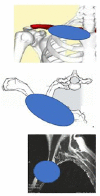 |
Wide resection with 1st rib resection, bone reconstruction (consider joined surgery with thoracic surgeon). |
4. Conclusion
Surgery is still the main treatment modality for primary clavicle tumors and tumorous lesions. This study confirmed that clavicle resection in management of medial third clavicle tumor is technically demanding. Preoperative planning to perform safe surgery is mandatory. We recommend three types of clavicular resection based on tumor extension. Reconstruction surgery following clavicle resection can be done in order to restore symmetry of the lower neck and upper chest, protect nearby neurovascular bundle, and rarely associated with the significant shoulder function loss.
Patients consent
Written informed consent was obtained from the patients for publication of this case report and accompanying images. A copy of the written consent is available for review by the Editor-in-Chief of this journal on request.
Ethical approval
Ethical approval is exempt at our institution.
Funding
The authors received no financial support for the research, authorship, and/or publication of this article.
Author contribution
Yogi Prabowo: study concept, data collection, data interpretation, providing revisions to scientific content of manuscript
M. Fajrin Armin: data collection, writing the paper
Sammy Saleh Alhuraiby: writing the paper, data interpretation
Anissa Feby Canintika: writing the paper, providing grammatical revisions of manuscript
Guarantor
Yogi Prabowo
Research registration number
Not applicable.
Conflict of interest statement
The authors certify that they have no affiliations with or involvement in any organization or entity with any financial interest or non-financial interest in the subject matter or materials discussed in this manuscript.
References
- 1.Ren K., Wu S., Shi X., Zhao J., Liu X. Primary clavicle tumors and tumorous lesions: a review of 206 cases in East Asia. Arch. Orthop. Trauma Surg. 2012 doi: 10.1007/s00402-012-1462-2. Published online. [DOI] [PubMed] [Google Scholar]
- 2.Abdehgah A.G., Molavi B., Mehrpour S.R., et al. Clavicular chondrosarcoma: a case report and brief review of the literature. Int. J. Hematol. -Oncol. 2016;10(3):191–194. Published online. [PMC free article] [PubMed] [Google Scholar]
- 3.Agha R.A., Franchi T., Sohrabi C., The S.C.A.R.E., et al. Guideline: updating consensus surgical CAse REport (SCARE) guidelines. Int. J. Surg. 2020;2020:84. doi: 10.1016/j.ijsu.2020.10.034. [DOI] [PubMed] [Google Scholar]
- 4.Kapoor S., Tiwari A., Kapoor S. Primary tumours and tumorous lesions of clavicle. Int. Orthop. 2008 doi: 10.1007/s00264-007-0397-7. Published online. [DOI] [PMC free article] [PubMed] [Google Scholar]
- 5.Liu Y., Huang X.Y., Feng W.Y., et al. Analysis of the clinical efficacy of tumor resection methods used in 20 patients with clavicular tumor. World J. Surg. Oncol. 2019;17(1):9–11. doi: 10.1186/s12957-019-1642-4. [DOI] [PMC free article] [PubMed] [Google Scholar]
- 6.Lin B., He Y., Xu Y., Sha M. Outcome of bone defect reconstruction with clavicle bone cement prosthesis after tumor resection: a case series study. BMC Musculoskelet. Disord. 2014 doi: 10.1186/1471-2474-15-183. Published online. [DOI] [PMC free article] [PubMed] [Google Scholar]
- 7.Gurd F.B. The treatment of complete dislocation of the outer end of the clavicle: an hitherto undescribed operation. Ann. Surg. 1941 doi: 10.1097/00000658-194106000-00045. Published online. [DOI] [PMC free article] [PubMed] [Google Scholar]
- 8.Cahueque M., Macias D., Moreno G. Reconstruction with non-vascularized fibular autograft after resection of clavicular benign tumor. J. Orthop. 2015 doi: 10.1016/j.jor.2015.10.008. Published online. [DOI] [PMC free article] [PubMed] [Google Scholar]
- 9.Faber K.J., Patterson S.D., Heathcote J.G., Richard R.R. Osteoblastoma of the clavicle. J. South. Orthop. Assoc. 2003;12(2):66–70. Published online. [PubMed] [Google Scholar]
- 10.Basarir K., Polat O., Saglik Y., Yildiz Y. Bone tumors of the clavicle: risk of malignancy in the elderly and safe needle biopsy. Orthopedics. 2010 doi: 10.3928/01477447-20100429-16. Published online. [DOI] [PubMed] [Google Scholar]
- 11.Abbott L.C., Lucas D.B. The function of the clavicle; its surgical significance. 1954;4(140):583–599. doi: 10.1097/00000658-195410000-00014. [DOI] [PMC free article] [PubMed] [Google Scholar]
- 12.Abbott L.C., Lucas D.B. The function of the clavicle; its surgical significance. Ann. Surg. 1954;140(4):583–599. doi: 10.1097/00000658-195410000-00014. [DOI] [PMC free article] [PubMed] [Google Scholar]
- 13.Sugarbaker D., Bueno R., Colson Y. second ed. McGraw-Hill; 2015. Adult Chest Surgery. [Google Scholar]
- 14.Wood V.E. The results of total claviculectomy. Clin. Orthop. Relat. Res. 1986;207:186–190. [PubMed] [Google Scholar]
- 15.Moseley H.F. The clavicle - its anatomy and function. Clin. Orthop. 1968;58:17. [PubMed] [Google Scholar]
- 16.Abbott L.C., Lucas D.B. The function of the clavicle; its surgical significance. 1954;4(140):583–599. doi: 10.1097/00000658-195410000-00014. [DOI] [PMC free article] [PubMed] [Google Scholar]
- 17.Lewis M.M., Ballet F.L., Kroll P.G., Bloom N. En bloc clavicular resection: operative procedure and postoperative testing of function. Case reports. Clin. Orthop. Relat. Res. 1985;193):214-220 [PubMed] [Google Scholar]
- 18.Krishnan S.G., Schiffern S.C., Pennington S.D., Rimlawi M., Burkhead W.Z. Functional outcomes after total claviculectomy as a salvage procedure. A series of six cases. J. Bone Joint Surg. Am. 2007;89(6):1215–1219. doi: 10.2106/JBJS.E.01436. [DOI] [PubMed] [Google Scholar]