Abstract
Cell-free genetically encoded biosensors have been developed to detect small molecules and nucleic acids, but they have yet to be reliably engineered to detect proteins. Here we develop an automated platform to convert protein-binding RNA aptamers into riboswitch sensors that operate within low-cost cell-free assays. We demonstrate the platform by engineering 35 protein-sensing riboswitches for human monomeric C-reactive protein, human interleukin-32γ, and phage MS2 coat protein. The riboswitch sensors regulate output expression levels by up to 16-fold with input protein concentrations within the human serum range. We identify two distinct mechanisms governing riboswitch-mediated regulation of translation rates and leverage computational analysis to refine the protein-binding aptamer regions, improving design accuracy. Overall, we expand the cell-free sensor toolbox and demonstrate how computational design is used to develop protein-sensing riboswitches with future applications as low-cost medical diagnostics.
Subject terms: Riboswitches, Synthetic biology, Computational biophysics
Cell-free genetically encoded biosensors have been developed to detect small molecules and nucleic acids, but they have yet to be reliably engineered to detect proteins. Here the authors develop an automated platform to convert protein-binding RNA aptamers into riboswitch sensors that operate within low-cost cell-free assays.
Introduction
Synthetic biologists have created a wide variety of sensor systems to detect small molecules and nucleic acids1–13. Several of these sensors have been developed for usage in cell-free expression systems, where there is no barrier between the expression machinery and exogenously added bulky macromolecules that would otherwise be unable to pass through a cellular membrane14–16. Cell-free sensors are particularly useful as low-cost, portable diagnostic and field assays as they genetically encode their own detection machinery and do not require a cold chain during storage and distribution14,15,17–22. However, even though protein detection is a cornerstone of both modern medical diagnostics and biological research23, there are only a few cell-free sensors that utilize gene regulation to detect proteins of interest24–26.
Currently, measuring protein titers is widely carried out using immunoassays (e.g., enzyme-linked immunosorbent assays) or liquid chromatography–mass spectrometry analytics, which can offer high sensitivity and specificity across a diverse range of protein targets27,28. More recently, another class of nucleic acid-based recognition elements, called aptamers, have been harnessed for protein detection and diagnostics29,30. Protein-binding aptamers are now available for specific binding to a wide variety of targets, including human proteins31–34, HIV viral proteins35,36, and bacterial toxins37. However, these assays require expensive detection reagents (e.g., purified antibodies or synthesized aptamers), expensive and bulky instruments, sample cold chain storage and distribution, and trained personnel. Instead, it is possible to utilize RNA-based aptamers to develop low-cost, genetically encoded riboswitch biosensors that carry out in situ protein detection within cell-free expression systems (TX-TL)38. Past efforts to engineer such riboswitch sensors have largely relied on trial-and-error experimentation, for example, constructing and characterizing large random libraries to identify riboswitch variants that work best. Instead, it is possible to apply biophysical modeling and computational design to engineer riboswitch sensors to directly couple protein binding to gene regulation, thereby creating a sense-and-respond capability without trial-and-error experimentation.
Here, we applied biophysical modeling and computational design to engineer protein-detecting riboswitches that directly regulate the expression of a desired output protein within the TX-TL cell-free expression system, utilizing our Riboswitch Calculator algorithm to automatically convert RNA aptamers into designed riboswitch sequences39. We initially engineered riboswitches to detect the phage MS2 coat protein as a proof-of-principle, followed by engineering riboswitches to detect human monomeric C-reactive protein (mCRP) and interleukin-32 gamma (IL-32γ) as examples of medically relevant biomarkers. The best riboswitch sensors regulated reporter expression levels by 13.8, 15.9, and 2.5-fold when sensing the MS2, mCRP, and IL-32γ proteins, respectively, at biomarker concentrations of 1.25 μM mCRP and 0.78 μM IL-32γ. We demonstrated that these riboswitches controlled gene expression levels via two distinct mechanisms: (i) protein-induced conformational changes to RNA structure, which modifies the ribosome’s ability to initiate translation; and (ii) protein-dependent steric repression, which blocks the ribosome from binding to the 5′ untranslated region. We critically tested the accuracy of the Riboswitch Calculator model predictions and found that improving the specification of the protein-aptamer interaction led to higher model accuracy. Overall, our automated design approach can be applied to convert any protein-binding RNA aptamer into a protein-detecting, cell-free biosensor with potential applications as portable, low-cost diagnostics.
Results
Riboswitch design and characterization platform
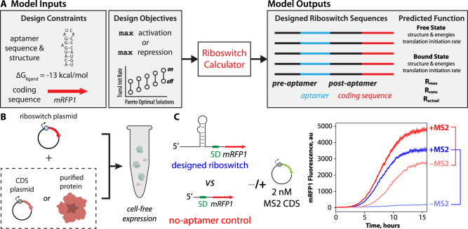
We created protein-binding riboswitch sequences using a biophysical model of translation-regulating riboswitches called the Riboswitch Calculator, which combines statistical thermodynamics with computational optimization to design synthetic riboswitches according to inputted specifications39. The design specifications include (i) the sequence of an RNA aptamer that binds to the protein of interest; (ii) the secondary structure of the RNA aptamer when it is bound by the protein; (iii) the protein’s binding free energy (or binding affinity) to the RNA aptamer; and (iv) the coding sequence of the protein whose expression level is regulated by the riboswitch (Fig. 1A). The Riboswitch Calculator then identifies synthetic pre-aptamer and post-aptamer sequences that maximize the riboswitch’s dynamic range, utilizing a genetic algorithm to carry out computational multi-objective sequence optimization. Together, these pre-aptamer and post-aptamer sequences vary in length from 44 to 55 nucleotides, creating an overall searchable sequence space of 1026 to 1033 sequences. When designing riboswitches that activate translation (ON switches), the activation ratio is Rmax = TIRbound/TIRunbound, where TIRbound and TIRunbound are the mRNA’s translation initiation rates in the protein-bound and unbound states, respectively. When designing riboswitches that repress the translation rate (OFF switches), the repression ratio is Rmax = TIRunbound/TIRbound. Multiple equally optimal riboswitch sequences are plausible. The Riboswitch Calculator identifies the Pareto-optimal set of synthetic riboswitch sequences that are all predicted to maximize Rmax with varying magnitudes of TIRbound and TIRunbound.
Fig. 1. Design of protein-sensing riboswitches and cell-free riboswitch characterization.
A A model of translation-regulating riboswitches takes as inputs the sequence, structure, and binding free energy of a protein-binding RNA aptamer. The model designs candidate riboswitch sequences that maximize activation or repression of translation initiation in response to changing protein concentrations. B The riboswitch sensor is tested in a cell-free expression system (TX-TL), adding either protein expression plasmid or purified protein. C Riboswitch sensor function is characterized by measuring reporter protein expression levels in response to changing protein-ligand concentrations alongside the same measurements on a no-aptamer control. The riboswitch activation or repression ratio is determined by comparing reporter expression levels, including the no-aptamer control measurements, to exclude non-specific interactions. Example cell-free assay measurements include (light blue), an MS2-sensing riboswitch without added MS2 protein; (dark blue) an MS2-sensing riboswitch with added MS2 expression plasmid; (light red) the no-aptamer control without added MS2 protein; and (dark red) the no-aptamer control with added MS2 expression plasmid. Lines are the mean mRFP1 fluorescence levels at each time point. Shaded regions are the 95% confidence interval at each time point (N = 6 biological replicates).
To do this, the Riboswitch Calculator uses a statistical thermodynamic model, called the RBS Calculator, to calculate the interaction energies between the ribosome and mRNA that control its translation initiation rate40,41. The strengths of these interactions are determined by a 5-term Gibbs free energy model, including (i) conformational distortions when ribosomes initially bind to upstream standby sites in the mRNA; (ii) hybridization between the last 9 nucleotides of the 16S rRNA and the mRNA at the Shine–Dalgarno (SD) sequence; (iii) the unfolding of inhibitory mRNA structures that overlap with the ribosome’s footprint, spanning the region from the 5′ end of the SD sequence to 13 nucleotides past the start codon; (iv) base pairing between the start codon and the initiating tRNAfMet; and (v) ribosomal stretching or compression due to non-optimal spacer sequences between the SD and start codon. The overall result is the binding free energy of the ribosome to the mRNA (ΔGtotal). By convention, a stronger interaction is denoted by a more negative binding free energy. According to Boltzmann’s relationship, the mRNA’s translation initiation rate is proportional to exp(−β ΔGtotal), where beta is a constant that relates free energies to state probabilities40.
To predict the riboswitch function, the Riboswitch Calculator carries out free energy calculations on the protein-bound and unbound states of the mRNA (ΔGtotal,bound and ΔGtotal,unbound). In the unbound state, the RNA aptamer is indistinguishable from other parts of the mRNA and is allowed to form any secondary structure as part of the overall free energy minimization procedure, which utilizes the ViennaRNA suite of RNA folding algorithms42. In the protein-bound state, the RNA aptamer is locked into its protein-bound structure, which can alter the accessibility of the standby site, the folding free energies of inhibitory mRNA structures, and the overall binding free energy of the ribosome to the mRNA. The switching free energy is also calculated to determine the thermodynamic stability of the protein-bound state, using the protein’s binding free energy to the RNA aptamer (ΔGligand). The model predicts that the maximum fold-change in the mRNA’s translation rate is exp(−β [ΔGtotal,bound −ΔGtotal,unbound]) when the protein-bound state is stable and when an excess amount of protein is added. Riboswitch Calculator predictions were previously applied to engineer 62 synthetic riboswitches that detected a variety of small molecules (theophylline, tetramethylrosamineluoride, dopamine, thyroxine, 2,4-dinitrotoluene) and activated a protein reporter’s translation rate by up to 383-fold39. However, the model has not yet been applied to convert protein-binding RNA aptamers into cell-free biosensors.
We characterized riboswitch function using the cell-free TX-TL expression system43, adding plasmid-encoded genetic circuits that utilize each riboswitch to regulate the expression of the mRFP1 fluorescent protein reporter (Methods). We used two different approaches to add varying concentrations of protein ligands to the cell-free expression system: (i) co-expression or (ii) co-addition. For co-expression, we added varying amounts of a second expression plasmid to the cell-free expression system to constitutively produce the protein of interest. For co-addition, we directly added the purified protein of interest to the cell-free expression system. mRFP1 fluorescence levels were measured every 10 min using spectrophotometry (TECAN Spark), followed by endpoint analysis to quantify the overall change in reporter expression levels (Fig. 1B).
Prior work has demonstrated that gene expression in both the cellular and cell-free context is sensitive to added components or additional genetic load44–49. We carried out two types of controls to eliminate such confounding factors. In the first set of no-mRFP1 controls, we measured red fluorescence levels without any mRFP1 expression to quantify the autofluorescence of the cell-free assay and protein ligands. During endpoint analysis, we subtracted no-mRFP1 autofluorescence from all measurements. In the second set of no-aptamer controls, we measured red fluorescence levels from a genetic circuit that expresses mRFP1 using a standard 5′ UTR without any protein-binding aptamer (UTR-13641). According to the RBS Calculator v2.1 model, UTR-136 binds to the ribosome with a moderately high translation initiation rate (36,400 au or ΔGtotal = −7.52 kcal/mol). The purpose of this no-aptamer control is to measure any non-specific changes in red fluorescence levels when co-expressing or co-adding a protein-ligand at varying concentrations. If we detect non-specific activation or repression from the no-aptamer control, we remove this confounding factor by dividing the riboswitch’s measured activation or repression ratio by the no-aptamer control’s activation or repression ratio, respectively (Fig. 1C). If these riboswitch sensors are to be used as sensors in a future device, it would be expected that the same controls would be performed on the device, in parallel, to carry out these same measurements and analysis.
Design and characterization of MS2, mCRP, and IL32γ Riboswitch Sensors
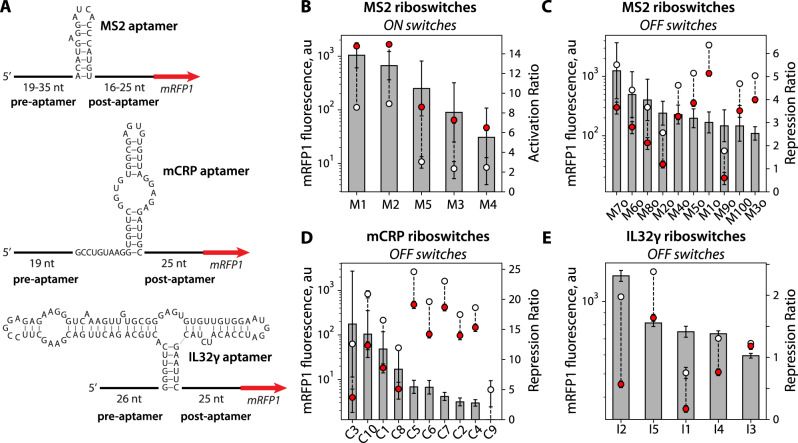
For the first proof-of-principle example, we engineered riboswitches to detect the phage MS2 coat protein, utilizing an RNA aptamer that folds into a well-defined hairpin structure and binds to MS2 protein with very high affinity (Kd = 0.7 nM)50,51 (Fig. 2A). We then selected two medically relevant protein biomarkers, engineering riboswitches to detect changes in the levels of monomeric C-reactive protein (mCRP) and interleukin-32 gamma (IL-32γ). CRP is found in human plasma and natively forms a homopentameric complex, but will irreversibly dissociate into its monomeric form during a pro-inflammatory response (e.g., tissue damage, heart disease, cancer, or bacterial infection)52,53. An mCRP concentration of 10 mg/L (430 nM) is considered elevated, and depending on the disease severity, mCRP concentrations can readily exceed 100 mg/L (4.3 μM). To develop mCRP-sensing riboswitches, we harnessed a hairpin RNA aptamer that binds specifically to the monomeric form of CRP (mCRP) with high affinity (Kd = 187.7 nM) and does not bind to pentameric CRP54. IL-32 is a cytokine that regulates the NF-κB pathway and acts during early host defense against pathogen infections55–57. IL-32γ is the longest and most pro-inflammatory isoform of IL-32. Elevated levels of IL-32γ in human serum have been found to be associated with heart failure, COPD, and multiple myeloma58 (up to around 61 pM or 1.6 ng/ml). To develop IL-32γ sensing riboswitches, we utilized a highly structured RNA aptamer that binds specifically to the gamma isoform with a high affinity (Kd = 78 nM)59. Altogether, we initially designed, constructed, and characterized 30 riboswitches to detect MS2, mCRP, and IL-32γ protein, including ON switches that activated mRFP1 expression and OFF switches that repressed mRFP1 expression (Fig. 2A). All sequences, model calculations, experimental and control measurements, and statistical significance tests (two-tailed t-tests with unequal variances) are available in the Source Data.
Fig. 2. Design and function of protein-sensing riboswitches.
A Riboswitch sequences designed for detecting the MS2, mCRP, and IL32γ protein ligands. B MS2 ON switches, induced with 8 nM MS2 CDS. C Characterization of MS2 OFF switches, induced with 16 nM MS2 CDS. D Characterization of mCRP OFF switches, induced with 1.25 μM of mCRP. E IL32γ OFF switches, induced with 780 nM IL32γ. Gray bars are the mean activation or repression ratios for designed riboswitches. White circles are mean mRFP1 fluorescence levels in the OFF state. Red circles are mean mRFP1 fluorescence levels in the ON state. Error bars are the 95% confidence intervals (N = 6 biological replicates for MS2 ON switches; N = 8 biological replicates for mCRP and IL32γ OFF switches).
We first designed and tested five MS2-sensing riboswitches to activate mRFP1 expression (ON switches) and carried out TX-TL assays using the co-expression approach to produce the MS2 protein. When adding 8 nM pFTV1-MS2, we found that the designed MS2-sensing ON switches all increased mRFP1 fluorescence levels by significant amounts (between 10.8- to 27-fold). We carried out the same measurements using our no-aptamer control and found that co-expression of MS2 non-specifically increased mRFP1 fluorescence levels by 1.9-fold (Source Data). We then used the no-aptamer control measurement to remove the non-specific effect of MS2 on mRFP1 expression (Methods). After removing the non-specific effect, we found that all five MS2-sensing ON switches activated mRFP1 expression between 5.5- and 13.8-fold (Fig. 2B). These results show that the Riboswitch Calculator was able to design MS2-sensing ON switches that significantly activated output protein expression with the highest activated ratios reported to date, compared to prior efforts26, though the dynamic ranges varied across the small number of designs tested.
We then designed and tested ten MS2-sensing riboswitches to repress mRFP1 expression (OFF switches), now adding 16 nM pFTV1-MS2 plasmid to the cell-free expression system as translation-repressing riboswitches are predicted to require higher protein concentrations to achieve a similar fold-change in output as compared to translation-activating riboswitches (Methods). Under the same conditions, the no-aptamer control increased mRFP1 fluorescence levels by 2.3-fold. In this scenario, a non-functional OFF switch would also cause mRFP1 fluorescence levels to increase by 2.3-fold due to this non-specific effect. Instead, we found that adding 16 nM pFTV1-MS2 plasmid caused all of the 10 MS2-sensing OFF switches to produce reduced mRFP1 fluorescence levels. After removing the non-specific effect, we found that their repression ratios varied from 2.5- to 5.3-fold (Fig. 2C). These results show that the Riboswitch Calculator can design MS2-sensing OFF switches by simply flipping its objective function during sequence optimization while utilizing the same biophysical calculations.
We next designed ten mCRP-sensing riboswitches and characterized their ability to repress mRFP1 expression (OFF switches). We initially added 16 nM of a plasmid expressing mCRP (co-expression), but observed no significant change in mRFP1 regulation. Instead, when we directly added 1250 nM of purified mCRP (co-addition), we found that the OFF switches decreased mRFP1 fluorescence levels by up to 6.7-fold. As the purified mCRP was produced using HEK 293 cells, it is possible that chaperone-assisted protein folding or glycosylation is needed to produce an mCRP that binds to the RNA aptamer. Similar to the MS2 protein, we found from our no-aptamer controls that the addition of 1250 nM mCRP non-specifically activated mRFP1 expression by about 2.3-fold (Source Data). Once we removed this non-specific effect, we found that 90% of the mCRP-sensing OFF switches successfully repressed mRFP1 expression with repression ratios from 2.8-fold to 15.9-fold (Fig. 2D).
Finally, we designed five IL32γ-sensing riboswitches and characterized their ability to repress mRFP1 expression (OFF switches). We directly added 780 nM of purified IL32γ to the cell-free expression system (co-addition). Using our no-aptamer controls, we found no significant non-specific effect on mRFP1 expression levels (Source Data), precluding the need to remove any non-specific effect. After characterizing the OFF switches, we found that 4 out of the 5 riboswitches were able to significantly repress mRFP1 fluorescence levels from 1.4- to 2.5-fold (Fig. 2E). These results show that the Riboswitch Calculator was able to harness RNA aptamers that bind to human protein biomarkers to successfully design translation-repressing riboswitches.
Structural and energetic contributions to riboswitch function
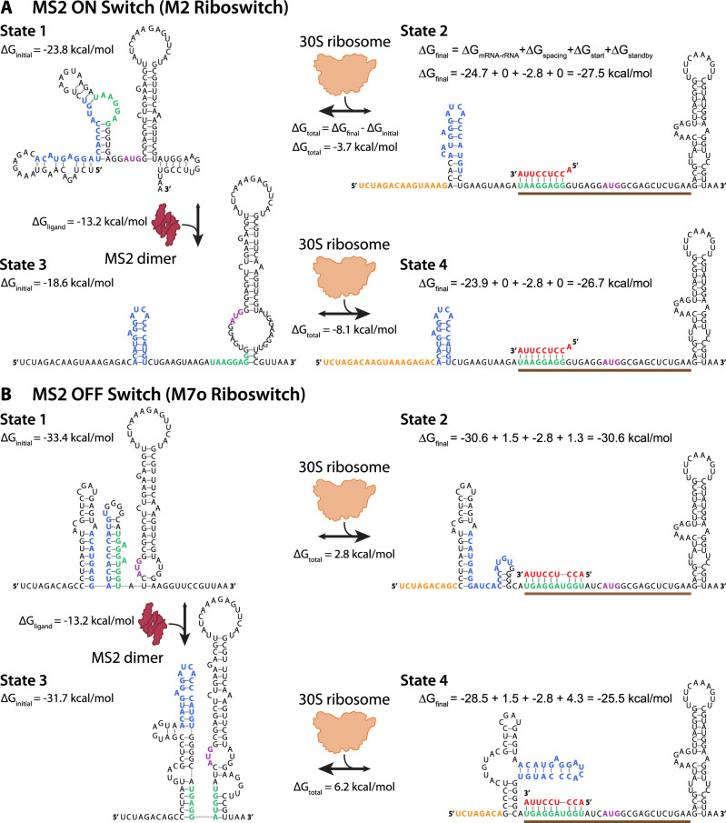
We next investigated the mechanisms responsible for riboswitch function to explain how protein binding to an aptamer domain can cause the riboswitch’s translation rate to be either activated or repressed. As examples, we focus on an MS2-binding ON switch (M2 riboswitch) and an MS2-binding OFF switch (M7o riboswitch), applying the Riboswitch Calculator model’s structural and thermodynamic calculations to visualize and quantify the process39. In Fig. 3A, we show the sequence, structure, and interactions of the M2 riboswitch in its free state (state 1) and in its ribosome-bound state (state 2) in the absence of the MS2 protein. Before the ribosome has bound, the riboswitch mRNA is predicted to fold into a stable structure where the aptamer domain and SD sequence are both partly sequestered by base pairing. After the ribosome has bound to form a pre-initiation complex, there are significant structural re-arrangements, including unfolding an inhibitory structure within the N-terminal CDS region40,60. While the structures provide visual cues, the ribosome’s ability to bind to the mRNA and initiate translation rate is actually controlled by the difference in Gibbs free energy between the initial and final states. In the absence of MS2, this difference is −3.7 kcal/mol.
Fig. 3. Sequence, structure, and interactions controlling riboswitch function.
The model-predicted mRNA structures and ribosome-mRNA binding free energies for the A MS2 ON switch (M2 riboswitch) and the B MS2 OFF switch (M7o riboswitch) across its four most relevant states. State 1 shows the initial mRNA structure and its Gibbs free energy of folding (ΔGinitial) when the MS2 coat protein is not bound. State 2 shows the change in mRNA structure and the final Gibbs free energy (ΔGfinal) when the ribosome binds to the mRNA in State 1. The free energy model for calculating ΔGfinal includes hybridization between the mRNA and ribosomal RNA (ΔGmRNA-rRNA), base pairing between the tRNA and start codon (ΔGstart), and energetic penalties for non-optimal spacing (ΔGspacing) and an inaccessible standby site (ΔGstandby). State 3 shows how the mRNA structure changes when the MS2 coat protein binds to its cognate RNA aptamer with binding free energy ΔGligand, which results in a change in mRNA folding free energy (ΔGinitial). State 4 shows the change in mRNA structure and final Gibbs free energy (ΔGfinal) when the ribosome binds to the mRNA in State 3. The translation initiation rates are predicted based on the difference in initial and final Gibbs free energies (ΔGtotal) according to Boltzmann’s relationship. Nucleotides are colored according to their interactions, including (blue) the RNA aptamer domain, (red) the last 9 nucleotides of the 16S ribosomal RNA, (green) the Shine–Dalgarno sequence, (purple) the start codon, and (orange) the standby site. The brown bar is the ribosomal footprint for initiation.
We then illustrate how the mRNA’s structure and ribosomal interactions are altered when the riboswitch is bound by MS2. When MS2 binds to its aptamer domain, the model predicts a substantial refolding of the 5′ UTR, almost completely exposing the SD sequence and creating a highly accessible standby site for ribosome binding (Fig. 3A, state 3). This structural re-arrangement requires an input of at least 5.2 kcal/mol energy to push the transition forward, which is provided by the −13.2 kcal/mol energy released when MS2 binds to its aptamer domain. The MS2-bound mRNA can now bind to the ribosome to form a more stable pre-initiation complex (Fig. 3A, state 4) with a more negative binding free energy (−8.1 kcal/mol), leading to activation of the translation rate. Like all models, the calculations provide a simplified version of reality that nonetheless enable riboswitch prediction and design. For example, the model considers only the four most predominant states of the riboswitch, though there exists an ensemble of states with varied mRNA structures and MS2 binding occupancies. Notably, MS2 binds to its aptamer while the mRNA is being transcribed, which can eliminate kinetic traps.
We next focus on the mechanism of an OFF switch, illustrating how MS2 binding triggers refolding of the mRNA to make it more energetically unfavorable for the ribosome to bind. In the absence of MS2, the model predicts that the initial state of the M7o riboswitch folds into highly stable mRNA structures that sequester the aptamer domain and the SD sequence (Fig. 3B, state 1). This mRNA structure does not favorably bind to the ribosome (2.8 kcal/mol), particularly due to the short distance between the SD and starts codon and the presence of mRNA structures that lower the accessibility of the standby site (Fig. 3B, state 2). However, once MS2 binds to the mRNA, the model predicts that the mRNA will bind even less favorably to the ribosome, due to a reconfiguring of both the initial and final mRNA states (Fig. 3B, states 3 and 4). The MS2-bound mRNA contains a highly stable structure that occludes the ribosome’s standby site41 and adds a penalty of 3 kcal/mol to the ribosome’s total binding free energy (6.2 kcal/mol). Overall, using the model, we can quantitatively understand how MS2 binds to the aptamer domain and causes large-scale re-arrangements in the riboswitch’s mRNA structure to control ribosome binding free energies and translation initiation rates.
Dose–response characterization of designed riboswitch sensors
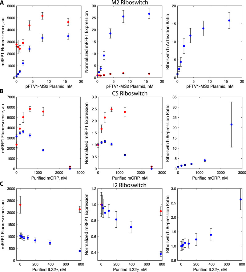
We next selected high-functioning riboswitches (M2, C5, and I2) and measured how systematically increasing the MS2, mCRP, and IL32γ concentrations affected their output expression levels (dose–response) as compared to the no-aptamer control (UTR-136). All measurements and statistical tests are available in the Source Data. We first reconfirmed that the cell-free expression system had sufficient capacity to express large amounts of mRFP1 in proportion to the mRFP1 expression plasmid across a wide range (0–32 nM plasmid added) (Supplementary Fig. 1). We then characterized the dose–response of the M2 riboswitch by keeping the riboswitch plasmid concentration constant and systematically increasing the concentration of the MS2 expression plasmid from 0 to 16 nM. The M2 riboswitch activated mRFP1 fluorescence levels with a dose-dependent sigmoidal behavior, increasing mRFP1 expression levels by up to 26.6-fold (Fig. 4A). Under the same conditions, the mRFP1 fluorescence levels from the no-aptamer control increased by up to 1.86-fold. When correcting for this non-specific effect, the M5 riboswitch activated mRFP1 expression levels by 15.9-fold. Activation of mRFP1 expression was detectable with statistical significance (p = 0.0008, two-tailed t-test) using only 0.5 nM of expression plasmid. These results confirm the expected sigmoidal dose response for a translation-activating riboswitch, consistent with previously engineered riboswitches39.
Fig. 4. Dose–response of MS2, CRP, and IL32γ riboswitches.
The measured mRFP1 fluorescence levels, normalized mRFP1 expression levels, and activation or repression ratios of the A M2, B C5, and C I2 riboswitch sensors (blue dots) in response to varied concentrations of the MS2 expression plasmid, purified human mCRP protein, and purified human IL32γ protein, respectively. The measured mRFP1 fluorescence levels and normalized mRFP1 expression levels of the no-aptamer control (UTR-136) under the same conditions (red dots). Dots and bars are the mean and standard deviation of replicate cell-free assays (A N = 8 biological replicates, B N = 8 biological replicates, C N = 6 biological replicates).
We then carried out the same dose–response characterization on the C5 riboswitch, directly adding up to 2500 nM of purified mCRP to cell-free assays. We found that the C5 riboswitch lowered mRFP1 fluorescence levels by 1.74-fold at 1250 nM mCRP and by 237-fold at 2500 nM mCRP (Fig. 4B). However, we found that the no-aptamer control exhibited a non-linear dosage response such that mRFP1 fluorescence levels were non-specifically increased by 2.37-fold at 1250 nM mCRP and non-specifically decreased by 11-fold at 2500 nM mCRP. Based on these measurements, mCRP non-specifically activates mRFP1 expression at lower concentrations, but then interferes with cell-free expression at higher concentrations. After taking into account these non-specific effects, the C5 riboswitch was found to repress mRFP1 expression by 4.1-fold at 1250 nM mCRP and by 21.5-fold at 2500 nM mCRP, though the interference with the cell-free assay at 2500 nM mCRP substantially increased the variability of the measurement. Repression was detectable with statistical significance at 156 nM mCRP, which is the lowest tested concentration above zero (p = 0.0005, two-tailed t-test), while half-maximal repression was achieved at 403 nM mCRP. These results show that the C5 riboswitch is sensitive enough to detect and quantify mCRP concentrations across the physiological range from normal levels (100–430 nM) to elevated levels (above 430 nM), though levels above 2500 nM will inhibit cell-free expression.
We repeated the same cell-free assays using the I2 riboswitch, directly adding up to 780 nM of purified IL32γ. We found that the I2 riboswitch lowered mRFP1 fluorescence levels by 2.6-fold at the highest IL32γ concentration (Fig. 4C). In comparison, the no-aptamer control exhibited non-specific repression of only 1.09-fold, which was statistically indistinguishable from the baseline (p = 0.18, two-tailed t-test). Using the I2 riboswitch, repression of mRFP1 expression levels was detectable with statistical significance at a concentration of 195 nM IL32γ with half-maximal repression taking place at about 548 nM IL32γ (p = 0.015, two-tailed t-test). However, using the current IL32γ aptamer (Kd = 78 nM), the I2 riboswitch could not sense IL32γ levels within the picomolar range, which would be needed to distinguish between normal and elevated levels in human serum.
Translation repression by steric hindrance and structural switching
We next investigated the mechanisms responsible for protein-induced translational repression with the goal of distinguishing between two types of interactions. When a protein binds specifically to the aptamer region of the mRNA, we anticipate that translational repression could be exerted by steric hindrance alone; a stably bound protein could prevent the 30S ribosomal subunit from associating with the mRNA, for example, at upstream standby sites, or it could block the 30S ribosomal subunit’s 16S rRNA from hybridizing to the SD sequence to form a stable ternary complex. As a second mechanism, when a protein binds specifically to the aptamer region, it can induce changes in the mRNA structure, including the formation or removal of inhibitory mRNA structures that the ribosome must unfold prior to initiating translation. These mechanisms are layered on top of any non-specific interactions when adding protein to the cell-free system. Using a learn-by-design approach to distinguish these mechanisms, we designed and characterized several mCRP-binding riboswitches that employ either the first mechanism alone (“steric switches”) or a combination of both mechanisms together (“OFF switches”) alongside no-aptamer controls that measure the effects of non-specific interactions. For these experiments, we began using a second batch of purified mCRP that, through comparative testing on the same riboswitch (C10), exhibited about 4.1-fold less activity, though the cell-free composition remained the same (Source Data).
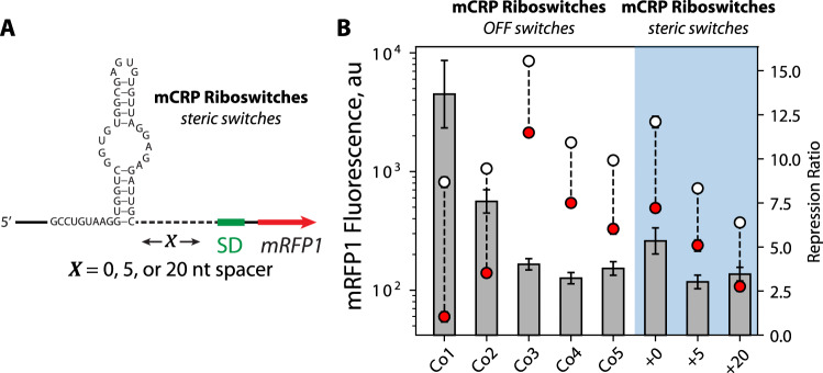
For the steric switch designs, we inserted the mCRP-binding aptamer upstream of a consensus SD sequence at varying distances (0–20 nt upstream of the 5′ ends of the SD sequence). We also designed the pre-aptamer and post-aptamer so that the mRNA’s structure remained the same in both the free and mCRP-bound states, eliminating the second mechanism as a source of translation regulation (Fig. 5A). We then characterized how well these riboswitches regulate mRFP1 expression levels, adding either 0 or 1250 nM mCRP. We found that placing the mCRP-binding aptamer directly upstream of the SD sequence lowered mRFP1 fluorescence levels by 3.2-fold. After using our no-aptamer controls to remove the effect of the non-specific interactions (1250 nM mCRP activated mRFP1 fluorescence levels by 1.65-fold), we found that specific binding of mCRP repressed mRFP1 expression by 5.3-fold, showing that steric hindrance alone is greatly contributing to protein-induced translational repression (Fig. 5B). When the aptamer was placed farther upstream of the SD sequence (5 or 20 nt), mRFP1 expression was repressed by smaller, but similar magnitudes, of 3.0 and 3.4-fold, respectively, again removing the non-specific interactions’ contributions. These results show that steric hindrance can repress translation even when the aptamer is farther from the SD sequence.
Fig. 5. Placement of mCRP aptamer in the standby site of the 5′UTR represses gene expression.
A The sequence and structure of the mCRP-binding steric switches, showing the spacer length (X) separating the mCRP aptamer and a consensus Shine–Dalgarno sequence. B The repression ratios of the designed mCRP OFF switches (Co1–Co5) and mCRP steric switches (+0, +5, +20) are compared. Gray bars are mean repression ratios. White circles are mean mRFP1 fluorescence levels in the OFF state. Red circles are the mean mRFP1 fluorescence levels in the ON state. Error bars are the 95% confidence intervals (N = 8 biological replicates).
In the second set of OFF switch designs, we applied the Riboswitch Calculator algorithm to design 5 additional mCRP-binding riboswitches (Co1 to Co5) while varying the distance between the aptamer region and the SD-like sequence from 4 to 13 nt. We found that mRFP1 expression levels were repressed by 3.2- to 13.6-fold (Fig. 5C) while taking into account the no-aptamer controls. Two of the OFF switches (Co1, Co2) had repression ratios far greater than the steric switches, showing that some of the designed riboswitches are utilizing both mechanisms to regulate translation rate. Overall, these results show that translation repression can take place by either steric inhibition alone or through a combination of both steric inhibition and protein-induced structural re-arrangements that block ribosome binding.
Accuracy analysis and improvement by modifying aptamer structural constraints
We next critically analyzed the Riboswitch Calculator’s model accuracy for protein-sensing riboswitches. We first considered the riboswitches’ OFF states where no protein-ligand is added. The Riboswitch Calculator predicts how well the ribosome binds to the mRNA as quantified by a change in binding free energy (ΔGtotal), which is expected to be related to the mRNA’s translation initiation rate according to the log–linear Boltzmann relationship. We compared the riboswitches’ model-predicted ΔGtotal free energies to the natural logarithm of the end-point mRFP1 expression levels and found statistically significant and quantitative agreement (R2 = 0.47, p = 5.8 × 10⁻⁶, N = 34) (Supplementary Fig. 2). However, we noticed that the expected dynamic range in translation rates was compacted in TX-TL assays as compared to equivalent in vivo assays. In other words, changing the ribosome’s binding free energy to an mRNA had a smaller-than-expected effect on its translation rate. Quantitatively, the apparent Boltzmann factor for these TX-TL assays was β = 0.23 as compared to β = 0.45 in equivalent in vivo assays. This phenomenon has been previously observed in prior comparisons between cell-free and in vivo assays49,61,62.
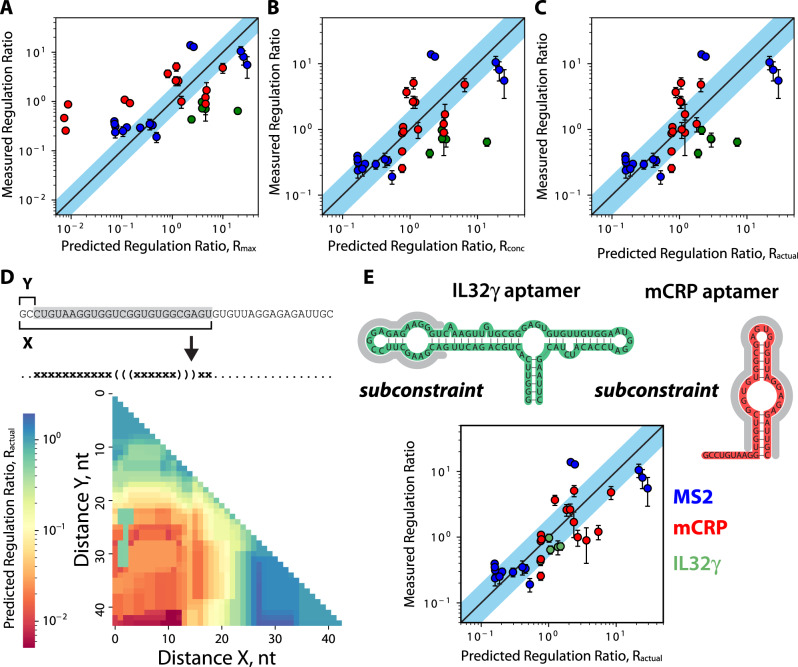
We then analyzed how well the Riboswitch Calculator predicted the activation or repression of mRFP1 expression levels, using three related calculations39: Rmax, Rconc, and Ractual. Rmax is the maximum possible fold-change in translation rate when an excess amount of protein is added, and 100% of the riboswitch is bound by protein, while Rconc is the fold-change in translation rate when a specified concentration of protein is added. Ractual is an extension of the Rconc calculation that additionally takes into account the thermodynamic stability of the riboswitch’s protein-bound state; if the protein’s binding free energy is insufficient to stabilize that state, the riboswitch can spontaneously transition back to its free state. The Rmax and Rconc calculations make simplifying assumptions, whereas the Ractual calculation is the complete version of the current model. In the first comparison, the Rmax prediction had a measurable but weaker correlation with the measured activation or repression ratios (R2 = 0.38, p = 9.3 × 10⁻⁹, N = 34, linear regression) with only 15% of riboswitch variants regulating expression levels to within 2-fold of the predicted ratios (Fig. 6A). However, when we consider the proteins’ concentrations using the Rconc prediction, we find that the model more accurately predicted the riboswitches’ activation or repression ratios (R2 = 0.46, p = 1 × 10⁻⁵, N = 34, linear regression) with 38% of variants regulating expression to within 2-fold of the predicted ratio (Fig. 6B). Finally, using the Ractual prediction to account for differences in the proteins’ binding free energies to their respective RNA aptamers, the model accuracy increased even further (R2 = 0.48, p = 4.7 × 10⁻⁶, N = 34, linear regression) with 44% of the riboswitch variants falling within the 2-fold tolerance (Fig. 6C).
Fig. 6. Optimizing Riboswitch Calculator predictions for in vitro protein riboswitches.
Model predictions were compared to measured riboswitch regulation (activation or repression) ratios, utilizing three different equations that take into account additional interactions for improved accuracy. Model predictions include A the maximum predicted regulation ratio, Rmax, B the predicted regulation ratio when considering the protein concentration, Rconc, and C the predicted actual regulation ratio, Ractual, when considering both the protein concentration and the thermodynamics of the mRNA-protein complex. D Model predictions were recalculated while systematically varying the length of the aptamer subconstraint region, showing riboswitch Co1 as an example. E The optimal aptamer subconstraints for the MS2, mCRP, and IL32γ aptamers were identified and utilized to predict the actual regulation ratio, Ractual, for all riboswitches. Overall accuracy is R2 = 0.64, with 56% of riboswitches predicted to be within 2-fold of their measured activation ratio. Dots and error bars are the mean and standard deviation of data with 6–8 biological replicates.
A key input into the model calculations is the mRNA structure of the aptamer region when it is bound to the protein. When calculating the translation initiation rate of the riboswitch’s protein-bound state, this mRNA structure is “locked” into place, which we call the aptamer structural constraint. In our initial model predictions, we extracted the aptamer region from the studies that developed it and assumed that the entire region is important to protein binding. However, it is possible that only a portion of the aptamer binds to its protein. For example, only 13 nucleotides of the 19-nt long MS2 aptamer are fully resolved in a crystal structure of the aptamer in complex with a MS2 coat protein dimer50. Considering that the mCRP and IL32γ aptamers have longer sequences (44-nt and 90-nt, respectively), it is likely that only a portion of these aptamers is truly “locked” into place when bound by protein.
We therefore investigated how changing the aptamer structural constraint alters the model’s calculations and its predictive accuracy. To do this, we systematically varied the portion of the aptamer region that is “locked” in the protein-bound state, leaving a shorter contiguous region (Fig. 6D). For each of these shorter aptamer subregions, we recalculated its mRNA structure and used that structure as the aptamer’s structural constraint. Here, any flanking unpaired nucleotides were left as unconstrained positions (dots) in the structural constraint, allowing them to refold with adjacent mRNA regions in the riboswitch’s protein-bound state. We then repeated the model’s Ractual calculations. Throughout, all other riboswitch sequences and model parameters remain the same. Overall, changing the aptamer structural constraint varied the model’s predicted Ractual by up to 385-fold with a very rugged response surface, indicating that small changes to this input could have large effects on the model’s predictions (Fig. 6D, Supplementary Fig. 3). We then identified the structural constraint for the mCRP and IL32γ aptamers that achieved the highest overall model accuracy across all riboswitches (Methods). Higher model accuracies were achieved by allowing more of the 5′ portion of the mCRP aptamer to freely fold and by only “locking” a single bulged hairpin structure of the IL32γ aptamer. When inputting these aptamer structural constraints into the model (Source Data), we found that overall accuracy greatly improved (R2 = 0.64, p = 1.2 × 10⁻⁸, N = 34, linear regression) with 56% of the riboswitch variants achieving less than 2-fold prediction error (Fig. 6E). Altogether, this analysis shows that accurately predicting riboswitch function requires correctly inputting the concentration of the protein-ligand, the binding free energy of the protein to its respective aptamer, and the actual aptamer structure that becomes “locked” when in its protein-bound state.
Discussion
We engineered 35 protein-sensing riboswitches that operate within cell-free expression assays, demonstrating an automated model-predictive workflow for converting protein-binding RNA aptamers into high-performance sensors with potential medical diagnostic applications. As demonstrations of our approach, we developed sensors for two human biomarkers—monomeric C-reactive protein and interleukin-32γ protein—that have been previously used as serum-accessible proxies for cardiovascular disease, pro-inflammatory conditions, and pathogen infection. As RNA aptamers can be developed to bind many proteins of interest, our design platform provides a reliable and versatile route to developing larger toolboxes of protein diagnostic assays using a variety of low-cost input and output formats.
We designed these genetically encoded protein sensors using a forward engineering approach, applying our Riboswitch Calculator algorithm to predict the riboswitches’ sequence–structure–function relationship and computationally optimizing riboswitch sequences toward maximum translation activation or repression. Our use of computational design enabled us to reduce the large riboswitch sequence space to just a few designs that have a high chance of success. This approach yielded protein-sensing riboswitches with high activation or repression ratios—13.9-fold, 15.9, and 2.6-fold when detecting the MS2, mCRP, and IL-32γ proteins, respectively—at protein concentrations within the relevant physiological range. Notably, the biophysical model equations and parameters were fixed and constant throughout this study, demonstrating that a quantitative & predictive knowledge of biophysics from one realm can be applied to solve challenges in another realm without requiring new training or validation datasets.
We also investigated the key determinants of protein-sensing riboswitch function and identified two distinct mechanisms responsible for translation regulation. We began by engineering MS2-binding riboswitches that act as either ON and OFF switches, changing only their pre-aptamer and post-aptamer sequences, to show that protein-induced changes to mRNA structure can be harnessed for either translation activation or repression. However, when designing mCRP-sensing OFF switches, we found that steric hindrance alone could explain how mCRP could bind, block the ribosome binding site, and repress the translation rate. While it was possible to engineer mCRP OFF switches that combined both steric inhibition and protein-induced structural inhibition at the same time to repress translation rate, we were not able to engineer mCRP ON switches after testing a small number of candidate designs. In practice, when developing a new protein-sensing riboswitch, it is worthwhile to design both ON and OFF switches to best determine how each protein interacts with the ribosome and whether steric inhibition plays an overriding role.
With our automated platform, it is now feasible to design large toolboxes of riboswitch sensors able to detect small molecule and protein ligands across a range of medical applications. For example, rather than using a fluorescent protein reporter, protein-sensing riboswitches can regulate the expression of enzymes, such as glucose oxidase, enabling electrical current generation as a measurable output signal22. Notably, cell-free expression systems have already been harnessed for such medical diagnostic applications. Though the addition of complex sample matrices (plasma, serum, urine, saliva) has been found to inhibit cell-free expression due to the presence of RNases, it has been shown that co-expression of RNase inhibitors can mitigate this inhibition, unlocking their potential63. A platform combining genetically encoded biosensors and cell-free expression could finally enable multiplexed protein detection in a compact, low-cost device that would provide longitudinal biomarker measurements in non-clinical settings for data-driven medicine.
Methods
Construction of riboswitch and protein-expressing plasmids
To construct riboswitch and CDS plasmids for this study, we started with the pFTV1 vector backbone, which contains mRFP1 modified to contain an N-terminal SacI restriction site41. We used the Riboswitch Calculator to design candidate riboswitch sequences and the Operon Calculator to codon-optimize the MS2 coat protein CDS and design an optimal RBS sequence (Source Data)39,64. We designed and ordered gBlocks, containing primer binding sites and additional restriction sites, and PCR primers for both the riboswitches and MS2 coat protein CDS (Integrated DNA Technologies). We PCR amplified the gBlocks using Phusion or Q5 DNA polymerase (New England Biolabs). For the riboswitches, we digested the riboswitch amplicons and pFTV1 vector backbone with XbaI and SacI-HF (New England Biolabs). For the MS2 coat protein CDS, we digested the CDS amplicon and pFTV1 vector backbone with XbaI and NotI-HF (New England Biolabs). For both the riboswitches and CDS, we ligated the digested inserts with digested backbone using T4 DNA ligase (New England Biolabs), and heat-shock transformed the ligation product into chemically competent DH10B. We then performed Sanger sequencing to verify that the insert had been cloned correctly.
Crude cell lysate preparation
The crude cell lysate was prepared using the following protocol43. Twenty liters of Escherichia coli BL21 with the Rosetta2 plasmid encoding rare tRNAs were cultured in a Micros 30-l fermentor (New Brunswick) in 2XYT + P medium until the cells reached an OD600 of 1.5–2.0. The cell pellet was then collected in a T-1-P Laboratory continuous flow centrifuge (Sharples) and resuspended in 1 mL S30A buffer per gram of cell pellet. The resuspended cells were run through an M110-EH-30 microfluidizer (Microfluidics Corp.) at 20,000 PSI twice to ensure complete lysis. The lysate was clarified by centrifugation at 12,000 × g for 30 min at 4 °C. The clarified lysate was then incubated for 80 min at 37 min while undergoing orbital shaking to perform the runoff reaction. After incubation, the lysate was centrifuged again at 12,000 × g for 30 min at 4 °C. Following lysis, clarification, and the runoff reaction, the lysate was diafiltered with a Pellicon Biomax 10 kDa MWCO 0.005 m2 ultrafiltration module. Six retentate volumes of buffer S30B were run against the lysate at 4 °C. After diafiltration, the retentate was centrifuged for 30 min at 12,000 × g at 4 °C. The protein concentration of the retentate was quantified using a Bradford BSA Protein Assay Kit assay (Bio-Rad). The retentate was aliquoted and flash-frozen in liquid nitrogen and stored at −80 °C.
Cell-free expression reactions
Cell-free expression reactions were assembled on ice using the following protocol43,46. Amino acid and energy solutions were prepared separately and combined with crude cell extract to reach the following final concentrations: 7.4 mg/mL protein (1/3rd total reaction volume), 1.5 mM each amino acid (except for leucine at 1.25 mM), 50 mM HEPES, 1.5 mM ATP and GTP, 0.9 mM CTP and UTP, 0.2 mg/ml tRNA, 0.26 mM CoA, 0.33 mM NAD, 0.75 mM cAMP, 0.068 mM folinic acid, 1 mM putrescine, and 30 mM PEP. 4 mM additional magnesium glutamate (8.67 mM total), 80 mM additional potassium glutamate (100 mM total), and 2% w/v PEG-8000 were added to each reaction. Plasmid DNA was either miniprepped and ethanol precipitated or midiprepped and isopropanol precipitated and added to the reaction to a final concentration of 2 nM. Where protein was directly added to the reaction, mCRP (R&D Systems) or IL-32γ (Biotechne) was added at the specified concentration. Five microlitre reactions were incubated at 29 °C for 16 h in a 96-well polypropylene conical bottom plate sealed with a plate storage mat (Corning) in a TECAN Spark microplate reader. mRFP1 fluorescence was measured every 10 min, using 584 nm/607 nm ex/em with a 5 nm bandwidth.
Endpoint mRFP1 data analysis
The endpoint was taken as the average of the last approximately 15 mRFP1 fluorescence data points of each reaction, during which active mRFP1 production had ceased. The following corrections were applied: the background for each reaction was calculated as the average of the first 15 data points, approximately, of each reaction, prior to the onset of mature mRFP1 accumulation. We observed that, even in the absence of plasmid DNA, there was a slight increase in the fluorescence between the beginning and end of each reaction, so each reaction was also corrected for the non-specific fluorescence increase by performing the same endpoint fluorescent calculation as above on a no-DNA reaction, and subtracting that non-specific fluorescence increase.
Design of protein-detecting riboswitch sequences using the Riboswitch Calculator
We obtained the sequence and binding affinity for each aptamer used in this study from the literature. Aptamer secondary structures were determined using RNAfold (Vienna RNA v2.5), using the Turner 2004 nearest-neighbor parameter set, with no dangling end free energies42. To design the riboswitches, we used the design mode of the Riboswitch Calculator model of translation initiation regulation (Python v2.7 and v3.7.7), which builds on the RBS Calculator v2.1 model to predict the translation initiation states of each riboswitch in the uninduced and induced states39,40. From the output list of riboswitch sequences, we sub-selected sequences based on their predicted maximum regulation ratios and on- and off-state expression levels. Structural schematics were partly made using Forna diagrams65.
Alternate constraint analysis
To perform the alternate constraint analysis on the selected riboswitches, we iterated through every possible aptamer substring and re-calculated the predicted Ractual for each substring. Briefly, for subconstraints i (first subconstrained nucleotide position) and j (last subconstrained nucleotide position), we re-folded the aptamer subconstraint from position i to j using RNAfold as described above. We then appended the sequence from the beginning of the full aptamer constraint to i to the existing pre-aptamer sequence to generate a new pre-aptamer sequence and prepended the sequence from the end of the subconstraint to the end of the full aptamer sequence to the existing post-aptamer sequence to generate a new post-aptamer sequence. With the new pre-aptamer, post-aptamer, and aptamer sequences and structural constraint, we then calculated the predicted Ractual at maximum induction using the Riboswitch Calculator.
Statistical analysis
For pairwise comparisons, we used two-tailed, two-sample t-tests. For determining correlations, we used linear regression to calculate the Pearson squared correlation coefficient (R2) and a hypothesis test to calculate the test statistic and p-value for the regression slope. All sample means standard deviations, replicate numbers, t-values, and p-values are provided in Source Data. For all tests, the significance level was set to α = 0.05.
Reporting summary
Further information on research design is available in the Nature Portfolio Reporting Summary linked to this article.
Supplementary information
Acknowledgements
This project was supported by funds from the Defense Advanced Research Projects Agency (HR001117C0095) and the Air Force Office of Scientific Research (FA9550-14-1-0089).
Source data
Author contributions
G.E.V. and H.M.S. conceived the study, carried out designs and analyses, and wrote the paper. G.E.V. and L.R.G. carried out the experiments.
Peer review
Peer review information
Nature Communications thanks the anonymous reviewer(s) for their contribution to the peer review of this work. Peer reviewer reports are available.
Data availability
All sequences, model calculations, experimental & control measurements, and statistical significance tests are available in the Source Data. Source data are provided in this paper.
Competing interests
A.U.S. patent application was filed on December 21, 2022, on protein-sensing riboswitches (application No. 63/434,212 assigned to Penn State University). H.M.S. is also a founder of De Novo DNA. G.E.V. and L.R.G. declare no competing interests.
Footnotes
Publisher’s note Springer Nature remains neutral with regard to jurisdictional claims in published maps and institutional affiliations.
Supplementary information
The online version contains supplementary material available at 10.1038/s41467-023-38098-0.
References
- 1.Tsao CY, Hooshangi S, Wu HC, Valdes JJ, Bentley WE. Autonomous induction of recombinant proteins by minimally rewiring native quorum sensing regulon of E. coli. Metab. Eng. 2010;12:291–297. doi: 10.1016/j.ymben.2010.01.002. [DOI] [PubMed] [Google Scholar]
- 2.Branco R, Cristovao A, Morais PV. Highly sensitive, highly specific whole-cell bioreporters for the detection of chromate in environmental samples. PLoS ONE. 2013;8:e54005. doi: 10.1371/journal.pone.0054005. [DOI] [PMC free article] [PubMed] [Google Scholar]
- 3.King JM, et al. Rapid, sensitive bioluminescent reporter technology for naphthalene exposure and biodegradation. Science. 1990;249:778–781. doi: 10.1126/science.249.4970.778. [DOI] [PubMed] [Google Scholar]
- 4.Rogers JK, et al. Synthetic biosensors for precise gene control and real-time monitoring of metabolites. Nucleic Acids Res. 2015;43:7648–7660. doi: 10.1093/nar/gkv616. [DOI] [PMC free article] [PubMed] [Google Scholar]
- 5.Green AA, Silver PA, Collins JJ, Yin P. Toehold switches: de-novo-designed regulators of gene expression. Cell. 2014;159:925–939. doi: 10.1016/j.cell.2014.10.002. [DOI] [PMC free article] [PubMed] [Google Scholar]
- 6.Chappell J, Westbrook A, Verosloff M, Lucks JB. Computational design of small transcription activating RNAs for versatile and dynamic gene regulation. Nat. Commun. 2017;8:1051. doi: 10.1038/s41467-017-01082-6. [DOI] [PMC free article] [PubMed] [Google Scholar]
- 7.Davidson ME, Harbaugh SV, Chushak YG, Stone MO, Kelley-Loughnane N. Development of a 2,4-dinitrotoluene-responsive synthetic riboswitch in E. coli cells. ACS Chem. Biol. 2013;8:234–241. doi: 10.1021/cb300274g. [DOI] [PubMed] [Google Scholar]
- 8.Joe MH, et al. Pigment-based whole-cell biosensor system for cadmium detection using genetically engineered Deinococcus radiodurans. Bioprocess Biosyst. Eng. 2012;35:265–272. doi: 10.1007/s00449-011-0610-3. [DOI] [PubMed] [Google Scholar]
- 9.Kylilis N, et al. Whole-cell biosensor with tunable limit of detection enables low-cost agglutination assays for medical diagnostic applications. ACS Sens. 2019;4:370–378. doi: 10.1021/acssensors.8b01163. [DOI] [PubMed] [Google Scholar]
- 10.Riangrungroj P, Bever CS, Hammock BD, Polizzi KM. A label-free optical whole-cell Escherichia coli biosensor for the detection of pyrethroid insecticide exposure. Sci. Rep. 2019;9:12466. doi: 10.1038/s41598-019-48907-6. [DOI] [PMC free article] [PubMed] [Google Scholar]
- 11.Shaw WM, et al. Engineering a model cell for rational tuning of GPCR signaling. Cell. 2019;177:782–796.e727. doi: 10.1016/j.cell.2019.02.023. [DOI] [PMC free article] [PubMed] [Google Scholar]
- 12.Beltrán, J. et al. Rapid biosensor development using plant hormone receptors as reprogrammable scaffolds. Nat. Biotechnol.40, 1855–1861 (2022). [DOI] [PMC free article] [PubMed]
- 13.Wu MJ, Andreasson JO, Kladwang W, Greenleaf W, Das R. Automated design of diverse stand-alone riboswitches. ACS Synth. Biol. 2019;8:1838–1846. doi: 10.1021/acssynbio.9b00142. [DOI] [PMC free article] [PubMed] [Google Scholar]
- 14.Jung JK, et al. Cell-free biosensors for rapid detection of water contaminants. Nat. Biotechnol. 2020;38:1451–1459. doi: 10.1038/s41587-020-0571-7. [DOI] [PMC free article] [PubMed] [Google Scholar]
- 15.Pardee K, et al. Paper-based synthetic gene networks. Cell. 2014;159:940–954. doi: 10.1016/j.cell.2014.10.004. [DOI] [PMC free article] [PubMed] [Google Scholar]
- 16.Voyvodic PL, et al. Plug-and-play metabolic transducers expand the chemical detection space of cell-free biosensors. Nat. Commun. 2019;10:1697. doi: 10.1038/s41467-019-09722-9. [DOI] [PMC free article] [PubMed] [Google Scholar]
- 17.Thavarajah W, et al. Point-of-use detection of environmental fluoride via a cell-free riboswitch-based biosensor. ACS Synth. Biol. 2020;9:10–18. doi: 10.1021/acssynbio.9b00347. [DOI] [PMC free article] [PubMed] [Google Scholar]
- 18.Levine, M. Z., Gregorio, N. E., Jewett, M. C., Watts, K. R. & Oza, J. P. Escherichia coli-based cell-free protein synthesis: protocols for a robust, flexible, and accessible platform technology. J. Vis. Exp.144, e58882 (2019). [DOI] [PubMed]
- 19.Pardee K. Perspective: Solidifying the impact of cell-free synthetic biology through lyophilization. Biochem. Eng. J. 2018;138:91–97. doi: 10.1016/j.bej.2018.07.008. [DOI] [PMC free article] [PubMed] [Google Scholar]
- 20.Silverman AD, Kelley-Loughnane N, Lucks JB, Jewett MC. Deconstructing cell-free extract preparation for in vitro activation of transcriptional genetic circuitry. ACS Synth. Biol. 2019;8:403–414. doi: 10.1021/acssynbio.8b00430. [DOI] [PMC free article] [PubMed] [Google Scholar]
- 21.Chushak, Y. et al. Characterization of synthetic riboswitch in cell-free protein expression systems. RNA Biol.18, 1727–1738 (2021). [DOI] [PMC free article] [PubMed]
- 22.Amalfitano E, et al. A glucose meter interface for point-of-care gene circuit-based diagnostics. Nat. Commun. 2021;12:1–10. doi: 10.1038/s41467-020-20639-6. [DOI] [PMC free article] [PubMed] [Google Scholar]
- 23.Leca‐Bouvier B, Blum LJ. Biosensors for protein detection: a review. Anal. Lett. 2005;38:1491–1517. doi: 10.1081/AL-200065780. [DOI] [Google Scholar]
- 24.Iyer S, Doktycz MJ. Thrombin-mediated transcriptional regulation using DNA aptamers in DNA-based cell-free protein synthesis. ACS Synth. Biol. 2014;3:340–346. doi: 10.1021/sb4000756. [DOI] [PMC free article] [PubMed] [Google Scholar]
- 25.Byun JY, Lee KH, Shin YB, Kim DM. Cascading amplification of immunoassay signal by cell-free expression of firefly luciferase from detection antibody-conjugated DNA in an Escherichia coli extract. ACS Sens. 2019;4:93–99. doi: 10.1021/acssensors.8b00949. [DOI] [PubMed] [Google Scholar]
- 26.Katz N, et al. Synthetic 5′ UTRs can either up- or downregulate expression upon RNA-binding protein binding. Cell Syst. 2019;9:93–106.e108. doi: 10.1016/j.cels.2019.04.007. [DOI] [PubMed] [Google Scholar]
- 27.Lequin RM. Enzyme immunoassay (EIA)/enzyme-linked immunosorbent assay (ELISA) Clin. Chem. 2005;51:2415–2418. doi: 10.1373/clinchem.2005.051532. [DOI] [PubMed] [Google Scholar]
- 28.Jannetto PJ, Fitzgerald RL. Effective use of mass spectrometry in the clinical laboratory. Clin. Chem. 2016;62:92–98. doi: 10.1373/clinchem.2015.248146. [DOI] [PubMed] [Google Scholar]
- 29.Cox JC, Ellington AD. Automated selection of anti-protein aptamers. Bioorg. Med. Chem. 2001;9:2525–2531. doi: 10.1016/S0968-0896(01)00028-1. [DOI] [PubMed] [Google Scholar]
- 30.Dunn MR, Jimenez RM, Chaput JC. Analysis of aptamer discovery and technology. Nat. Rev. Chem. 2017;1:0076. doi: 10.1038/s41570-017-0076. [DOI] [Google Scholar]
- 31.Gold, L. et al. Aptamer-based multiplexed proteomic technology for biomarker discovery. Nat. Prec.10.1038/npre.2010.4538.1 (2010). [DOI] [PMC free article] [PubMed]
- 32.Cox JC, et al. Automated selection of aptamers against protein targets translated in vitro: from gene to aptamer. Nucleic Acids Res. 2002;30:e108–e108. doi: 10.1093/nar/gnf107. [DOI] [PMC free article] [PubMed] [Google Scholar]
- 33.Chen C-HB, Chernis GA, Hoang VQ, Landgraf R. Inhibition of heregulin signaling by an aptamer that preferentially binds to the oligomeric form of human epidermal growth factor receptor-3. Proc. Natl Acad. Sci. USA. 2003;100:9226–9231. doi: 10.1073/pnas.1332660100. [DOI] [PMC free article] [PubMed] [Google Scholar]
- 34.Wang E, et al. Aptamer-based fluorescent biosensors. Curr. Med. Chem. 2011;18:4175–4184. doi: 10.2174/092986711797189637. [DOI] [PMC free article] [PubMed] [Google Scholar]
- 35.Kensch O, et al. HIV-1 reverse transcriptase-pseudoknot RNA aptamer interaction has a binding affinity in the low picomolar range coupled with high specificity *. J. Biol. Chem. 2000;275:18271–18278. doi: 10.1074/jbc.M001309200. [DOI] [PubMed] [Google Scholar]
- 36.Matsugami A, et al. Structural basis of the highly efficient trapping of the HIV Tat protein by an RNA aptamer. Structure. 2003;11:533–545. doi: 10.1016/S0969-2126(03)00069-8. [DOI] [PubMed] [Google Scholar]
- 37.Frohnmeyer E, Frisch F, Falke S, Betzel C, Fischer M. Highly affine and selective aptamers against cholera toxin as capture elements in magnetic bead-based sandwich ELAA. J. Biotechnol. 2018;269:35–42. doi: 10.1016/j.jbiotec.2018.01.012. [DOI] [PubMed] [Google Scholar]
- 38.Garenne D, et al. Cell-free gene expression. Nat. Rev. Methods Prim. 2021;1:1–18. [Google Scholar]
- 39.Espah Borujeni A, Mishler DM, Wang J, Huso W, Salis HM. Automated physics-based design of synthetic riboswitches from diverse RNA aptamers. Nucleic Acids Res. 2016;44:1–13. doi: 10.1093/nar/gkv1289. [DOI] [PMC free article] [PubMed] [Google Scholar]
- 40.Reis AC, Salis HM. An automated model test system for systematic development and improvement of gene expression models. ACS Synth. Biol. 2020;9:3145–3156. doi: 10.1021/acssynbio.0c00394. [DOI] [PubMed] [Google Scholar]
- 41.Espah Borujeni A, Channarasappa AS, Salis HM. Translation rate is controlled by coupled trade-offs between site accessibility, selective RNA unfolding and sliding at upstream standby sites. Nucleic Acids Res. 2014;42:2646–2659. doi: 10.1093/nar/gkt1139. [DOI] [PMC free article] [PubMed] [Google Scholar]
- 42.Gruber AR, Lorenz R, Bernhart SH, Neubock R, Hofacker IL. The Vienna RNA websuite. Nucleic Acids Res. 2008;36:W70–W74. doi: 10.1093/nar/gkn188. [DOI] [PMC free article] [PubMed] [Google Scholar]
- 43.Sun, Z. Z. et al. Protocols for implementing an Escherichia coli based TX-TL cell-free expression system for synthetic biology. J. Vis. Exp.79, e50762 (2013). [DOI] [PMC free article] [PubMed]
- 44.Cai Q, et al. A simplified and robust protocol for immunoglobulin expression in Escherichia coli cell-free protein synthesis systems. Biotechnol. Prog. 2015;31:823–831. doi: 10.1002/btpr.2082. [DOI] [PMC free article] [PubMed] [Google Scholar]
- 45.Kai L, Dotsch V, Kaldenhoff R, Bernhard F. Artificial environments for the co-translational stabilization of cell-free expressed proteins. PLoS ONE. 2013;8:e56637. doi: 10.1371/journal.pone.0056637. [DOI] [PMC free article] [PubMed] [Google Scholar]
- 46.Shin J, Noireaux V. Efficient cell-free expression with the endogenous E. coli RNA polymerase and sigma factor 70. J. Biol. Eng. 2010;4:8. doi: 10.1186/1754-1611-4-8. [DOI] [PMC free article] [PubMed] [Google Scholar]
- 47.Siegal-Gaskins D, Tuza ZA, Kim J, Noireaux V, Murray RM. Gene circuit performance characterization and resource usage in a cell-free “breadboard”. ACS Synth. Biol. 2014;3:416–425. doi: 10.1021/sb400203p. [DOI] [PubMed] [Google Scholar]
- 48.Takahashi MK, et al. Rapidly characterizing the fast dynamics of RNA genetic circuitry with cell-free transcription-translation (TX-TL) systems. ACS Synth. Biol. 2015;4:503–515. doi: 10.1021/sb400206c. [DOI] [PMC free article] [PubMed] [Google Scholar]
- 49.Vezeau GE, Salis HM. Tuning cell-free composition controls the time delay, dynamics, and productivity of TX-TL expression. ACS Synth. Biol. 2021;10:2508–2519. doi: 10.1021/acssynbio.1c00136. [DOI] [PubMed] [Google Scholar]
- 50.Valegard K, Murray JB, Stockley PG, Stonehouse NJ, Liljas L. Crystal structure of an RNA bacteriophage coat protein-operator complex. Nature. 1994;371:623–626. doi: 10.1038/371623a0. [DOI] [PubMed] [Google Scholar]
- 51.Lowary PT, Uhlenbeck OC. An RNA mutation that increases the affinity of an RNA-protein interaction. Nucleic Acids Res. 1987;15:10483–10493. doi: 10.1093/nar/15.24.10483. [DOI] [PMC free article] [PubMed] [Google Scholar]
- 52.Sproston, N. R. & Ashworth, J. J. Role of C-reactive protein at sites of inflammation and infection. Front. Immunol.9, 754–765 (2018). [DOI] [PMC free article] [PubMed]
- 53.Black S, Kushner I, Samols D. C-reactive Protein. J. Biol. Chem. 2004;279:48487–48490. doi: 10.1074/jbc.R400025200. [DOI] [PubMed] [Google Scholar]
- 54.Wang MS, Black JC, Knowles MK, Reed SM. C-reactive protein (CRP) aptamer binds to monomeric but not pentameric form of CRP. Anal. Bioanal. Chem. 2011;401:1309–1318. doi: 10.1007/s00216-011-5174-1. [DOI] [PMC free article] [PubMed] [Google Scholar]
- 55.Heinhuis B, Netea MG, van den Berg WB, Dinarello CA, Joosten LA. Interleukin-32: a predominantly intracellular proinflammatory mediator that controls cell activation and cell death. Cytokine. 2012;60:321–327. doi: 10.1016/j.cyto.2012.07.010. [DOI] [PubMed] [Google Scholar]
- 56.El-Far M, et al. Proinflammatory isoforms of IL-32 as novel and robust biomarkers for control failure in HIV-infected slow progressors. Sci. Rep. 2016;6:22902. doi: 10.1038/srep22902. [DOI] [PMC free article] [PubMed] [Google Scholar]
- 57.Gui M, et al. Clinical significance of interleukin-32 expression in patients with rheumatoid arthritis. Asian Pac. J. Allergy Immunol. 2013;31:73–78. [PubMed] [Google Scholar]
- 58.Xin T, Chen M, Duan L, Xu Y, Gao P. Interleukin-32: its role in asthma and potential as a therapeutic agent. Respir. Res. 2018;19:124. doi: 10.1186/s12931-018-0832-x. [DOI] [PMC free article] [PubMed] [Google Scholar]
- 59.Kim S-H, et al. Generation of antagonistic RNA aptamers specific to proinflammatory cytokine interleukin-32. Bull. Korean Chem. Soc. 2010;31:3561–3566. doi: 10.5012/bkcs.2010.31.12.3561. [DOI] [Google Scholar]
- 60.Espah Borujeni A, et al. Precise quantification of translation inhibition by mRNA structures that overlap with the ribosomal footprint in N-terminal coding sequences. Nucleic Acids Res. 2017;45:5437–5448. doi: 10.1093/nar/gkx061. [DOI] [PMC free article] [PubMed] [Google Scholar]
- 61.Moore SJ, et al. Rapid acquisition and model-based analysis of cell-free transcription–translation reactions from nonmodel bacteria. Proc. Natl Acad. Sci. USA. 2018;115:E4340. doi: 10.1073/pnas.1715806115. [DOI] [PMC free article] [PubMed] [Google Scholar]
- 62.Underwood KA, Swartz JR, Puglisi JD. Quantitative polysome analysis identifies limitations in bacterial cell-free protein synthesis. Biotechnol. Bioeng. 2005;91:425–435. doi: 10.1002/bit.20529. [DOI] [PubMed] [Google Scholar]
- 63.Voyvodic PL, et al. Evaluating and mitigating clinical samples matrix effects on TX-TL cell-free performance. Sci. Rep. 2022;12:13785. doi: 10.1038/s41598-022-17583-4. [DOI] [PMC free article] [PubMed] [Google Scholar]
- 64.Halper SM, Cetnar DP, Salis HM. An automated pipeline for engineering many-enzyme pathways: computational sequence design, pathway expression-flux mapping, and scalable pathway optimization. Methods Mol. Biol. 2018;1671:39–61. doi: 10.1007/978-1-4939-7295-1_4. [DOI] [PubMed] [Google Scholar]
- 65.Kerpedjiev P, Hammer S, Hofacker IL. Forna (force-directed RNA): simple and effective online RNA secondary structure diagrams. Bioinformatics. 2015;31:3377–3379. doi: 10.1093/bioinformatics/btv372. [DOI] [PMC free article] [PubMed] [Google Scholar]
Associated Data
This section collects any data citations, data availability statements, or supplementary materials included in this article.
Supplementary Materials
Data Availability Statement
All sequences, model calculations, experimental & control measurements, and statistical significance tests are available in the Source Data. Source data are provided in this paper.


