Abstract
Background
To determine the accuracy of serum-effusion albumin gradient (SEAG) and pleural fluid to serum albumin ratio (ALBR) in the diagnostic separation of pleural effusion into transudate and exudate and to compare SEAG and ALBR with pleural fluid LDH (FLDH) the most widely used test.
Methods
Data collected from 200 consecutive patients with a known cause of pleural effusion in a United Kingdom district general hospital.
Results
The median and inter quartile ranges (IQR) for SEAG 93.5 (33.8 to 122.5) g/dl, ALBR 0.49 (0.42 to 0.62) and FLDH 98.5 IU/L(76.8 to 127.5) in transudates were significantly lower than the corresponding values for exudates 308.5 (171 to 692), 0.77 (0.63 to 0.85), 344 (216 to 695) all p < 0.0001. The Area Under the Curve (AUC) with 95% confidence intervals (Cl) for SEAG, ALBR and FLDH were 0.81 (0.75 to 0.87), 0.79 (0.72 to 0.86) and 0.9 (0.87 to 0.96) respectively. The positive likelihood ratios with 95%CI for FLDH, SEAG, and ALBR were: 7.3(3.5–17), 6.3(3–15) 6.2(3–14) respectively. There was a significant negative correlation between SEAG and ALBR (r= -0.89, p < 0.0001).
Conclusion
The discriminative value for SEAG and ALBR appears to be similar in the diagnostic separation of transudates and exudates. FLDH is a superior test compared to SEAG and ALBR.
Background
Pleural effusion is a common clinical problem resulting from thoracic or systemic diseases. An effusion is termed as transudate when its formation is due to alterations in the mechanical factors such as increased pulmonary hydrostatic pressure or decreased plasma oncotic pressure [1,2]. An effusion is classified as an exudate when the pleural fluid is formed because of loss of pleural surface integrity by inflammatory or infiltrative processes causing an increase in microvascular permeability [1,3].
Based on this principle pleural fluid is termed an exudate if fluid to serum total protein ratio (TPR) is ≥ 0.5, or the pleural fluid absolute value of FLDH is ≥ 200 IU/L, or the fluid to serum ratio of LDH value (LDHR) is ≥ 0.6. An effusion is classified as a transudate if the TPR is <0.5, FLDH is <200 IU/L, and the LDHR is <0.6 [4]. Roth and associates have documented that serum-effusion gradient of albumin is a better discriminator than Light's criteria in the diagnostic separation of transudates and exudates [5]. On the other hand, Burgess et. al. [6] using an albumin gradient of 12 g/L found the sensitivity and specificity to be 87% and 92% respectively and concluded that the criteria by Light et. al [4] remained the best method for distinguishing exudates from transudates. It is worth noting here that both studies used conventional analysis and not Receiver Operating Characteristics (ROC).
Recently, we have established that FLDH and TPR perform better and that fluid to serum ratio of LDH (LDHR) had no role in the diagnostic separation of transudates and exudates using ROC analysis [7]. Therefore, this study was undertaken to compare the accuracy of serum-effusion albumin gradient (SEAG) and fluid to serum albumin ratio (ALBR) with the best available criteria i.e. FLDH from our previous report using ROC [7].
Materials & Methods
Patients referred for evaluation of pleural effusion to the respiratory unit at Rotherham Trust hospital, United Kingdom, from January 1989 to June 1991 for a prospective investigation of the dynamics of pleural effusion formation and removal were included in the study [8]. The cause of pleural effusion and their diagnostic separation was determined using clinical criteria set by Light and associates [4]. All patients were followed for at least 3 months or till the final cause of the pleural effusion was established. During the study, pleural effusion samples from two hundred and twelve (212) consecutive patients were collected. However, eight patients with uncertain diagnosis and four patients with possible multiple causes for the pleural effusion were excluded from the analysis. Effusions due to pulmonary embolism were classified as exudates according to Burgess et al [6].
Blood and pleural effusion samples collected and stored at -80°C were analyzed within a period of six months for albumin, total protein and LDH level. The pleural fluid and the corresponding serum samples were drawn simultaneously at the time of admission before any treatment was administered. LDH was measured with Boehringer Mannheim kit according to established methods and results expressed in IU/L [9]. The upper limit for the normal serum LDH value in our laboratory during the study was 200 IU/L. SEAG was calculated by subtracting the pleural fluid albumin value from serum albumin value.
Statistical analysis
The median values for SEAG, ALBR and FLDH were compared in transudates and exudates using Mann-Whitney U test. A p-value of <0.05 was considered significant. To compare the performance of SEAG, ALBR and FLDH in the diagnostic separation of transudates and exudates, ROC curves were generated for each of the criteria by plotting the true positive value (sensitivity) against the false positive value (1- specificity) for multiple test results. Area under the curve (AUC) with 95% confidence intervals was calculated using Wilcoxon estimate. The AUC for the three tests were compared using the method described by Hanley and McNeil [10]. Furthermore, to account for multiple comparisons, we performed Bonferoni's correction of the final p value. Optimum cut-off points for SEAG, ALBR and FLDH were computed by using the statistical software StatsDirect (www.statsdirect.com) taking the best possible values for sensitivity and 1-specificity on the ROC plot. Finally, the correlation between SEAG and ALBR was estimated using Pearson's correlation. Data on age is presented as mean ± standard deviation and other test variables are presented as median and interquartile range (IQR) as they are not distributed normally.
Results
Of the 200 patients with pleural effusion, 116 were males and 84 were females. The mean age ± standard deviation was 62 ± 15. One hundred and fifty six (78%) effusions were exudates. The causes for the pleural effusion were: thoracic malignancy 90, thoracic infection including empyema 37, pulmonary embolism 11, pancreatitis 6, autoimmune disease 5, other exudates 7, cardiac failure 37, chronic liver disease 5, and nephrotic syndrome 2. The median and IQR for SEAG, ALBR and FLDH are given in table 1. The median value for all three tests in transudates and exudates were significantly different.
Table 1.
Median and Interquartile range (IQR) for the three tests
| Test | Transudate (n = 44) | IQR | Exudate (n = 156) | IQR | P value * |
| SEAG (g/L) | 93.5 | 33.8–122.5 | 308.5 | 171–692 | 0.0001 |
| ALBR | 0.49 | 0.42–0.62 | 0.77 | 0.63–0.85 | 0.0001 |
| FLDH (IU/L) | 98.5 | 76.8–127.5 | 344 | 216–695 | 0.0001 |
* Mann-Whitney U test
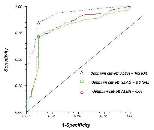
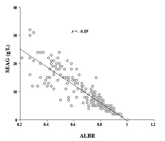
The ROC plots for SEAG, ALBR and FLDH are shown in figure-1. At all test values, FLDH was more sensitive and specific than SEAG and ALBR. The optimum cut-off levels were 163IU/L for FLDH, 9 g/L for SEAG, and 0.68 for ALBR respectively. FLDH was the best of the three tests. The AUC and the corresponding 95% Cl for SEAG, ALBR and FLDH were 0.81 (0.75 to 0.87), 0.79 (0.72 to 0.86) and 0.9 (0.87 to 0.96) respectively. Although the AUC for SEAG and ALBR were of similar magnitude, the AUC for FLDH was significantly more than the AUC for ALBR (z = 2.75, p < 0.01). Of the 156 exudates, FLDH correctly classified 132 (85%) 95% Cl 0.78–0.90, SEAG 112 (72%) 0.64–0.79, and ALBR 111 (71%) 0.63–0.78 respectively as exudates and all three tests classified 39 (89%) 0.75–0.96 of the 44 transudates correctly. Table 2 lists the positive likelihood ratios (LR +ve) with 95% Cl for the three tests at the optimum cut-off level. Figure-2 shows the correlation between SEAG and ALBR. There was a significant negative correlation between the two tests (r = -0.89, p < 0.0001).
Figure 1.

Receiver Operating Characteristic plots of the pleural fluid FLDH, ALBR, and SEAG. The optimum cut-off level was determined by selecting the points of test values that provided the greatest sum of sensitivity and specificity. The optimum cutoff for FLDH was 163 IU/L, ALBR = 0.68, SEAG = 9.0.
Table 2.
Positive Likelihood Ratios (LR) at optimum cut-off points
| Test | LR | 95% CI |
| FLDH | 7.3 | 3.5 to 17 |
| SEAG | 6.3 | 3 to 15 |
| ALBR | 6.2 | 3 to 14 |
Figure 2.

Association between ALBR and SEAG in pleural effusion
Discussion
Pleural effusion is a common clinical problem and it's diagnostic approach has been simplified by characterizing effusion into transudate and exudate [11]. Several biochemical markers have been used in the past for this purpose [12-14]. However, TPR and FLDH have remained the most widely used criteria so far. Therefore, this study was undertaken to assess the global performance of SEAG and ALBR with the existing best criteria i.e. FLDH [7]. The strength of our study is that we have utilized ROC analysis for comparing different tests. Furthermore, ROC analysis provides an opportunity to calculate likelihood ratios at various cutoff points of the diagnostic test. The upper limit of the normal LDH varies according to the methods used for estimating lactate dehydrogenase. In this study we have used the Boehringer Mannheim kit and the upper normal for this method is 200IU//L [9].
In our present study, the mean age and causes of pleural effusion were similar to that of Roth et al. [5]. However, the mean SEAG value of 14 g/L for transudate and 8 g/L for exudate in our series were different from that provided by Roth and associates about 20 g/L for transudates and 0.75 g/L for exudates. From ROC analysis it is clear that FLDH performed better compared to SEAG and ALBR as documented by the highest value for the AUC. ALBR has been reported to be inferior to SEAG in the diagnostic separation of transudates and exudates [5]. However, in our analysis the accuracy of ALBR and SEAG were similar as evidenced by similar AUC and positive LR. Furthermore, correlation analysis between SEAG and ALBR has shown a significant negative correlation (0.89).
As the level of pleural fluid albumin increases depending on the degree of pleural microvacular injury in exudative processes, the ALBR increases correspondingly. Whereas, pleural fluid to SEAG is highest in transustive effusions as less albumin is filtered through the relatively normal pleural microvasculature. However, as pleural microvasculature is damaged in exudative processes, the level of albumin leaking into the pleural cavity would progressively increase depending on the degree of injury. Thus a very low SEAG can be expected in a trasudative processes with a correspondingly higher ALBR. This would partly explain the significant negative correlation observed between SEAG and ALBR thus indicating that these two tests likely to be operating through the same mechanism [15].
In our study using 9 g/L as the optimum cutoff level for SEAG, 51 of the 156 exudates were incorrectly labeled as transudates (false negative) and 5 of the 44 transudates were labeled as exudates (false positives). There were no trends for any specific diseases clustering in either of the groups.
The pathophysiology of transudative or exudative pleural effusion in humans is not well studied. We have previously documented that the rate of formation and absorption are different in different disease states causing pleural effusion [8]. The decreased concentration of total protein and albumin in transudates can be explained by the selective sieving of these macromolecules by the normal intact microvasculature in transudates [3]. However, animal and human studies have shown that increase in hydrostatic pressure can increase the protein content in effusions [16,17]. This is partly explained by the opening up of large pores in endothelium with elevated hydrostatic pressure [18,19]. Thus in a patient with pleural effusion from congestive heart failure, it is possible that the amount of protein in the pleural fluid can increase significantly to exudative levels if the degree of pulmonary artery pressure causing the formation of effusion was very high. Such a linear correlation between the hydrostatic pressure and the protein content of ascites in patients with portal hypertension has been documented [17]. Therefore, this phenomenon may help explain the pathogenesis of exudative effusions documented in congestive heart failure when total protein in pleural effusion was used as a diagnostic criteria [5,20,21].
The dynamics of LDH in the pleural cavity is not well studied. Our recent investigation into the dynamics of pleural fluid LDH has documented that there was no correlation between serum and pleural fluid LDH concentration in transudates or exudates [7]. This compartmentalized elevation of LDH concentration in pleural cavity in exudative processes help explain the superior performance of FLDH as a diagnostic test in the separation of pleural effusion into transudate and exudate compared to SEAG, ALBR. We have reported earlier that LDHR has no role in the diagnostic separation of transudates and exudates. Furthermore, FLDH is the most accurate marker and combining TPR with FLDH improves the diagnostic utility marginally [7].
Conclusion
The discriminative value for SEAG and ALBR appears to be similar in the diagnostic separation of transudates and exudates. FLDH is a superior test compared to SEAG and ALBR. Based on these results, SEAG and ALBR appear to have limited diagnostic utility in clinical practice.
Competing interests
None declared
Author contributorship
Author one JJ conceived the idea, designed the study, collected the data, took part in analysis of data and drafted the manuscript.
Author two PB analyzed the data, interpreted the results and contributed in writing the manuscript.
Author three GSB designed the study, supervised the data collection and contributed in writing the manuscript.
Author four SS participated in the data analysis, interpreting results and contributed in writing and editing the manuscript.
All authors read and approved the final manuscript.
Pre-publication history
The pre-publication history for this paper can be accessed here:
Acknowledgments
Acknowledgement
The authors would like to thank Mr. Steve Viney for carrying out the pleural fluid biochemical analysis.
Contributor Information
Jose Joseph, Email: jjoseph@uaeu.ac.ae.
Padmanabhan Badrinath, Email: pbadrinath@uaeu.ac.ae.
Gurnam S Basran, Email: basran.gs@rgh-tr.trent.nhs.uk.
Steven A Sahn, Email: sahnsa@musc.edu.
References
- Black LF. The pleural space and pleural fluid. Mayo Clin Proc. 1972;47:493–506. [PubMed] [Google Scholar]
- Sahn SA. State of the art. The pleura. Am Rev Respir Dis. 1988;138:184–234. doi: 10.1164/ajrccm/138.1.184. [DOI] [PubMed] [Google Scholar]
- Sahn SA. The pathophysiology of pleural effusions. Annu Rev Med. 1990;41:7–13. doi: 10.1146/annurev.med.41.1.7. [DOI] [PubMed] [Google Scholar]
- Light RW, Macgregor Ml, Luchsinger PC, Ball WCJ. Pleural effusions: the diagnostic separation of transudates and exudates. Ann Intern Med. 1972;77:507–13. doi: 10.7326/0003-4819-77-4-507. [DOI] [PubMed] [Google Scholar]
- Roth BJ, O'Meara TF, Cragun WH. The serum-effusion albumin gradient in the evaluation of pleural effusions. Chest. 1990;98:546–49. doi: 10.1378/chest.98.3.546. [DOI] [PubMed] [Google Scholar]
- Burgess LJ, Maritz FJ, Taljaard JJ. Comparative analysis of the biochemical parameters used to distinguish between pleural transudates and exudates. Chest. 1995;107:1604–9. doi: 10.1378/chest.107.6.1604. [DOI] [PubMed] [Google Scholar]
- Joseph J, Badrinath P, Basran GS, Sahn SA. Is the pleural fluid transudate or exudate? A revisit of the diagnostic criteria. Thorax. 2001;56:867–70. doi: 10.1136/thorax.56.11.867. [DOI] [PMC free article] [PubMed] [Google Scholar]
- Smith MJ, Joseph J, Flatman WD, Basran GS. A dual-isotope method for studying protein kinetics in pleural effusions in humans. Nucl Med Commun. 1992;13:432–39. doi: 10.1097/00006231-199206000-00043. [DOI] [PubMed] [Google Scholar]
- Recommended methods for the determination of four enzymes in blood. Scand J Clin Lab Invest. 1974;33:291–306. doi: 10.1080/00365517409082499. [DOI] [PubMed] [Google Scholar]
- Hanley JA, McNeil BJ. A method of comparing the areas under receiver operating characteristic curves derived from the same cases. Radiology. 1983;148:839–43. doi: 10.1148/radiology.148.3.6878708. [DOI] [PubMed] [Google Scholar]
- Jay SJ. Diagnostic procedures for pleural disease. Clin Chest Med. 1985;6:33–48. [PubMed] [Google Scholar]
- Chandrasekhar AJ, Palatao A, Dubin A, Levine H. Pleural fluid lactic acid dehydrogenase activity and protein content. Value in diagnosis. Arch Intern Med. 1969;123:48–50. doi: 10.1001/archinte.123.1.48. [DOI] [PubMed] [Google Scholar]
- Costa M, Quiroga T, Cruz E. Measurement of pleural fluid cholesterol and lactate dehydrogenase. A simple and accurate set of indicators for separating exudates from transudates. Chest. 1995;108:1260–1263. doi: 10.1378/chest.108.5.1260. [DOI] [PubMed] [Google Scholar]
- Meisel S, Shamiss A, Thaler M, Nussinovitch N, Rosenthal T. Pleural fluid to serum bilirubin concentration ratio for the separation of transudates from exudates. Chest. 1990;98:141–44. doi: 10.1378/chest.98.1.141. [DOI] [PubMed] [Google Scholar]
- Heffner JE, Brown LK, Barbieri CA. Diagnostic value of tests that discriminate between exudative and transudative pleural effusions. Primary Study Investigators. Chest. 1997;111:970–980. doi: 10.1378/chest.111.4.970. [DOI] [PubMed] [Google Scholar]
- Broaddus VC, Araya M. Liquid and protein dynamics using a new minimally invasive pleural catheter in rabbits. J Appl Physiol. 1992;72:851–57. doi: 10.1152/jappl.1992.72.3.851. [DOI] [PubMed] [Google Scholar]
- Hoefs JC. Serum protein concentration and portal pressure determine the ascitic fluid protein concentration in patients with chronic liver disease. J Lab Clin Med. 1983;102:260–273. [PubMed] [Google Scholar]
- Haraldsson B. Effects of noradrenaline on the transcapillary passage of albumin, fluid and CrEDTA in the perfused rat hind limb. Acta Physiol Scand. 1985;125:561–71. doi: 10.1111/j.1748-1716.1985.tb07758.x. [DOI] [PubMed] [Google Scholar]
- Wolf MB, Porter LP, Watson PD. Effects of elevated venous pressure on capillary permeability in cat hindlimbs. Am J Physiol. 1989;257:H2025–H2032. doi: 10.1152/ajpheart.1989.257.6.H2025. [DOI] [PubMed] [Google Scholar]
- Chakko SC, Caldwell SH, Sforza PP. Treatment of congestive heart failure. Its effect on pleural fluid chemistry. Chest. 1989;95:798–802. doi: 10.1378/chest.95.4.798. [DOI] [PubMed] [Google Scholar]
- Melsom RD. Diagnostic reliability of pleural fluid protein estimation. J R Soc Med. 1979;72:823–25. doi: 10.1177/014107687907201106. [DOI] [PMC free article] [PubMed] [Google Scholar]


