Abstract
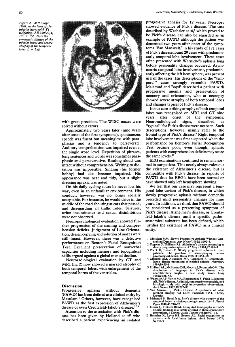
We report a patient who suffered from progressive aphasia for nine years, before developing mild behavioural disturbances. Sequential computed tomography (CT) scanning and magnetic resonance (MRI) imaging showed progressive bilateral temporal atrophy. The case is thought to be a temporal form of Pick's disease, in which isolated progressive aphasia was the only symptom over many years.
Full text
PDF

Images in this article
Selected References
These references are in PubMed. This may not be the complete list of references from this article.
- Groen J. J., Hekster R. E. Computed tomography in Pick's disease: findings in a family affected in three consecutive generations. J Comput Assist Tomogr. 1982 Oct;6(5):907–911. [PubMed] [Google Scholar]
- Hamsher K. d., Levin H. S., Benton A. L. Facial recognition in patients with focal brain lesions. Arch Neurol. 1979 Dec;36(13):837–839. doi: 10.1001/archneur.1979.00500490051008. [DOI] [PubMed] [Google Scholar]
- Holland A. L., McBurney D. H., Moossy J., Reinmuth O. M. The dissolution of language in Pick's disease with neurofibrillary tangles: a case study. Brain Lang. 1985 Jan;24(1):36–58. doi: 10.1016/0093-934x(85)90096-3. [DOI] [PubMed] [Google Scholar]
- Mandell A. M., Alexander M. P., Carpenter S. Creutzfeldt-Jakob disease presenting as isolated aphasia. Neurology. 1989 Jan;39(1):55–58. doi: 10.1212/wnl.39.1.55. [DOI] [PubMed] [Google Scholar]
- Mesulam M. M. Slowly progressive aphasia without generalized dementia. Ann Neurol. 1982 Jun;11(6):592–598. doi: 10.1002/ana.410110607. [DOI] [PubMed] [Google Scholar]
- Poeck K., Luzzatti C. Slowly progressive aphasia in three patients. The problem of accompanying neuropsychological deficit. Brain. 1988 Feb;111(Pt 1):151–168. doi: 10.1093/brain/111.1.151. [DOI] [PubMed] [Google Scholar]
- Pogacar S., Williams R. S. Alzheimer's disease presenting as slowly progressive aphasia. R I Med J. 1984 Apr;67(4):181–185. [PubMed] [Google Scholar]
- Wechsler A. F., Verity M. A., Rosenschein S., Fried I., Scheibel A. B. Pick's disease. A clinical, computed tomographic, and histologic study with golgi impregnation observations. Arch Neurol. 1982 May;39(5):287–290. doi: 10.1001/archneur.1982.00510170029008. [DOI] [PubMed] [Google Scholar]


