



SUMMARY
Timely completion of genome replication is a prerequisite for mitosis, genome integrity, and cell survival. A challenge to this timely completion comes from the need to replicate the hundreds of untranscribed copies of rDNA that organisms maintain in addition to the copies required for ribosome biogenesis. Replication of these rDNA arrays is relegated to late S phase despite their large size, repetitive nature, and essentiality. Here, we show that, in Saccharomyces cerevisiae, reducing the number of rDNA repeats leads to early rDNA replication, which results in delaying replication elsewhere in the genome. Moreover, cells with early-replicating rDNA arrays and delayed genome-wide replication aberrantly release the mitotic phosphatase Cdc14 from the nucleolus and enter anaphase prematurely. We propose that rDNA copy number determines the replication time of the rDNA locus and that the release of Cdc14 upon completion of rDNA replication is a signal for cell cycle progression.
In brief
Kwan et al. show that the end of genome replication and cell cycle progression are coordinated by the replication time of the ribosomal DNA (rDNA) locus in S. cerevisiae. Changing rDNA’s high copy number or late-replicating characteristics results in both genome-wide replication delays and premature anaphase entry.
Graphical Abstract

INTRODUCTION
Ribosomal DNA (rDNA) is uniquely situated at the intersection of ribosome biogenesis and genome replication, fundamental processes required for cell growth and proliferation. The rDNA sequence is arranged in tandem to form large arrays of ribosomal RNA genes, which encode the core components of the ribosome. Many species have hundreds of rDNA copies per haploid genome, e.g., 90–300 in Saccharomyces cerevisiae, 70–400 in Caenorhabditis elegans, 80–600 in Drosophila melanogaster, 500–2,500 in Arabidopsis thaliana, and 30–800 in humans.1–4 As the rDNA copy number often substantially exceeds what is required to meet ribosome demands, the majority of rDNA repeats are silenced.5–10 Nevertheless, organisms maintain a seemingly excess number of rDNA copies,11 hinting that high rDNA copy number may have roles beyond ribosome production.
Variation in rDNA copy number affects a broad range of cellular processes. If rDNA copy number drops below a certain threshold, ribosome deficiencies result.8,12–14 However, even variants with sufficient rDNA copies for ribosome biogenesis show deleterious phenotypes.15–22 Furthermore, the plastic nature of the rDNA locus allows rDNA copy number to fluctuate in response to stresses such as nutrient availability23 or replication defects.14,24–26 In particular, cells with mutations in key replication factors or that experience replication stress often reduce their rDNA copy number.
Because cells that survive replication stress frequently have fewer rDNA copies, we reasoned that pre-emptively reducing rDNA copy number might alleviate replication stress by facilitating genome replication. Given the vast length of rDNA arrays, replication initiation must occur within the rDNA sequence.27–32 In the budding yeast S. cerevisiae, each 9.1-kb repeat contains a potential origin of replication (rDNA autonomously replicating sequence or rARS33,34). For an rDNA array of 150 copies (within the wild-type range for yeast), only 30–40 of the 150 replication origins are estimated to initiate replication (“fire”) within an S phase.28,35 Origin firing across the genome is limited by the low abundance of initiation factors that promote the temporal staggering of origin activation.25,36–38 When rDNA origins are hyperactivated, replication factors become diverted from unique regions of the genome,38–40 leading to persistent under-replication of certain genomic regions.41 We therefore reasoned that reduction of rDNA copy number from the wild-type size of 100–200 copies would alleviate the competition for limiting initiation factors at the other ~300 replication origins across the genome.42
Here, we assess genome replication in isogenic yeast strains with rDNA arrays that are either wild-type in size (100–180 copies) or reduced (35 copies). We discovered that this reduced rDNA array does not alleviate competition with non-rDNA origins, and instead it drastically advances rDNA replication time and increases the density of active rDNA origins. A consequence of this large burst of early rDNA initiations from the 35-copy rDNA array is delayed replication across the genome. However, this genome replication delay does not trigger a compensatory delay in cell cycle progression. Instead, the 35-copy rDNA strain mislocalizes and prematurely releases Cdc14, the phosphatase associated with completion of mitotic anaphase, from the nucleolus. Our findings demonstrate that the rDNA array actively coordinates completion of genome replication with cell cycle progression.
RESULTS
Reduction of rDNA copy number advances initiation of rDNA replication
To investigate how rDNA copy number affects genome-wide replication, we generated isogenic strains with reduced rDNA copy number in a fob1Δ background to prevent rDNA expansion.39,43,44 rDNA copy number was confirmed using clamped homogeneous electric field (CHEF) gel electrophoresis (Figures 1A, S1A, and S1B). We isolated strains with 35 rDNA copies, just above the threshold rDNA copy number required for normal ribosome biogenesis in S. cerevisiae.8,16,45,46 We assessed ribosome sufficiency by measuring growth rates, cycloheximide sensitivity, and ribosomal RNA (rRNA) abundance (Figures S1C–S1E). By all measures, strains with reduced rDNA copy number behaved similarly to both fob1Δ and FOB1 strains with wild-type rDNA copy number, confirming that rDNA reduction to 35 rDNA copies does not generate significant ribosome biogenesis defects.
Figure 1. rDNA arrays with fewer copies replicate early.
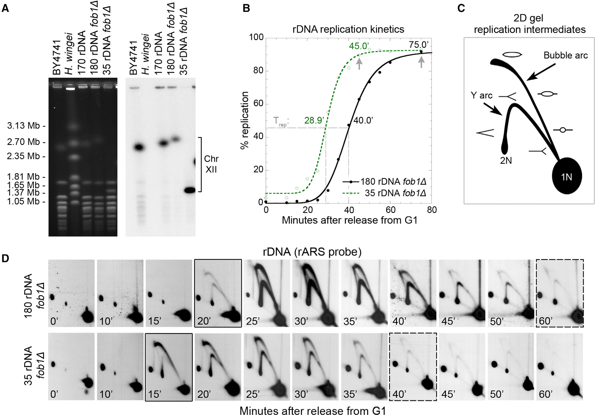
(A) CHEF gel estimation of rDNA copy number using chromosome XII size. Ethidium bromide-stained gel (left) and the resulting Southern blot (right) hybridized with a single copy Chr. XII probe (CDC45).
(B) Replication kinetic curves generated from density transfer slot blot analysis show that the minimal rDNA array replicates earlier than the 180-copy rDNA array. Dashed lines indicate time at half-maximal replication (Trep). Arrows indicate the estimated times at which rDNA replication has neared completion.
(C) 2D gel diagram of positions for replication intermediates that contain an active origin (bubble arc) or are passively replicated (Y arc).
(D) 2D gel analysis of replication across a synchronous S phase for the NheI fragment containing the rDNA ARS (rARS). One of three sets of biological replicates, all with similar results, is presented here. The black borders indicate times of first perceptible replication intermediates. The dashed borders indicate the times when rDNA replication is essentially completed.
We first asked whether rDNA copy number reduction alters replication of the rDNA locus itself, which is late replicating in S. cerevisiae and several eukaryotic species.41,47–50 Replication kinetics can be examined by density transfer experiments, which exploit the semi-conservative nature of DNA replication to distinguish newly replicated from unreplicated DNA.51–53 Analysis of rDNA replication kinetics revealed that reducing rDNA copy number dramatically shifted rDNA replication from late S phase to early S phase (Figure 1B). We calculated rDNA Trep, the time at which half-maximal replication was achieved, for each strain. While the rDNA locus was late replicating in the 180-copy rDNA strain (Trep = 40.0 min) as expected, the 35-copy rDNA array replicated 11 min earlier (Trep = 28.9 min), a substantial shift considering that S phase in S. cerevisiae is only 20–30 min at 30°C.54 Furthermore, the time to complete rDNA replication was reduced by 20–30 min (Figure 1B). Early replication time of reduced rDNA arrays was reproducible, even in strains with a weak rDNA origin sequence (Figures S1F and S1G).
Although the rDNA replication kinetic curves suggested that the 35-copy rDNA array begins replicating earlier than the wild-type array, we examined replication initiation directly using 2D gel electrophoresis of synchronized cells sampled across S phase. If the time of replication initiation were altered, we would observe differences in when the replication “bubble” arc becomes visible among our strains (Figure 1C). In the 35-copy rDNA locus, rDNA origin (rARS) initiation began robustly at 15 min into S phase, whereas in the wild-type rDNA locus, initiation was first detected at 20 min, and robust initiation was not seen until 25 min (Figure 1D, black borders). These results confirm that early rDNA origin initiation is responsible for the earlier replication of the 35-copy rDNA locus seen by density transfer. The persistence of other intermediates on 2D gel confirms the 20-min difference in the time for the rDNA locus to complete replication (Figure 1D, dashed borders).
How many rDNA origins initiate replication during early S phase?
Approximately one in five of the rDNA origins is thought to serve as a replication initiation site in cells with wild-type rDNA.28,35 By this estimate, a strain with 180 rDNA copies would have ~36 active origins in the rDNA locus—close to the maximum number of possible rDNA origin initiations in the 35-copy rDNA strains. We asked how many rDNA origins fire in early S phase in both strains and whether increased early rDNA firing alters replication timing genome-wide.
To answer these questions, we performed another set of 2D gels under conditions designed to quantify early replication dynamics. Digestion of replicating rDNA by the restriction enzyme NheI generates a variety of distinct molecular intermediates/structures, such as “bubbles” from active initiation, passively replicated “Y” fragments, and “X” fragments produced by converging replication forks (Figure 2A), all of which can be resolved by 2D gel electrophoresis and detected by Southern blotting.28,55 To limit passive replication of rARSs by forks established at a flanking active rARS, we released G1-synchronized cultures into S phase in the presence of the ribonucleotide reductase inhibitor hydroxyurea (HU). Fork rates in HU are 1/15 the wild-type rate, and HU also limits initiation events to those origins that are activated early in S phase.51,56–58 In addition to the 4.7-kb rARS fragments from every rDNA repeat, the NheI digest also generates a single 24.4-kb rARS fragment at the telomere proximal end of the rDNA array (Figures 2A and 2B) that can be used to deduce a “per cell” estimation of active rDNA initiation in early S phase.
Figure 2. Quantification of rDNA replication initiations in early S phase.
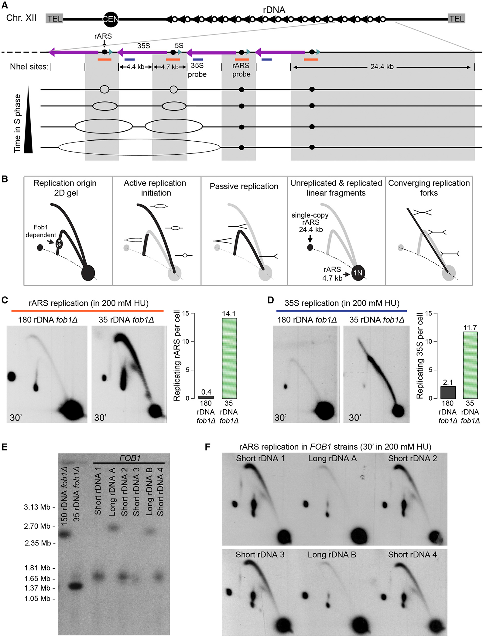
(A) Schematic of rDNA locus organization, NheI restriction sites, Southern blot probe locations (orange and blue bars), and example of replication intermediates throughout S phase. The 24.4-kb NheI fragment on the telomere proximal edge of the rDNA array is present in a single copy per cell and hybridizes to the rARS probe (orange bar).
(B) Diagram of replication intermediates resolved by 2D gel electrophoresis.
(C and D) 2D gels of synchronized cells released into S phase in the presence of 200 mM HU, sequentially hybridized with the rARS probe (C) and the 35S probe (D). rARS replication bubbles per cell were quantified and normalized to the signal in the single-copy 24.4-kb linear spot. Estimation of the non-rARS 35S replication fork intermediates was normalized to the 4.4-kb 1N spot and adjusted for rDNA copy number.
(E) Meiotic spores from a cross between a FOB1, 170-copy rDNA strain and a fob1Δ, 35-rDNA copy strain were analyzed by CHEF gel electrophoresis to measure rDNA repeat number.
(F) 2D gels of DNA harvested as in (B) for the same six spores (from E) were analyzed for rARS initiation.
Compared with a strain with wild-type rDNA copy number, the 35-copy rDNA strain showed a far stronger rDNA bubble arc signal (Figure 2C), which reflects the shift in rDNA origin initiations to early S phase we had observed in the absence of HU (Figures 1B and 1D). Comparing the signal in the bubble arc to that in the 24.4-kb NheI fragment, we estimate that the 180-copy rDNA strain had 0.4 early rDNA initiations per cell, while the 35-copy rDNA strain had 14.1, a 35-fold increase. These values are likely an underestimate due to the movement of replication forks off the NheI fragment containing the rDNA origin and into the adjacent non-rARS NheI fragment where the forks generate Y and convergent double-Y intermediates (Figure 2D). The replication intermediates in the adjacent fragment represent an additional 2.1 early initiations per cell in the 180-copy rDNA strain, and 11.7 in the 35-copy rDNA strain, for a total of 2.5 and 25.8 rDNA initiations per cell, respectively. In addition, the presence of converging forks in the 35-copy rDNA strain indicates that adjacent rDNA origin firing is common. Since the subset of earliest replicating origins across the S. cerevisiae genome is fewer than ~60 origins in a population of cells,51,59 the additional early-replicating rDNA origins in a cell could easily generate significant competition for limiting replication factors or nucleotides in early S phase37,40 and establish a cascade of origin initiation delays later in S phase.
Early rDNA initiation in reduced rDNA arrays is not fob1Δ dependent
Because of the important role that Fob1 plays at the rDNA locus, we wanted to address the possibility that early rDNA replication was an artifact of the fob1Δ background. The fob1Δ mutation prevents expansion of reduced rDNA arrays44; by contrast, in a FOB1 wild-type background, this expansion can make it challenging to assess the consequences of short rDNA arrays. To overcome this challenge, we examined spores from a cross of the 35-copy rDNA strain (fob1Δ) strain with a FOB1 strain with 150 rDNA copies. Each spore was allowed to form a colony, which was immediately inoculated into culture, and FOB1 status, rDNA copy number, and early S phase rDNA replication were assessed using samples from the same culture. We were able to capture multiple FOB1 strains with reduced rDNA copy number (short rDNA; ~50 rDNA copies) as well as corresponding FOB1 strains with wild-type rDNA arrays (long rDNA; ~150 rDNA copies) (Figure 2E). Analyzing early S phase samples collected in 200 mM HU, we found that the FOB1 short rDNA strains exhibited the same precocious rDNA origin initiation that we found for the fob1Δ, 35-copy rDNA strain (Figure 2F). The presence of the replication fork barrier (RFB, Figure 2B) spot indicated that Fob1 was active in its fork-blocking function in these short rDNA and long rDNA strains. We conclude that early rDNA replication from rDNA reduction is independent of FOB1.
Early replication at reduced rDNA arrays delays genome replication
We predicted that an additional burden of 23 or more early rDNA initiations per cell would generate cascading delays across the genome by diverting a large fraction of the limiting replication factors away from unique genomic origins. To test this prediction, we examined replication of several non-rDNA genomic loci using the density transfer experiments discussed previously and compared Trep changes between the 180-copy and 35-copy rDNA strains (Figure 3A). The time of S phase entry was similar between the two strains, and ARS305 and ARS607, two of the earliest and most robust replication origins in S. cerevisiae,60,61 did not differ greatly in their time of initiation. However, the late-replicating loci ARS501 (renamed ARS522), ARS735.5, and a late-replicating non-ARS fragment on chromosome V (R1162 on ChrV:534,000) were delayed in the 35-copy rDNA strain, while their relative replication order was maintained.
Figure 3. rDNA copy number reduction generates genome replication delays and defects.
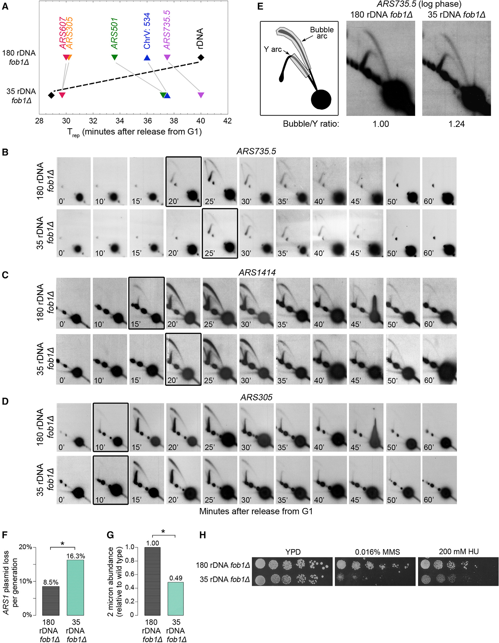
(A) Comparison of the Trep (time of half-maximal replication derived from a density transfer experiment) for five different genomic origins and an origin-free genomic region (Chr. V: 534) in strains with 180 vs. 35 rDNA copies. Additional biological replicates are presented in Figures S1F and S2G.
(B–D) 2D gel analysis of replication across a synchronous S phase for time of initiation at the late-replicating genomic origins ARS735.5 and ARS1414 and the early-replicating origin ARS305. The boxed panels denote time of first perceptible bubble replication intermediates.
(E) Relative origin efficiency for ARS735.5 was estimated by comparing the signal ratio of bubbles vs. Ys.
(F) Measurement of plasmid loss rates of an ARS1-based plasmid in the 180-rDNA and 35-rDNA copy number strains. p = 0.03; one representative biological replicate of two is shown.
(G) Natural abundance of the parasitic 2-micron plasmid in the 180-rDNA and 35-rDNA copy number strains. Paired two-tailed t test p = 0.007.
(H) Spot assays to measure sensitivity to DNA damage from0.016%MMSand replication stress from200mMHUfor the 180-rDNAand 35-rDNAcopy number strains.
Given that late-replicating genomic loci were impacted the most by early rDNA replication, we asked if any regions of the genome were particularly sensitive to rDNA-induced replication delays. Density transfer samples were hybridized to microarrays to generate chromosome replication profiles at 5-min intervals across S phase (Figure S3). We found that replication was broadly delayed by 5 min across chromosomes in the 35-copy rDNA strain (Figure S4A), but we did not find any genomic sites that showed increased sensitivity to replication delays (Figure S4B). The general replication delays likely contributed to the delayed completion of chromosomal replication in the 35-copy rDNA strain (Figures S5A–S5C). We conclude that rDNA copy number reduction generates broad genome-wide replication delays without targeting specific regions.
Replication time delays are due to delayed origin initiation and not reduced origin efficiency
Delayed replication time can result from delayed replication initiation, reduction of origin efficiency, or a combination of the two. Using 2D gel electrophoresis of synchronized cell samples, we found that both ARS735.5 and ARS1414 initiated replication approximately 5 min later in the 35-copy rDNA strain (Figures 3B and 3C), while ARS305 showed no difference in initiation time between the two strains (Figure 3D), a result consistent with the density transfer data for ARS305. To examine efficiency of two origins delayed in the 35-copy rDNA strain, we compared the intensities of the active bubble arc to the passively replicating Y arc of 2D gels from asynchronous cultures.24,61,63 Neither ARS735.5 (Figure 3E) nor ARS501 (Figure S5D) had reduced efficiency in the 35-copy rDNA strain. We conclude that the changes to genome replication timing in the 35-copy rDNA strain are not due to changes in initiation efficiency but to altered time of replication initiation at later non-rDNA origins of replication.
Strains with reduced rDNA arrays exhibit replication defects
Many S. cerevisiae replication mutants have difficulty maintaining non-chromosomal elements such as plasmids14,64,65 and are sensitive to drugs that induce DNA damage and replication stress.66–68 In addition to delayed genome replication and delayed chromosomal completion, 35-copy rDNA strains exhibited these classic replication defects. First, the 35-copy rDNA strain exhibited high loss rates of the ARS1 (autonomously replicating sequence 169) test plasmid: 16.3%/generation for the 35-copy rDNA strain, almost double that of the control fob1Δ strain with wild-type rDNA (8.5%/generation, p = 0.03, Figure 3F). Second, as a proxy for plasmid maintenance, we examined the abundance of the 2-micron plasmid, which is reduced in strains with replication defects.70,71 The 35-copy rDNA strain had reduced 2-micron abundance—approximately 50% that of wild-type control strains (Figure 3G). Finally, the 35-copy rDNA strain showed greater sensitivity to the DNA-damaging agent methyl methanesulfonate (MMS)16 and replication stress-inducing hydroxyurea (Figure 3H).
Early rDNA replication delays genome replication more than increased rDNA origin initiations
Increased rDNA origin initiations and reduced replication at non-rDNA sites during early S phase have been reported for rif1Δ and sir2Δ mutants,38,40,72,73 but the remainder of S phase has not been investigated. It thus remained unclear whether the competition between rDNA and genome replication is caused by increases in rDNA origin initiation, early rDNA replication, or both. However, even if all 35 rDNA origins in the 35-copy rDNA strain initiate replication, this number does not exceed the estimated ~36 active rDNA origins in the 180-copy rDNA strain. Therefore, it appeared likely that precocious rDNA replication would be the major contributor to delayed genome replication. To further investigate the relative contributions to genome replication delays of early rDNA replication versus increased rDNA origin initiations, we tested other mutants that exhibit early or altered rDNA replication.
Because previous reports focused on replication that occurred in early S phase only, we pursued the question of timing vs. number of rDNA origin initiations using 2D gel electrophoresis across a synchronous S phase. We analyzed rDNA origin initiation kinetics in sir2Δ, fob1Δ, rif1Δ, and sir2Δ fob1Δ mutants (Figures 4A and 4B), all reported to have early or altered rDNA replication initiation but wild-type rDNA copy number. For the sir2Δ, rif1Δ, and sir2Δ fob1Δ mutants, the rDNA replication intermediates appeared and peaked in activity 5–10 min earlier than in the wild-type strain. In contrast, the fob1Δ mutant had slightly delayed rDNA replication time compared with wild type (Figure 4B). Thus, sir2Δ, fob1Δ, and rif1Δ mutations alter the window of S phase in which rDNA replication initiation occurs.
Figure 4. Characterization of altered rDNA replication time and rDNA origin efficiency in mutant strains.
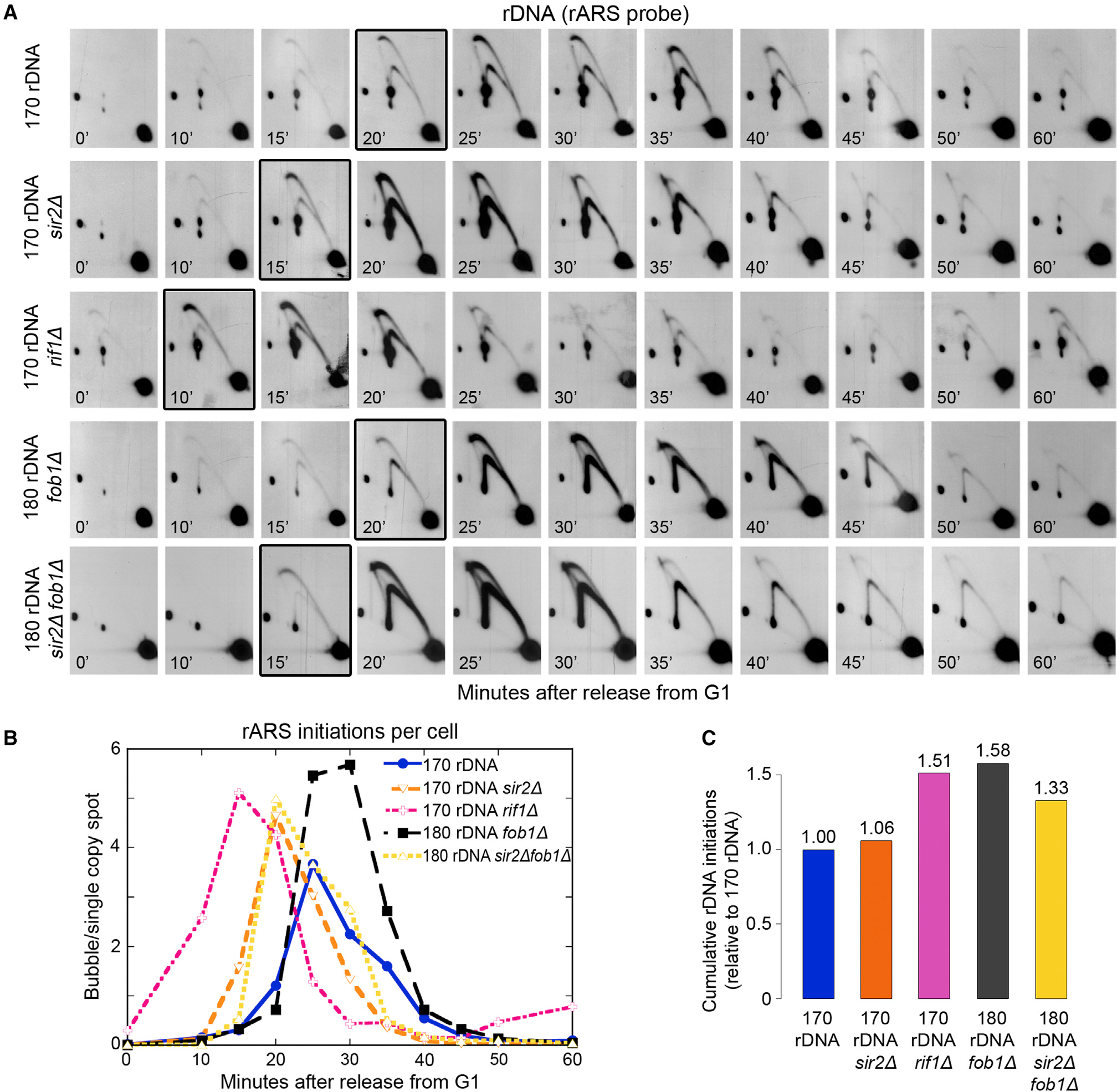
(A) Comparison of rDNA replication initiation time in synchronized cells using 2D gel electrophoresis for four mutants (sir2Δ, rif1Δ, fob1Δ, or sir2Δ fob1Δ) relative to wild-type cells—all with wild-type rDNA copy numbers. The boxed panels indicate the first appearance of more than 0.5 rARS bubble intermediates per cell.
(B) Quantification of bubbles per cell, based on the ratio of bubbles to the 24.4-kb single-copy fragment for the five strains in (A).
(C) Total number of rDNA origins fired in the three mutant strains in (A) compared with the wild-type strain.
To determine whether these mutants affect the number of active rDNA origins, we quantified cumulative rDNA initiations relative to the wild-type strain across S phase (Figure 4C). sir2Δ cells exhibited only a 6% increase in rDNA origin initiations over wild type, which is equivalent to an increase of two active rDNA origins per cell. This modest increase in active rDNA origins contrasts with the dramatic genome replication delays seen in sir2Δ mutants38,41 and supports our hypothesis that it is early rDNA replication and not the number of active rDNA origins that delays replication of the rest of the genome. In contrast, both fob1Δ and rif1Δ strains showed ~50% more active rDNA origins across S phase (Figure 4C); however, fob1Δ cells exhibited improved non-rDNA replication38 (Figures S5E and S5F), while Lian et al. and Shyian et al. showed that rif1Δ cells compromise non-rDNA replication.40,74 Thus, although both rif1Δ and fob1Δ strains show increased rDNA origin activity, only the rif1Δ strain, where rDNA origins are activated earlier than wild type, shows compromised non-rDNA replication. Non-rDNA replication is not compromised in fob1Δ, where rDNA origin firing is not advanced. The difference in non-rDNA replication between these mutants can therefore be attributed to the early rDNA replication in the rif1Δ mutant. We conclude that early rDNA replication rather than the increased number of rDNA origin initiations delays non-rDNA genome replication.
Early rDNA replication uncouples the completion of genome replication from anaphase entry and increases DNA damage sensitivity
Genome replication delays may be tolerated if cells can also delay progression through anaphase. To test whether early rDNA replication and the concomitant delayed genome replication causes delayed anaphase, we examined DAPI-stained nuclear morphology (Figure 5A) across S phase as a proxy for entry into anaphase.75,76 The sir2Δ mutant entered anaphase later than the wild type, consistent with this strain’s genome replication delays (Figure 5B). However, both the 35-copy rDNA strain and the rif1Δ strain entered anaphase at the same time (35-copy rDNA) or earlier (rif1Δ) than their wild-type control strains (Figure 5C), though they did not display premature cohesin cleavage (Figures S6C and S6D). These mutants are unable to delay anaphase in response to delayed genome replication. The link between genome replication completion and anaphase entry appears to be partially dependent on FOB1, as no difference was observed in anaphase entry between sir2Δ and SIR2 strains in the absence of FOB1 (Figure 5D).
Figure 5. Uncoupling of anaphase entry from delayed genome replication increases DNA damage sensitivity in strains with early rDNA replication.
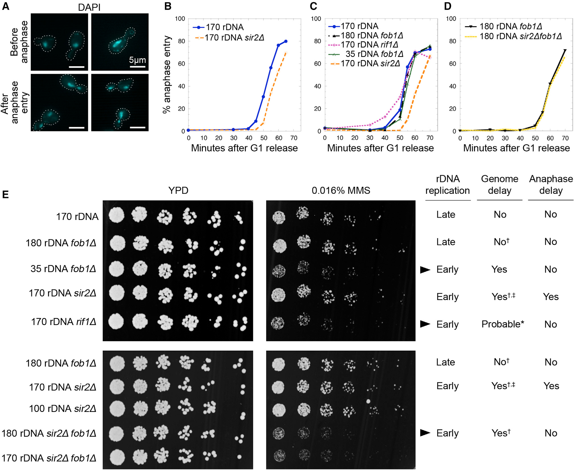
(A) Examples of DAPI-stained nuclei representing cells scored as either being “before anaphase” (no nuclear migration) or “after anaphase entry” (with nuclear migration into the daughter cell). Two representative images for each stage are presented, and cell outlines are indicated by white dotted lines. White scale bar indicates 5 μm.
(B–D) Quantification of percentage of cells undergoing anaphase over time after release from G1 with n ≥ 200 cells for each timed sample in each replicate. One of two biological replicates is shown here; see Figure S6 for the other replicate.
(E) Spot assays comparing growth on YPD vs. YPD +0.016% MMS. Each strain’s rDNA replication time (early or late), known genome replication delays (†38; ‡41; *40,74,77), and anaphase entry delays (from B–D) are indicated. Arrowheads highlight strains for which anaphase entry is earlier than expected based on rDNA/genome replication times.
Premature entry into anaphase before completion of DNA replication is detrimental for cells, especially in the presence of DNA damage.78–81 We found that strains with early rDNA replication and premature anaphase—the rif1Δ single mutant, the sir2Δ fob1Δ double mutant, and the 35-copy rDNA strain—were more sensitive to MMS (Figure 5E). In contrast, the sir2Δ single mutant, which had both genome replication delays and correspondingly delayed anaphase entry, behaved like wild type in response to MMS. These results suggest that the increased DNA damage sensitivity of strains with early rDNA replication is linked to their failure to coordinate genome replication with anaphase entry.
Strains with reduced rDNA copy number have Cdc14 localization defects
That the premature anaphase entry of the sir2Δ fob1Δ double mutant was dependent on FOB1 suggested involvement of the mitotic phosphatase Cdc14. Fob1 not only blocks replication forks, it is also involved in recruitment of Cdc14 to the nucleolus.45,82,83 Cdc14 is sequestered in the nucleolus until early anaphase,84–87 upon which it is released into the nucleus and cytoplasm to prime the cell for completion of mitosis. Since the rDNA replicates in late S, near the anaphase boundary, we hypothesized that replication time of the rDNA locus may regulate the release of Cdc14 from the nucleolus. We therefore tested cells with early rDNA replication for aberrant Cdc14 localization.
We examined localization of Cdc14-GFP in comparison to DAPI-stained nuclei in both the 35-copy and wild-type rDNA strains arrested in G1. Almost 90% of wild-type rDNA cells showed Cdc14-GFP sequestered to the nucleolus, which excludes DAPI (Figures 6A and 6B); the remaining cells with Cdc14-GFP overlapping the DAPI-stained nucleus are likely a consequence of their nucleoli positioned above or below the bulk of the nucleus during microscopy. However, the 35-copy rDNA strain exhibited defects in Cdc14 localization even in G1-arrested cells. In the 35-copy rDNA strain, only a third of G1 cells showed nucleolar Cdc14-GFP, with the remaining two-thirds of G1 cells showing diffuse nuclear Cdc14-GFP. This aberrant Cdc14 localization is not due to a loss of nucleolar integrity. We examined nucleolar structure using a GFP fusion of Utp13, a nucleolar protein involved in ribosome biogenesis.88,89 Utp13-GFP localization was identical in the two strains (Figures 6B and 6C), indicating that nucleolar structure was not altered, a result consistent with previous findings.45 These data suggest that wild-type rDNA copy number is required to maintain nucleolar Cdc14 localization.
Figure 6. Strains with early rDNA replication have defects in Cdc14 localization.

(A) C-terminally tagged Cdc14-GFP was visualized in G1-arrested cells with either 180 or 35 rDNA copies and compared with DAPI nuclear staining.
(B) Quantification of the percentage of G1 cells with nucleolar localization of Cdc14-GFP or Utp13-GFP.
(C) Utp13-GFP, a nucleolar protein that is involved in rRNA processing, was used to evaluate nucleolar structure in G1 arrested cells.
(D and E) Examples of cells scored as being “before anaphase” or “after anaphase entry” using DAPI staining of nuclei, Utp13-mCherry, and Cdc14-GFP morphology. Quantification of cell fractions that (F and G) had lost nucleolar localization of Cdc14-GFP or (H and I) displayed nuclear migration indicative of anaphase entry. Two representative images are presented. Cell outlines are indicated by white dotted lines. White scale bar indicates 5 μm. For all samples, n ≥ 200.
To investigate whether mislocalized Cdc14-GFP is still bound to its primary inhibitor Net1,87,90 we tagged Net1 with C-terminal mCherry. In both the 180-copy and 35-rDNA copy number strains, Net1-mCherry co-localized with Cdc14-GFP (Figure S6E), suggesting that Net1 remains bound to Cdc14 despite mislocalization. However, we noticed that the growth rate of the double-tagged Cdc14-GFP Net1-mCherry strain with 35 rDNA copies was 30% slower than the strain with 180 rDNA copies (Figures S6F and S6G). No rDNA-related growth rate difference was seen in the untagged or single-tagged Cdc14-GFP strains (Figures S1C and S6F). We postulate that steric hindrance from the mCherry tag may create a partial loss of Net1 function, which would have no discernible effect in a 180-copy rDNA strain that can additionally regulate Cdc14 through nucleolar sequestration. In a 35-copy rDNA strain, reduced Net1 function is combined with aberrant localization of Cdc14 to the detriment of cell growth. These results suggest that Net1 still inhibits Cdc14 in the 35-copy rDNA strain, but also that full Cdc14 inhibition is reduced without nucleolar sequestration.
Early rDNA replication in the absence of FOB1 results in premature Cdc14p release
To further explore the link between early rDNA replication and premature anaphase entry, we scored S phase Cdc14-GFP localization and nuclear migration in the sir2Δ single mutant and the sir2Δ fob1Δ double mutant (Figures 6D and 6E). Both sir2Δ strains have been reported to exhibit genome replication delays,38 but we found that they differ in anaphase entry. The sir2Δ single mutant showed delayed Cdc14-GFP release from the nucleolus (Figure 6F). In contrast, the sir2Δ fob1Δ double mutant released Cdc14-GFP from the nucleolus at the same time as wild-type cells (Figure 6G). Cdc14-GFP release in both strains aligned with anaphase entry (Figures 6H and 6I). In sum, our results suggest that strains with early rDNA replication lose cell cycle control due to aberrant Cdc14 release from the nucleolus, exacerbating effects from delayed genome replication.
DISCUSSION
S. cerevisiae forced to maintain a low copy number of rDNA repeats advanced the replication time of these repeats from late to early S phase. This change in rDNA replication timing not only caused replication delays throughout the genome and increased DNA damage sensitivity, but it also resulted in premature release of the mitotic phosphatase Cdc14 from the nucleolus. The activity of Cdc14 led to premature entry of the cells into anaphase. We thus propose that S phase completion in yeast is coupled to anaphase entry by the release of Cdc14, signaled by the completion of replication of the very late-replicating rDNA.
Double peril from early-replicating rDNA arrays
Our data clarifies the mechanism by which early rDNA replication drives the rDNA’s competition with the rest of the replicating genome, unifying prior observations with several key mutants. In a rif1Δ mutant, the rDNA locus recruits more of the limiting factor Sld3 during G1 and shows earlier DNA polymerase loading than observed in wild-type cells, with a corresponding decreased recruitment at non-rDNA genome origins.40 Early S phase rDNA replication and reduced non-rDNA genome replication was also observed by Yoshida et al. using a variety of mutants, including sir2Δ, fob1Δ, and sir2Δ fob1Δ.38 Foss et al. found that rDNA competition in a sir2Δ mutant translates to problems with late S phase genome replication, complete with persistent under-replicated regions and resulting genome instability.41 Given the similarity of the 35-copy rDNA strain phenotypes to the phenotypes of rif1Δ and sir2Δ mutants, we propose that reduction of rDNA copy number similarly generates substantial early S phase competition for replication factors, resulting in the observed genome-wide replication delays.
The second and likely more detrimental effect of early rDNA replication comes from our discovery that the timing of rDNA replication has a direct link with the control of cell cycle progression. The phosphatase Cdc14 is recruited to the rDNA and sequestered in the nucleolus until it is released to carry out key roles in anaphase of mitosis.84,87,90–92 Cdc14 is recruited to two sites in an rDNA repeat via the RENT complex82,83: upstream of the 35S transcription start site and at the Fob1-bound RFB. We propose that replication of the rDNA drives release of Cdc14 through the replication machinery dislodging DNA-tethered proteins93–96 (Figure 7A). If so, Cdc14’s recruitment to the Fob1-tethered RFB will extend Cdc14’s nucleolar sequestration because of the slower rate of rDNA replication due to the unidirectional replication fork block and Fob1’s constant presence at the RFB until dislodged by replication from an oncoming fork. Early rDNA replication in the absence of Fob1 results in the premature release of Cdc14 and premature anaphase with respect to genome replication. Taken together, we posit that the conserved excess of rDNA copies in concert with their late replication act as a checkpoint for completion of genome replication via Cdc14 sequestration in the nucleolus.
Figure 7. Proposed models for early rDNA replication mechanisms and consequences.
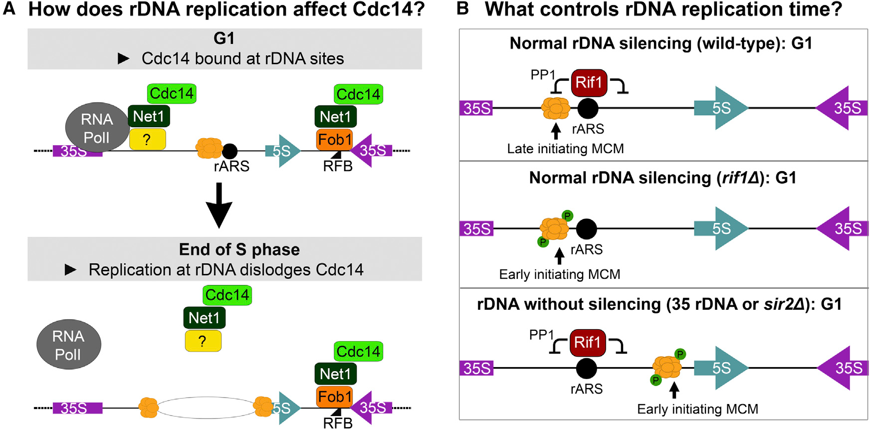
(A) In G1, Cdc14 is bound to the rDNA at the 35S rRNA transcription start site (TSS) and at the replication fork barrier (RFB). As rDNA begins replication in late S phase, Cdc14 is progressively released but persists at the RFB until replication forks stalled at the RFBs are resolved by oncoming forks and replication of the rDNA is completed. Thus, complete release of Cdc14 is coupled to completion of rDNA replication.
(B) Regulation of rDNA initiation time in wild-type and mutant cells. In wild-type cells, late S phase replication is enforced by two factors: (1) repressive chromatin established by Sir2 confines the MCM helicase to the vicinity of the rARS where (2) Rif1 keeps the helicase in an inactive (unphosphorylated) form (top). In the absence of Rif1 (middle), the MCM helicase is phosphorylated early leading to early origin activation. In sir2Δ or 35-copy rDNA strains (bottom), the open chromatin conformation allows the loaded MCM helicase to translocate away from the repressive environment of Rif1 so that initiation occurs early.
Our findings also are consistent with prior observations that rDNA copy number reduction both increases sensitivity to DNA damage and decreases sensitivity to replication stress. Reduction in rDNA copy number was previously shown to increase sensitivity to DNA damage.16 We are expanding the scope of this prior observation to encompass broader causes for the increased sensitivity of DNA damage: delayed replication genome-wide and premature anaphase plausibly explain the greater DNA damage sensitivity of strains with reduced rDNA copy number. Although rDNA copy number reduction has long been thought to act as a compensatory mechanism in cells surviving replication stress,14,24,26,39 the resulting reduced rDNA arrays remained late-replicating in two replication mutants examined thus far.25 Future studies will be necessary to determine the copy number threshold leading to early rDNA replication and whether this shift in replication time is gradual or precipitous.
How does rDNA initiation shift from late to early S phase?
Late replication of the rDNA locus is a conserved feature that has been linked to its heterochromatic status,48,49,97,98 which depends in part on Sir2 function in S. cerevisiae. Sir2 is a histone deacetylase that silences rDNA repeats.99 Without Sir2, the reduced nucleosome occupancy and increased transcription at active rDNA repeats facilitate the translocation of MCM helicases away from repressive chromatin environments100,101 that are likely generated by rARS-bound Rif1 (Figure 7B). Rif1 inhibits local replication initiation by recruiting protein phosphatase 1 (PP1, Glc7), which prevents the premature activation of the replication helicase subunit Mcm4.74,102,103 Because Rif1 binds at rDNA origins,40,104,105 wild-type rDNA arrays replicate early in the absence of Rif1. The 35-copy rDNA strain shares a critical feature with a sir2Δ mutant: its rDNA array is also euchromatic and highly transcribed,8,16 features which could allow loaded MCM helicases to translocate away from the rARS-bound Rif1. High rDNA copy number appears to be key in maintaining rDNA arrays as heterochromatic and late-replicating in an otherwise wild-type background.
How plausible is DNA replication as a gauge of whole genome replication status?
The decision to enter anaphase and exit mitosis must integrate several multifaceted inputs from the cell.106 The existence of a checkpoint that assesses genome replication completion has remained elusive, contentious, and unresolved.107–110 The late replication of the rDNA locus makes for a convenient checkpoint that signals the end of S phase: Cdc14, whose release from the nucleolus controls anaphase entry,84,87 is tethered to the rDNA through interactions with Fob1, Net1, and Sir2 (Figure 7A). The interaction of Cdc14 with Fob1 at the RFBs, the last positions within the rDNA repeat to be replicated, keeps Cdc14 within the nucleolus until rDNA replication has been completed. While it was originally considered that an exciting molecule like Cdc14 could not be in “a place as mundane as the nucleolus,”90 Cdc14 sequestration in the nucleolus is now recognized as an important hallmark of cell cycle regulation. Every cell with a nucleus contains rDNA and nucleoli, which are both highly responsive to cell and organismal physiology, including nutritional status, stress, and aging.17,23,111–114 The conservation of highly repetitive, heterochromatic, and late-replicating states of rDNA loci across a wide variety of species115 may both mitigate replication competition and coordinate genome replication status with cell cycle progression.
rDNA copy number and genome replication: Implications for disease
S. cerevisiae populations typically maintain strain-specific rDNA copy number,116 but the repetitive nature of rDNA arrays can allow for rare array contraction below the range of natural variation. Thus, rDNA copy number should always be taken into consideration as a background variable when interpreting the consequences of other genetic variants. In fact, rDNA copy number changes are frequently observed after standard S. cerevisiae genetic manipulation practices.116 For metazoans, rDNA copy number and replication time may have implications for health outcomes. A recent study reports that rDNA copy number reduction precedes pathogenesis in an mTOR-activated cancer mouse model, suggesting that rDNA reductions may act as driver mutations in certain cancers.21,22,111 Although the RFB protein Fob1 itself is not conserved, mutations affecting the metazoan equivalents generate developmental defects due to cell cycle misregulation.117 Finally, progeria in Hutchinson-Gilford cells is associated with bloated nucleoli, hyperactive ribosome biogenesis, and DNA damage that appears late in S phase,118,119 echoing the replication phenotypes observed in yeast strains with early-replicating rDNA.
Limitations of the study
While we have covered some limitations of our study in the appropriate results and discussion sections, we acknowledge some caveats that still remain. More precise timing markers for anaphase/cell cycle progression, such as spindle length measurements, may provide more accurate comparisons than the DAPI nuclear morphology used in this study. We would also like to note that the mechanism behind the anaphase delay seen in a sir2Δ strain is not known. Our use of the rif1Δ mutant as an early-replicating rDNA strain is tempered by previous reports of Rif1’s direct effects on local modulation of replication across the genome, although others have reported effects in line with replication delays at non-rDNA sites associated with rif1Δ’s early rDNA replication; to our knowledge, no genome-wide replication data for an S. cerevisiae rif1Δ strain have been published.40,74,77 Finally, our data show associations between rDNA replication and Cdc14 localization, but clear mechanistic experiments will be needed to define the molecular underpinnings of regulation of Cdc14, Net1, their direct binding interactions with rDNA, and anaphase progression.
STAR★METHODS
RESOURCE AVAILABILITY
Lead contact
Further information and requests for resources and reagents should be directed to and will be fulfilled by the Lead Contact, M. K. Raghuraman (raghu@uw.edu).
Materials availability
Strains generated for this manuscript are listed in Table S1 and the key resources table. Please request strains by contacting the lead contact. Unique reagents will be available without restrictions.
KEY RESOURCES TABLE.
| Antibodies | SOURCE | IDENTIFIER |
|---|---|---|
|
| ||
| Antibodies | ||
|
| ||
| Rat monoclonal anti-HA-Peroxidase | Sigma Aldrich | Roche Cat#12013819001; RRID:AB_390917 |
| Mouse monoclonal anti-PGK1 conjugated to HRP | Abcam | Abcam Cat#ab197960; RRID:AB_2756444 |
|
| ||
| Deposited data | ||
|
| ||
| Replication density transfer microarray data | This paper | GEO: GSE205068 |
|
| ||
| Experimental models: Organisms/strains | ||
|
| ||
| S288c MATa; BY rDNA (170 copies) | This paper | B150 |
| S288c MATa his3Δ0 leu2Δ0 met15Δ0 fob1 ::cloNAT; BY rDNA (35 copies) | This paper | B30 |
| S288c MATa fob1::cloNAT; BY rDNA (35 copies) | This paper | EK342 |
| S288c MATa fob1::cloNAT; BY rDNA (180 copies) | This paper | EK68 |
| S288c MATa; RM rDNA (100 copies) | Kwan et al.39 | R90 |
| S288c MATa his3Δ0 leu2Δ0 lys2Δ0 fob1 ::cloNAT; RM rDNA (30 copies) | This paper | R30 |
| S288c MATa his3Δ0 leu2Δ0 met15Δ0 ura3Δ0 fob1::cloNAT; BY rDNA (55 copies) | This paper | EK040 |
| S288c MATa his3Δ0 leu2Δ0 met15Δ0 fob1 ::cloNAT; BY rDNA (45 copies) | This paper | EK051 |
| S288c MATa his3Δ0 leu2Δ0 lys2Δ0 fob1 ::cloNAT; RM rDNA (45 copies) | This paper | EK057 |
| S288c MATa his3Δ0 leu2Δ0 met15Δ0 ura3Δ0 fob1::cloNAT; BY rDNA (80 copies) | This paper | EK100 |
| S288c MATa sir2::cloNAT HML::KanMX; BY rDNA (150 copies) | This paper | EK150 |
| S288c MATa sir2::cloNAT HML::KanMX; BY rDNA (100 copies) | This paper | EK151 |
| S288c MATa his3Δ0 leu2Δ0 met15Δ0 fob1 ::cloNAT; BY rDNA (35 copies) + pUC19-KanMX-KwCEN7-ARS1 | This paper | EK152 |
| S288c MATa his3Δ0 leu2Δ0 lys2Δ0 fob1 ::cloNAT; RM rDNA (35 copies) + pUC19-KanMX-KwCEN7-ARS1 | This paper | EK154 |
| S288c MATa BY rDNA fob1::cloNAT; BY rDNA (180 copies) + pUC19-KanMX-KwCEN7-ARS1 | This paper | EK156 |
| S288c MATa; BY rDNA (170 copies) + pUC19-KanMX-KwCEN7-ARS1 | This paper | EK184 |
| S288c MATa; RM rDNA (100 copies) + pUC19-KanMX-KwCEN7-ARS1 | This paper | EK185 |
| S288c MATa his3Δ0 leu2Δ0 met15Δ0 ura3Δ0 fob1::cloNAT CDC14-GFP:HIS3; BY rDNA (35 copies) | This paper | EK194 |
| S288c MATa his3Δ0 leu2Δ0 met15Δ0 ura3Δ0 fob1::cloNAT CDC14-GFP:HIS3; BY rDNA (170 copies) | This paper | EK202 |
| S288c MATa sir2::cloNAT fob1::cloNAT HML::KanMX; BY rDNA(180 copies) | This paper | EK360 |
| S288c MATa sir2::cloNAT fob1::cloNAT HML::KanMX; BY rDNA(170 copies) | This paper | EK361 |
| S288c MATa fob1::cloNAT rif1::KanMX; BY rDNA (35 copies) | This paper | EK375 |
| S288c MATa fob1::cloNAT sir2::cloNAT HML::KanMX; BY rDNA (35 copies) | This paper | EK376 |
| S288c MATa fob1::cloNAT rif1::KanMX; BY rDNA (180 copies) | This paper | EK379 |
| S288c MATa his3Δ0 leu2Δ0 met15Δ0 ura3Δ0 UTP13-GFP:HIS3 fob1::cloNAT; BY rDNA (35 copies) | This paper | EK396 |
| S288c MATa leu2Δ0 met15Δ0 ura3Δ0 fob1::NAT UTP13-GFP:HIS3; BY rDNA (~150 copies) | This paper | EK425 |
| S288c MATa fob1::NAT SLD2-HA:KanMX; BY rDNA (35 copies) | This paper | EK465 |
| S288c MATa fob1::NAT SLD2-HA:KanMX; BY rDNA (180 copies) | This paper | EK466 |
| S288c MATa URA3 rif1::KanMX; BY rDNA (180 copies) | This paper | EK468 |
| S288c MATa HIS3 leu2Δ0 met15Δ0 URA3 sir2::NAT hml::KanMX Cdc14-GFP:HIS3 UTP13-mCherry:HIS3; BY rDNA (150 copies) | This paper | EK604 |
| S288c MATa his3Δ0 leu2Δ0 met15Δ0 URA3 hml::KanMX Cdc14-GFP:HIS3 UTP13-mCherry:HIS3; BY rDNA (150 copies) | This paper | EK612 |
| S288c MATa his3Δ0 leu2Δ0 met15Δ0 URA3 sir2::NAT fob1::NAT hml::KanMX Cdc14-GFP:HIS3 UTP13-mCherry:HIS3; BY rDNA (150 copies) | This paper | EK615 |
| S288c MATa fob1::NAT; BY rDNA (180 copies), SCC1-HA-KanMX6 | This paper | EK626 |
| S288c MATa fob1::NAT; BY rDNA (35 copies), SCC1-HA-KanMX6 | This paper | EK628 |
| S288c MATa his3Δ0 leu2Δ0 met15Δ0 ura3Δ0 fob1::NAT CDC14-GFP:HIS3 NET1-mCherry-His3; BY rDNA (35 copies) | This paper | EK647 |
| S288c MATa his3Δ0 leu2Δ0 met15Δ0 ura3Δ0 fob1::NAT CDC14-GFP:HIS3 NET1-mCherry-His3; BY rDNA (35 copies) | This paper | EK648 |
| S288c MATa his3Δ0 leu2Δ0 met15Δ0 ura3Δ0 fob1::NAT CDC14-GFP:HIS3 NET1-mCherry-His3; BY rDNA (180 copies) | This paper | EK650 |
| S288c MATa his3Δ0 leu2Δ0 met15Δ0 ura3Δ0 fob1::NAT CDC14-GFP:HIS3 NET1-mCherry-His3; BY rDNA (180 copies) | This paper | EK651 |
|
| ||
| Oligonucleotides | ||
|
| ||
| Primers for Southern blot probes, see Table S1 | This paper | N/A |
|
| ||
| Recombinant DNA | ||
|
| ||
| pRDN1-Hyg plasmid | Chernoff et al.43 | pRDN1-Hyg |
| pUC19-KanMX-KwCEN7-ARS1 | Kwan et al.39 | pUC19-KanMX-KwCEN7-ARS1 |
|
| ||
| Software and algorithms | ||
|
| ||
| Quantity One® | Bio-Rad | https://www.bio-rad.com/en-us/sku/1709608-quantity-one-add-1-user-network-license?ID=1709608 |
| FlowJo | FlowJo, LLC | https://www.flowjo.com/ |
| R | The R Project for Statistical Computing | https://www.r-project.org/ |
Data and code availability
The density transfer microarray data are available at NCBI Gene Expression Omnibus (GEO) under accession number GEO: GSE205068.
This study did not generate new original code.
Any additional information required to reanalyze the data reported in this paper is available from the lead contact upon request.
EXPERIMENTAL MODELS AND SUBJECT DETAILS
Yeast strains
Genotypes of the S. cerevisiae strains used in this study are listed in the key resources table. Strains with deletions of SIR2 also have deletions of HML to maintain MATa function. Yeast strains were grown, unless noted otherwise, in synthetic complete media buffered with 1% succinic acid (per liter: 1.45 g yeast nitrogen base, 20 g glucose, 10 g succinic acid, 6 g NaOH, 5 g (NH4)2SO4, 2.8 g amino acid powder mix with pH adjusted to 5.8). When used, YPD medium contains per liter 20 g bacto peptone, 10 g yeast extract, and 20 g glucose. For each experiment comparing different strains, samples for all strains were collected concurrently in the same media.
METHOD DETAILS
rDNA reduction
S288c fob1Δ strains transformed with the pRDN1-Hyg plasmid were first isolated by selection for uracil prototrophy, then plated onto medium containing hygromycin B to select for rDNA copy number reduction.39,43,120 The pRDN1-Hyg plasmid was then removed by plating on medium containing 5-fluoroorotic acid (5-FOA). Individual colonies were picked for screening by CHEF gel electrophoresis to measure rDNA copy number (Figure S1). We identified and isolated strains with 35, 45, and 55 copies of rDNA and decided to focus on strains with 35 rDNA copies (“35 rDNA fob1Δ” and “35 rDNARM fob1Δ”), restoring endogenous URA3 to facilitate downstream replication assays. Genetic crosses were used to generate prototrophic strains and GFP-tagged strains.
During the isolation of strains with reduced rDNA copy number by this pRDN1-HYG plasmid method, we noticed that 20–25% of the isolates with rDNA reductions had either diploidized or tetraploidized (Figure S1), something we had not previously observed when constructing strains by transformation. Subsequently, we verified ploidy of each strain for each experiment by flow cytometry and used only confirmed haploid strains for each experiment. This frequent increase in ploidy may be related to rDNA reduction by this pRDN1-Hyg method. While interesting, we have not identified the biological mechanism involved and strongly suggest verifying ploidy when this rDNA reduction method is employed in the future.
Preparation of DNA in agarose plugs
DNA was isolated in agarose plugs according to previously published protocols.121 Each 90 mL plug contained either ~108 stationary phase cells for CHEF gels, ~5 × 10⁷ log phase cells for rDNA 2D gels, or ~108 log phase cells for single-copy origin 2D gels. Collected cells were washed with 50 mM EDTA, resuspended in 90 μL 0.5% SeaPlaque GTG agarose in 50 mM EDTA, and transferred into plug molds. Once solidified, plugs were incubated in 1 mL spheroplasting solution (1 M sorbitol, 20 mM EDTA, 10 mM Tris-HCl pH7.5, 14 mM β-mercaptoethanol, 0.5 mg/mL Zymolyase-20T (Amsbio)) for 2–5 h at 37°C. Plugs were washed once with LDS (1% lithium dodecyl sulfate, 100 mM EDTA, 10 mM Tris–HCl pH 8.0) and incubated overnight at 37°C in LDS overnight with gentle shaking. Plugs were then washed 3 × 30 min in 0.2X NDS (1X NDS pH 9.5: 0.5 M EDTA, 10 mM Tris base, 1% Sarkosyl) and 5 × 30 min in TE pH 8.0. Processed plugs were stored at 4°C in TE pH 8.0 until use.
CHEF gel analysis
We used contour-clamped homogeneous electric field (CHEF) gel electrophoresis to resolve intact S. cerevisiae chromosomes. A slice of each genomic DNA agarose plug was embedded in a 0.8% agarose gel (0.5X TBE) and each gel contained one wild-type sample as reference. For most CHEF gels, we ran the samples in 2.3L of 0.5X TBE using a Bio-Rad CHEF-DRII electrophoresis cell at 100V for 66 h (switch time = 300 to 900 s). The gels were then stained with ethidium bromide to visualize all chromosomes, including the rDNA-containing chromosome XII. To examine the size of the excised rDNA array, genomic DNA samples in plugs were digested with BamHI or FspI and then run on a 0.8% CHEF gel at 165 V for 64 h (switch time = 47 to 170 s). Chromosome XII size and rDNA copy number were further examined via Southern blotting. For size comparison, known standards (H. wingei and/or Yeast Ladder from New England BioLabs) were included in each CHEF gel run.
Chromosome replication completion assay
Cells were grown to mid-logarithmic phase (2.5 × 10⁶ cell/mL), arrested in G1 with 3 μM α-factor, and released into S phase (by the addition of 0.15 mg/mL Pronase (EMD Millipore)) in the presence of 0.008% MMS. Samples were collected every 20 min and prepared as described above in agarose plugs for CHEF gel electrophoresis. The same Southern blot membrane was probed for all measured chromosomes except for the FspI-excised rDNA. One experimental replicate was analyzed using multiple isolates of the 35 rDNA fob1Δ strains.
Southern blotting
Each gel was transferred to a GeneScreen Hybridization membrane using standard Southern blotting protocols.121 We then hybridized each sequence of interest using a 32P-labeled probe. The blots were exposed to X-ray film and to Bio-Rad Molecular Imaging FX phosphor screens for visualization and quantification of signal intensity. Southern blots were often stripped and re-probed with a different sequence of interest (CHEF gel blots, 2D gel blots, density transfer blots). To strip a Southern blot, it was subjected to two washes of 20 min each in 500 mL stripping buffer (0.1% SSC; 1% SDS) that had been heated to 100°C. Blot stripping efficacy was gauged before the next probe hybridization.
Density transfer
The density transfer protocol was adapted from Alvino et al. 2007.51 Dense medium composition was 0.5% 13C-labeled glucose, 0.5% 15(NH4)2SO4, 0.00145% yeast nitrogen base (YNB), and 1% succinic acid (isotopically light medium was the same composition with normal glucose and (NH4)2SO4). Cells were cultured in logarithmic phase for at least 10 generations in dense medium with the growth rate assessed for abnormalities. To collect synchronous S-phase cell samples, cultures of ~2.5 × 10⁶ cell/mL were arrested with 3 μM α-factor for 1.25 population doublings (approximately 2 h). Once the cell culture achieved >95% G1 arrest, cells were collected and washed 3 times with isotopically light medium containing α-factor. Cells were resuspended in the original volume of isotopically light medium containing 3 μM α-factor and a 100 mL G1 sample was taken for flow cytometry and DNA analysis. Cells were released from G1 into S phase by the addition of 0.15 mg/mL Pronase (EMD Millipore). 100 mL samples were collected and immediately transferred into vessels containing frozen pellets of 40 mL of 0.1% sodium azide in 0.2 M EDTA. The entire set of timed samples was collected before pelleting cells, taking a small aliquot for flow cytometry, and transferring the rest of the dry pellet to −20°C for storage until DNA isolation. DNA was extracted using a phenol:chloroform “Smash & Grab” protocol (see above) with an additional chloroform cleanup. Isolated DNA was digested overnight with EcoRI and then centrifuged in CsCl to separate replicated from unreplicated DNA. Cesium chloride gradients were drip-fractionated and the collected samples were analyzed using slot blots and hybridization to microarrays. All density transfer experimental replicates used in this manuscript are presented in the results and supplemental data. The density transfer microarray data are available at NCBI Gene Expression Omnibus (GEO) under accession number GSE205068.
Flow cytometry
Cells for flow cytometry were fixed in 70% ethanol before processing for flow cytometry. Fixed cells were washed with 50 mM sodium citrate, sonicated, and resuspended in 500 μL 50 mM sodium citrate. RNase A was added to a concentration of 0.25 mg/mL and the samples were incubated for 1 h at 50°C. Proteinase K (50 μL of 20 mg/mL) was then added and cells were incubated another hour at 50°C before staining with 1 μM Sytox Green. Cells were analyzed on a BD Canto II flow cytometer and flow cytometry data was analyzed using FlowJo software. All flow cytometry profiles are presented in Figure S2.
2D gel electrophoresis
Cells from the 180 rDNA fob1Δ strain, the 35 rDNA fob1Δ strain, and the 180 rDNA sir2Δ fob1Δ strain were grown in logarithmic phase to a culture density of ~2.5 × 10⁶ cells/mL. Cultures were then arrested in α-factor for 1.25 doublings before being released into S phase by addition of Pronase (0.15 mg/mL). Samples were collected every 5 min: 100 mL for analysis of single-copy genomic origins or 30 mL for analysis of rDNA origins. Collection vessels contained frozen pellets of 0.1% sodium azide in 0.2 M EDTA to halt growth. Cells were washed once with 50 mM EDTA, a small sample taken for flow cytometry, and the remaining dry cell pellets were stored at −20°C until preparation for 2D gel electrophoresis. To extract DNA for 2D gels, cells were embedded in three 90 μL 0.5% SeaPlaque agarose plugs and prepared as CHEF gel plugs. For each 2D gel, each plug was washed 3 × 20 min in the appropriate restriction buffer with 1X BSA (100 μg/mL). The solution was then removed and the DNA was digested for 5 h by addition of 3 μL restriction enzyme directly onto each plug, and then subjected to standard 2D gel electrophoresis methods (Brewer and Fangman, 1987),55 Southern blotted and hybridized for the sequence of interest. The number of rDNA initiations per cell was estimated for each sample by quantifying the “bubble arc” signal and normalizing to the “single-copy rDNA spot” from the NheI digest. Cumulative origin initiation was estimated by integrating the area under the curve generated from plotting “rDNA initiations per cell” across time. Replicates of the 2D gel “Release from G1” time course experiments were performed as follows: three replicates comparing 180 rDNA vs. 35 rDNA, one replicate comparing only sir2Δ fob1Δ vs. fob1Δ, and one replicate comparing wt, sir2Δ, rif1Δ, fob1Δ, and sir2Δ fob1Δ samples that were collected/analyzed at the same time.
For 2D gels of cells in hydroxyurea (HU), 20 mL of the culture was transferred to another flask for the “no HU” control to check that cells would have had normal release into S phase. Hydroxyurea was added to the remaining culture to a final concentration of 200 mM and 10 min later, 0.15 mg/mL Pronase was added to both cultures to release the cells into S phase. Samples were collected every 30 min and prepared as above.
Plasmid maintenance assay
Cells that contained plasmids were grown to logarithmic phase in selective medium (YPD +200 μg/mL G418) and then released into non-selective medium (YPD) for the plasmid maintenance assay. Cells were kept in logarithmic phase growth and samples were collected approximately every 4 h over the course of 48 h. The growth rate was monitored to ascertain the number of generations/divisions between samples. DNA was extracted from cells using the “Smash & grab” protocol, digested with XmnI, and run on an agarose gel to resolve the 5.4 kb plasmid ARS1 fragment from the 3.4 kb genomic ARS1 fragment (used as a “per cell” loading control). The gel was then Southern blotted and the membrane hybridized to a 32P-labeled ARS1 fragment. We quantified the amount of signal from plasmid ARS1 and genomic ARS1 for each sample and generated a plasmid maintenance curve for each strain, from which we were able to calculate the rate of plasmid loss per generation and estimate significance using linear regression. Two experimental replicates were analyzed for this manuscript.
Spot assays
Cells were grown to log-phase, diluted in sterile water in 3-fold dilutions, and 2.5 μL was spotted onto YPD plates containing either no drug (control), 0.016% methyl methanesulfonate (MMS), or 200 mM hydroxyurea (HU). Plates were scanned after 40–48 h of growth at 30°C.
Cycloheximide sensitivity assay
Cells were grown to log phase, upon which 3 × 10⁴ log-phase cells were transferred to each well in a 96-well plate containing 150 μL medium per well and the appropriate concentration of cycloheximide (0–200 ng/mL). Each condition was performed in triplicate and optical densities were measured at 30°C for 48 h using a Bio-Tek reader. Two separate experimental replicates were performed (Figure S1D). The maximum log-phase growth rate was manually calculated for each well.
Microscopy
Cells were fixed according to the protocol described on the Koshland lab web site (http://mcb.berkeley.edu/labs/koshland/Protocols/MICROSCOPY/gfpfix.html): collected cell pellets were resuspended in paraformaldehyde solution (4% paraformaldehyde, 3.4% sucrose) and incubated at room temperature for 15 min. Cells were then washed once with KPO4/sorbitol solution and resuspended in 50 μL KPO4/sorbitol solution (0.1 M KPO4 pH 7.5, 1.2 M sorbitol) and stored at 4°C until visualization. Before fluorescence microscopy, cells were sonicated and incubated with 0.5 μg/mL DAPI for at least an hour. Cells were visualized using a Leica DM4000 B fluorescence microscope and greater than 200 cells were scored for each sample. Multiple replicates performed for the DAPI anaphase entry experiments are presented in Figures 5, 6, and S6.
Western blotting
Log phase cells were grown to ~2.5 × 10⁶ cell/mL before α-factor G1 arrest and release. For each strain, 1.5 mL was collected for protein extraction and 1 mL was collected for flow cytometry. Collected cell pellets were resuspended in 200 μL SUMEB buffer (1% SDS, 8 M urea, 10 mM MOPS pH 6.8, 10 mM EDTA, 0.01% bromophenol blue) supplemented with protease inhibitors and 5% β-mercaptoethanol. Glass beads (~100 μL 0.5 mm acid-washed) were added and cells were vortexed for 3 min. Lysates were incubated at 65°C for 10 min with intermittent shaking and then centrifuged for 5 min at 4 °C at 20,000 × g. The clarified supernatant was transferred to a new tube and protein concentration was assessed using a Qubit (Thermo Fisher). For each sample, 15 μg of protein was run on a Novex Tris-acetate SDS-PAGE gel and transferred to a nitrocellulose membrane for immunoblotting. HRP-conjugated antibodies against HA (Sigma Aldrich #12013819001) and Pgk1 (Abcam #ab197960) were used in this work.
QUANTIFICATION AND STATISTICAL ANALYSIS
For Southern blot quantification, phosphor screens exposed to probed blots were scanned using a Bio-Rad Personal Molecular Imaging scanner and hybridization intensity values were extracted using Bio-Rad’s Quantity One software.
For the plasmid maintenance assays, we performed linear regression using the R statistical software package (https://cran.r-project.org) to calculate the rate of plasmid loss per generation and estimate significance. Statistical details can be found in the results section regarding Figure 3F. One representative biological replicate of two is presented. Methods were not employed to determine whether data met assumptions of the statistical approach. SD/SEM are not applicable to experiments presented.
rRNA quantification was performed as described.14 Asynchronous logarithmic phase cells were collected and nucleic acids (RNA and DNA) were isolated using a “Smash & Grab” phenol:chloroform extraction protocol.122 The RNA northern blot was hybridized to a 32P-labeled probe for the 25S rRNA sequence. To assess loading normalization, the DNA Southern blot portion was hybridized to a 32P-labeled probe for ACT1, a single copy gene. One replicate was performed in two separate strain backgrounds containing long and short rDNA arrays. The rRNA and ACT1 blots were separately exposed to Bio-Rad phosphor screens and 25S rRNA and ACT1 DNA hybridization intensities were quantified using a Bio-Rad Personal Molecular Imager and Bio-Rad Quantity One software.
For microscopy experiments, n values are reported in the figure legends (Figures 5 and 6).
Supplementary Material
Highlights.
The end of genome replication and anaphase are linked by ribosomal DNA replication
Early rDNA replication causes misregulation of Cdc14 and anaphase entry
Early rDNA replication also generates genome replication delays
High rDNA copy number delays rDNA replication until late S phase
ACKNOWLEDGMENTS
This work was supported by the following funding sources: University of Washington Genome Training Grant T32HG000035 to K.L.L., NIGMS R35 GM122497 to B.J.B. and M.K.R., NIGMS R01 GM122088 and R35 GM139532 and NHGRI grant RM1 HG010461 to C.Q., and NIGMS R01 GM117466 to A.B. We are grateful to the Kaeberlein Lab for generously providing us with the Cdc14-GFP and Utp13-GFP strains used to generate strains for this paper. We greatly appreciate Dr. Curran Oi for providing his mCherry tagging construct, Dr. Kerry Bubb for her statistics expertise and help with linear regression, and Dr. Trisha Davis for helpful feedback on microscopy. We would like to thank Dr. Elizabeth Morton, Dr. Ashley Hall, Dr. Matthew Crane, Mitsuhiro Tsuchiya, and Dr. Benjamin Blue for lively rDNA club discussions, critiques, and moral support. Special thanks to Dr. Stanley Fields for his critical reading of the manuscript and his many helpful suggestions.
INCLUSION AND DIVERSITY
We support inclusive, diverse, and equitable conduct of research.
Footnotes
DECLARATION OF INTERESTS
C.Q. is a Cell Reports Advisory Board member, Ecology and Evolution.
SUPPLEMENTAL INFORMATION
Supplemental information can be found online at https://doi.org/10.1016/j.celrep.2023.112161.
REFERENCES
- 1.Mohan J, and Ritossa FM (1970). Regulation of ribosomal RNA synthesis and its bearing on the bobbed phenotype in Drosophila melanogaster. Dev. Biol. 22, 495–512. 10.1016/0012-1606(70)90165-x. [DOI] [PubMed] [Google Scholar]
- 2.Morton EA, Hall AN, Kwan E, Mok C, Queitsch K, Nandakumar V, Stamatoyannopoulos J, Brewer BJ, Waterston R, and Queitsch C (2020). Challenges and approaches to genotyping repetitive DNA. G3 10, 417–430. 10.1534/g3.119.400771. [DOI] [PMC free article] [PubMed] [Google Scholar]
- 3.Parks MM, Kurylo CM, Dass RA, Bojmar L, Lyden D, Vincent CT, and Blanchard SC (2018). Variant ribosomal RNA alleles are conserved and exhibit tissue-specific expression. Sci. Adv. 4, eaao0665. 10.1126/sciadv.aao0665. [DOI] [PMC free article] [PubMed] [Google Scholar]
- 4.Thompson O, Edgley M, Strasbourger P, Flibotte S, Ewing B,Adair R, Au V, Chaudhry I, Fernando L, Hutter H, et al. (2013). The million mutation project: a new approach to genetics in Caenorhabditis elegans. Genome Res. 23, 1749–1762. 10.1101/gr.157651.113. [DOI] [PMC free article] [PubMed] [Google Scholar]
- 5.Conconi A, Widmer RM, Koller T, and Sogo JM (1989). Two different chromatin structures coexist in ribosomal RNA genes throughout the cell cycle. Cell 57, 753–761. 10.1016/0092-8674(89)90790-3. [DOI] [PubMed] [Google Scholar]
- 6.Conconi A, Sogo JM, and Ryan CA (1992). Ribosomal gene clusters are uniquely proportioned between open and closed chromatin structures in both tomato leaf cells and exponentially growing suspension cultures. Proc. Natl. Acad. Sci. USA 89, 5256–5260. 10.1073/pnas.89.12.5256. [DOI] [PMC free article] [PubMed] [Google Scholar]
- 7.Dammann R, Lucchini R, Koller T, and Sogo JM (1993). Chromatin structures and transcription of rDNA in yeast Saccharomyces cerevisiae. Nucleic Acids Res. 21, 2331–2338. 10.1093/nar/21.10.2331. [DOI] [PMC free article] [PubMed] [Google Scholar]
- 8.French SL, Osheim YN, Cioci F, Nomura M, and Beyer AL (2003). In exponentially growing Saccharomyces cerevisiae cells, rRNA synthesis is determined by the summed RNA polymerase I loading rate rather than by the number of active genes. Mol. Cell Biol. 23, 1558–1568. 10.1128/MCB.23.5.1558-1568.2003. [DOI] [PMC free article] [PubMed] [Google Scholar]
- 9.McStay B, and Grummt I (2008). The epigenetics of rRNA genes: from molecular to chromosome biology. Annu. Rev. Cell Dev. Biol. 24, 131–157. 10.1146/annurev.cellbio.24.110707.175259. [DOI] [PubMed] [Google Scholar]
- 10.Ye J, and Eickbush TH (2006). Chromatin structure and transcription of the R1- and R2-inserted rRNA genes of Drosophila melanogaster. Mol. Cell Biol. 26, 8781–8790. 10.1128/MCB.01409-06. [DOI] [PMC free article] [PubMed] [Google Scholar]
- 11.Nelson JO, Watase GJ, Warsinger-Pepe N, and Yamashita YM (2019). Mechanisms of rDNA copy number maintenance. Trends Genet. 35, 734–742. 10.1016/j.tig.2019.07.006. [DOI] [PMC free article] [PubMed] [Google Scholar]
- 12.Delany ME, Muscarella DE, and Bloom SE (1994). Effects of rRNA gene copy number and nucleolar variation on early development: inhibition of gastrulation in rDNA-deficient chick embryo. J. Hered. 85, 211–217. 10.1093/oxfordjournals.jhered.a111437. [DOI] [PubMed] [Google Scholar]
- 13.Ritossa FM, and Atwood KC (1966). Unequal proportions of DNA complementary to ribosomal RNA in males and females of Drosophila simulans. Proc. Natl. Acad. Sci. USA 56, 496–499. 10.1073/pnas.56.2.496. [DOI] [PMC free article] [PubMed] [Google Scholar]
- 14.Sanchez JC, Kwan EX, Pohl TJ, Amemiya HM, Raghuraman MK, and Brewer BJ (2017). Defective replication initiation results in locus specific chromosome breakage and a ribosomal RNA deficiency in yeast. PLoS Genet. 13, e1007041. 10.1371/journal.pgen.1007041. [DOI] [PMC free article] [PubMed] [Google Scholar]
- 15.Gibbons JG, Branco AT, Yu S, and Lemos B (2014). Ribosomal DNA copy number is coupled with gene expression variation and mitochondrial abundance in humans. Nat. Commun. 5, 4850. 10.1038/ncomms5850. [DOI] [PubMed] [Google Scholar]
- 16.Ide S, Miyazaki T, Maki H, and Kobayashi T (2010). Abundance of ribosomal RNA gene copies maintains genome integrity. Science 327, 693–696. 10.1126/science.1179044. [DOI] [PubMed] [Google Scholar]
- 17.Lu KL, Nelson JO, Watase GJ, Warsinger-Pepe N, and Yamashita YM (2018). Transgenerational dynamics of rDNA copy number in Drosophila male germline stem cells. Elife 7, e32421. 10.7554/eLife.32421. [DOI] [PMC free article] [PubMed] [Google Scholar]
- 18.Michel AH, Kornmann B, Dubrana K, and Shore D (2005). Spontaneous rDNA copy number variation modulates Sir2 levels and epigenetic gene silencing. Genes Dev. 19, 1199–1210. 10.1101/gad.340205. [DOI] [PMC free article] [PubMed] [Google Scholar]
- 19.Paredes S, Branco AT, Hartl DL, Maggert KA, and Lemos B (2011). Ribosomal DNA deletions modulate genome-wide gene expression: “rDNA-sensitive” genes and natural variation. PLoS Genet. 7, e1001376. 10.1371/journal.pgen.1001376. [DOI] [PMC free article] [PubMed] [Google Scholar]
- 20.Picart-Picolo A, Grob S, Picault N, Franek M, Llauro C, Halter T, Maier TR, Jobet E, Descombin J, Zhang P, et al. (2020). Large tandem duplications affect gene expression, 3D organization, and plant-pathogen response. Genome Res. 30, 1583–1592. 10.1101/gr.261586.120. [DOI] [PMC free article] [PubMed] [Google Scholar]
- 21.Wang M, and Lemos B (2017). Ribosomal DNA copy number amplification and loss in human cancers is linked to tumor genetic context, nucleolus activity, and proliferation. PLoS Genet. 13, e1006994. 10.1371/journal.pgen.1006994. [DOI] [PMC free article] [PubMed] [Google Scholar]
- 22.Xu B, Li H, Perry JM, Singh VP, Unruh J, Yu Z, Zakari M, McDowell W, Li L, and Gerton JL (2017). Ribosomal DNA copy number loss and sequence variation in cancer. PLoS Genet. 13, e1006771. 10.1371/journal.pgen.1006771. [DOI] [PMC free article] [PubMed] [Google Scholar]
- 23.Aldrich JC, and Maggert KA (2015). Transgenerational inheritance of diet-induced genome rearrangements in Drosophila. PLoS Genet. 11, e1005148. 10.1371/journal.pgen.1005148. [DOI] [PMC free article] [PubMed] [Google Scholar]
- 24.Ide S, Watanabe K, Watanabe H, Shirahige K, Kobayashi T, and Maki H (2007). Abnormality in initiation program of DNA replication is monitored by the highly repetitive rRNA gene array on chromosome XII in budding yeast. Mol. Cell Biol. 27, 568–578. 10.1128/MCB.00731-06. [DOI] [PMC free article] [PubMed] [Google Scholar]
- 25.Lynch KL, Alvino GM, Kwan EX, Brewer BJ, and Raghuraman MK (2019). The effects of manipulating levels of replication initiation factors on origin firing efficiency in yeast. PLoS Genet. 15, e1008430. 10.1371/journal.pgen.1008430. [DOI] [PMC free article] [PubMed] [Google Scholar]
- 26.Salim D, Bradford WD, Freeland A, Cady G, Wang J, Pruitt SC, and Gerton JL (2017). DNA replication stress restricts ribosomal DNA copy number. PLoS Genet. 13, e1007006. 10.1371/journal.pgen.1007006. [DOI] [PMC free article] [PubMed] [Google Scholar]
- 27.Bénard M, Lagnel C, and Pierron G (1995). Site-specific initiation of DNA replication within the non-transcribed spacer of Physarum rDNA. Nucleic Acids Res. 23, 1447–1453. 10.1093/nar/23.9.1447. [DOI] [PMC free article] [PubMed] [Google Scholar]
- 28.Brewer BJ, and Fangman WL (1988). A replication fork barrier at the3’ end of yeast ribosomal RNA genes. Cell 55, 637–643. 10.1016/0092-8674(88)90222-x. [DOI] [PubMed] [Google Scholar]
- 29.Coffman FD, He M, Diaz M-L, and Cohen S (2006). Multiple initiation sites within the human ribosomal RNA gene. Cell Cycle 5, 1223–1233. 10.4161/cc.5.11.2814. [DOI] [PubMed] [Google Scholar]
- 30.Hyrien O, and Méchali M (1992). Plasmid replication in Xenopus eggs and egg extracts: a 2D gel electrophoretic analysis. Nucleic Acids Res. 20, 1463–1469. 10.1093/nar/20.7.1463. [DOI] [PMC free article] [PubMed] [Google Scholar]
- 31.Little RD, Platt TH, and Schildkraut CL (1993). Initiation and termination of DNA replication in human rRNA genes. Mol. Cell Biol. 13, 6600–6613. 10.1128/mcb.13.10.6600. [DOI] [PMC free article] [PubMed] [Google Scholar]
- 32.López-estraño C, Schvartzman JB, Krimer DB, and Hernández P (1998). Co-localization of polar replication fork barriers and rRNA transcription terminators in mouse rDNA. J. Mol. Biol. 277, 249–256. 10.1006/jmbi.1997.1607. [DOI] [PubMed] [Google Scholar]
- 33.Brewer BJ, Lockshon D, and Fangman WL (1992). The arrest of replication forks in the rDNA of yeast occurs independently of transcription. Cell 71, 267–276. 10.1016/0092-8674(92)90355-g. [DOI] [PubMed] [Google Scholar]
- 34.Miller CA, and Kowalski D (1993). cis-acting components in the replication origin from ribosomal DNA of Saccharomyces cerevisiae. Mol. Cell Biol. 13, 5360–5369. 10.1128/mcb.13.9.5360. [DOI] [PMC free article] [PubMed] [Google Scholar]
- 35.Linskens MH, and Huberman JA (1988). Organization of replication of ribosomal DNA in Saccharomyces cerevisiae. Mol. Cell Biol. 8, 4927–4935. 10.1128/mcb.8.11.4927. [DOI] [PMC free article] [PubMed] [Google Scholar]
- 36.Collart C, Allen GE, Bradshaw CR, Smith JC, and Zegerman P(2013). Titration of four replication factors is essential for the Xenopus laevis midblastula transition. Science 341, 893–896. 10.1126/science.1241530. [DOI] [PMC free article] [PubMed] [Google Scholar]
- 37.Mantiero D, Mackenzie A, Donaldson A, and Zegerman P (2011). Limiting replication initiation factors execute the temporal programme of origin firing in budding yeast. EMBO J. 30, 4805–4814. 10.1038/emboj.2011.404. [DOI] [PMC free article] [PubMed] [Google Scholar]
- 38.Yoshida K, Bacal J, Desmarais D, Padioleau I, Tsaponina O, Chabes A, Pantesco V, Dubois E, Parrinello H, Skrzypczak M, et al. (2014). The histone deacetylases sir2 and rpd3 act on ribosomal DNA to control the replication program in budding yeast. Mol. Cell 54, 691–697. 10.1016/j.molcel.2014.04.032. [DOI] [PubMed] [Google Scholar]
- 39.Kwan EX, Foss EJ, Tsuchiyama S, Alvino GM, Kruglyak L, Kaeberlein M, Raghuraman MK, Brewer BJ, Kennedy BK, and Bedalov A (2013). A natural polymorphism in rDNA replication origins links origin activation with calorie restriction and lifespan. PLoS Genet. 9, e1003329. 10.1371/journal.pgen.1003329. [DOI] [PMC free article] [PubMed] [Google Scholar]
- 40.Shyian M, Mattarocci S, Albert B, Hafner L, Lezaja A, Costanzo M, Boone C, and Shore D (2016). Budding yeast Rif1 controls genome integrity by inhibiting rDNA replication. PLoS Genet. 12, e1006414. 10.1371/journal.pgen.1006414. [DOI] [PMC free article] [PubMed] [Google Scholar]
- 41.Foss EJ, Lao U, Dalrymple E, Adrianse RL, Loe T, and Bedalov A (2017). SIR2 suppresses replication gaps and genome instability by balancing replication between repetitive and unique sequences. Proc. Natl. Acad. Sci. USA 114, 552–557. 10.1073/pnas.1614781114. [DOI] [PMC free article] [PubMed] [Google Scholar]
- 42.Nieduszynski CA, Hiraga S.i., Ak P, Benham CJ, and Donaldson AD (2007). OriDB: a DNA replication origin database. Nucleic Acids Res. 35, D40–D46. 10.1093/nar/gkl758. [DOI] [PMC free article] [PubMed] [Google Scholar]
- 43.Chernoff YO, Vincent A, and Liebman SW (1994). Mutations in eukaryotic 18S ribosomal RNA affect translational fidelity and resistance to aminoglycoside antibiotics. EMBO J. 13, 906–913. 10.1002/j.1460-2075.1994.tb06334.x. [DOI] [PMC free article] [PubMed] [Google Scholar]
- 44.Kobayashi T, Heck DJ, Nomura M, and Horiuchi T (1998). Expansion and contraction of ribosomal DNA repeats in Saccharomyces cerevisiae: requirement of replication fork blocking (Fob1) protein and the role of RNA polymerase I. Genes Dev. 12, 3821–3830. 10.1101/gad.12.24.3821. [DOI] [PMC free article] [PubMed] [Google Scholar]
- 45.Dauban L, Kamgoué A, Wang R, Léger-Silvestre I, Beckouët F, Cantaloube S, and Gadal O (2019). Quantification of the dynamic behaviour of ribosomal DNA genes and nucleolus during yeast Saccharomyces cerevisiae cell cycle. J. Struct. Biol. 208, 152–164. 10.1016/j.jsb.2019.08.010. [DOI] [PubMed] [Google Scholar]
- 46.Kim Y-H, Ishikawa D, Ha HP, Sugiyama M, Kaneko Y, and Harashima S (2006). Chromosome XII context is important for rDNA function in yeast. Nucleic Acids Res. 34, 2914–2924. 10.1093/nar/gkl293. [DOI] [PMC free article] [PubMed] [Google Scholar]
- 47.Coffman FD, He M, Diaz M-L, and Cohen S (2005). DNA replication initiates at different sites in early and late S phase within human ribosomal RNA genes. Cell Cycle 4, 1223–1226. 10.4161/cc.4.9.1961. [DOI] [PubMed] [Google Scholar]
- 48.Concia L, Brooks AM, Wheeler E, Zynda GJ, Wear EE, LeBlanc C, Song J, Lee T-J, Pascuzzi PE, Martienssen RA, et al. (2018). Genome-wide analysis of the Arabidopsis replication timing program. Plant Physiol. 176, 2166–2185. 10.1104/pp.17.01537. [DOI] [PMC free article] [PubMed] [Google Scholar]
- 49.Labit H, Perewoska I, Germe T, Hyrien O, and Marheineke K (2008). DNA replication timing is deterministic at the level of chromosomal domains but stochastic at the level of replicons in Xenopus egg extracts. Nucleic Acids Res. 36, 5623–5634. 10.1093/nar/gkn533. [DOI] [PMC free article] [PubMed] [Google Scholar]
- 50.Schübeler D, Scalzo D, Kooperberg C, van Steensel B, Delrow J, and Groudine M (2002). Genome-wide DNA replication profile for Drosophila melanogaster: a link between transcription and replication timing. Nat. Genet. 32, 438–442. 10.1038/ng1005. [DOI] [PubMed] [Google Scholar]
- 51.Alvino GM, Collingwood D, Murphy JM, Delrow J, Brewer BJ, and Raghuraman MK (2007). Replication in hydroxyurea: it’s a matter of time. Mol. Cell Biol. 27, 6396–6406. 10.1128/MCB.00719-07. [DOI] [PMC free article] [PubMed] [Google Scholar]
- 52.Meselson M, and Stahl FW (1958). The replication of DNA in Escherichia coli. Proc. Natl. Acad. Sci. USA 44, 671–682. 10.1073/pnas.44.7.671. [DOI] [PMC free article] [PubMed] [Google Scholar]
- 53.Raghuraman MK, Winzeler EA, Collingwood D, Hunt S, Wodicka L, Conway A, Lockhart DJ, Davis RW, Brewer BJ, and Fangman WL (2001). Replication dynamics of the yeast genome. Science 294, 115–121. 10.1126/science.294.5540.115. [DOI] [PubMed] [Google Scholar]
- 54.Lengronne A, and Schwob E (2002). The yeast CDK inhibitor Sic1 prevents genomic instability by promoting replication origin licensing in late G(1). Mol. Cell 9, 1067–1078. 10.1016/s1097-2765(02)00513-0. [DOI] [PubMed] [Google Scholar]
- 55.Brewer BJ, and Fangman WL (1987). The localization of replication origins on ARS plasmids in S. cerevisiae. Cell 51, 463–471. 10.1016/0092-8674(87)90642-8. [DOI] [PubMed] [Google Scholar]
- 56.Alcasabas AA, Osborn AJ, Bachant J, Hu F, Werler PJ, Bousset K, Furuya K, Diffley JF, Carr AM, and Elledge SJ (2001). Mrc1 transduces signals of DNA replication stress to activate Rad53. Nat. Cell Biol. 3, 958–965. 10.1038/ncb1101-958. [DOI] [PubMed] [Google Scholar]
- 57.Feng W, Collingwood D, Boeck ME, Fox LA, Alvino GM, Fangman WL, Raghuraman MK, and Brewer BJ (2006). Genomic mapping of single-stranded DNA in hydroxyurea-challenged yeasts identifies origins of replication. Nat. Cell Biol. 8, 148–155. 10.1038/ncb1358. [DOI] [PMC free article] [PubMed] [Google Scholar]
- 58.Santocanale C, and Diffley JF (1998). A Mec1- and Rad53-dependent checkpoint controls late-firing origins of DNA replication. Nature 395, 615–618. 10.1038/27001. [DOI] [PubMed] [Google Scholar]
- 59.Yabuki N, Terashima H, and Kitada K (2002). Mapping of early firing origins on a replication profile of budding yeast. Gene Cell. 7, 781–789. 10.1046/j.1365-2443.2002.00559.x. [DOI] [PubMed] [Google Scholar]
- 60.Friedman KL, Brewer BJ, and Fangman WL (1997). Replication profile of Saccharomyces cerevisiae chromosome VI. Gene Cell. 2, 667–678. 10.1046/j.1365-2443.1997.1520350.x. [DOI] [PubMed] [Google Scholar]
- 61.Hoang ML, Leon RP, Pessoa-Brandao L, Hunt S, Raghuraman MK, Fangman WL, Brewer BJ, and Sclafani RA (2007). Structural changes in Mcm5 protein bypass Cdc7-Dbf4 function and reduce replication origin efficiency in Saccharomyces cerevisiae. Mol. Cell Biol. 27, 7594–7602. 10.1128/MCB.00997-07. [DOI] [PMC free article] [PubMed] [Google Scholar]
- 62.Ferguson BM, and Fangman WL (1992). A position effect on the time of replication origin activation in yeast. Cell 68, 333–339. 10.1016/0092-8674(92)90474-q. [DOI] [PubMed] [Google Scholar]
- 63.Pohl TJ, Kolor K, Fangman WL, Brewer BJ, and Raghuraman MK (2013). A DNA sequence element that advances replication origin activation time in Saccharomyces cerevisiae. G3 3, 1955–1963. 10.1534/g3.113.008250. [DOI] [PMC free article] [PubMed] [Google Scholar]
- 64.Foss M, McNally FJ, Laurenson P, and Rine J (1993). Origin recognition complex (ORC) in transcriptional silencing and DNA replication in S. cerevisiae. Science 262, 1838–1844. 10.1126/science.8266071. [DOI] [PubMed] [Google Scholar]
- 65.Tye BK (1999). Minichromosome maintenance as a genetic assay for defects in DNA replication. Methods 18, 329–334. 10.1006/meth.1999.0793. [DOI] [PubMed] [Google Scholar]
- 66.Bellaoui M, Chang M, Ou J, Xu H, Boone C, and Brown GW (2003). Elg1 forms an alternative RFC complex important for DNA replication and genome integrity. EMBO J. 22, 4304–4313. 10.1093/emboj/cdg406. [DOI] [PMC free article] [PubMed] [Google Scholar]
- 67.Lee L, Rodriguez J, and Tsukiyama T (2015). Chromatin remodeling factors Isw2 and Ino80 regulate checkpoint activity and chromatin structure in S phase. Genetics 199, 1077–1091. 10.1534/genetics.115.174730. [DOI] [PMC free article] [PubMed] [Google Scholar]
- 68.Ogi H, Wang C-Z, Nakai W, Kawasaki Y, and Masumoto H (2008). The role of the Saccharomyces cerevisiae Cdc7-Dbf4 complex in the replication checkpoint. Gene 414, 32–40. 10.1016/j.gene.2008.02.010. [DOI] [PubMed] [Google Scholar]
- 69.Stinchcomb DT, Struhl K, and Davis RW (1979). Isolation and characterisation of a yeast chromosomal replicator. Nature 282, 39–43. 10.1038/282039a0. [DOI] [PubMed] [Google Scholar]
- 70.Maiti AK, and Sinha P (1992). The mcm2 mutation of yeast affects replication, rather than segregation or amplification of the two micron plasmid. J. Mol. Biol. 224, 545–558. 10.1016/0022-2836(92)90543-s. [DOI] [PubMed] [Google Scholar]
- 71.Storici F, Oberto J, and Bruschi CV (1995). The CDC6 gene is required for centromeric, episomal, and 2-microns plasmid stability in the yeast Saccharomyces cerevisiae. Plasmid 34, 184–197. 10.1006/plas.1995.0004. [DOI] [PubMed] [Google Scholar]
- 72.Ivessa AS, and Zakian VA (2002). To fire or not to fire: origin activation in Saccharomyces cerevisiae ribosomal DNA. Genes Dev. 16, 2459–2464. 10.1101/gad.1033702. [DOI] [PubMed] [Google Scholar]
- 73.Pasero P, Bensimon A, and Schwob E (2002). Single-molecule analysis reveals clustering and epigenetic regulation of replication origins at the yeast rDNA locus. Genes Dev. 16, 2479–2484. 10.1101/gad.232902. [DOI] [PMC free article] [PubMed] [Google Scholar]
- 74.Lian H-Y, Robertson ED, Hiraga S.i., Alvino GM, Collingwood D, McCune HJ, Sridhar A, Brewer BJ, Raghuraman MK, and Donaldson AD (2011). The effect of Ku on telomere replication time is mediated by telomere length but is independent of histone tail acetylation. Mol. Biol. Cell 22, 1753–1765. 10.1091/mbc.E10-06-0549. [DOI] [PMC free article] [PubMed] [Google Scholar]
- 75.Hartwell LH, Culotti J, Pringle JR, and Reid BJ (1974). Genetic control of the cell division cycle in yeast. Science 183, 46–51. 10.1126/science.183.4120.46. [DOI] [PubMed] [Google Scholar]
- 76.Yellman CM, and Roeder GS (2015). Cdc14 early anaphase release, FEAR, is limited to the nucleus and dispensable for efficient mitotic exit. PLoS One 10, e0128604. 10.1371/journal.pone.0128604. [DOI] [PMC free article] [PubMed] [Google Scholar]
- 77.Hayano M, Kanoh Y, Matsumoto S, Renard-Guillet C, Shirahige K, and Masai H (2012). Rif1 is a global regulator of timing of replication origin firing in fission yeast. Genes Dev. 26, 137–150. 10.1101/gad.178491.111. [DOI] [PMC free article] [PubMed] [Google Scholar]
- 78.Li R, and Murray AW (1991). Feedback control of mitosis in budding yeast. Cell 66, 519–531. 10.1016/0092-8674(81)90015-5. [DOI] [PubMed] [Google Scholar]
- 79.Michel LS, Liberal V, Chatterjee A, Kirchwegger R, Pasche B, Gerald W, Dobles M, Sorger PK, Murty VV, and Benezra R (2001). MAD2 haplo-insufficiency causes premature anaphase and chromosome instability in mammalian cells. Nature 409, 355–359. 10.1038/35053094. [DOI] [PubMed] [Google Scholar]
- 80.Villoria MT, Ramos F, Dueñas E, Faull P, Cutillas PR, and Clemente-Blanco A (2017). Stabilization of the metaphase spindle by Cdc14 is required for recombinational DNA repair. EMBO J. 36, 79–101. 10.15252/embj.201593540. [DOI] [PMC free article] [PubMed] [Google Scholar]
- 81.Yamamoto A, Guacci V, and Koshland D (1996). Pds1p, an inhibitor of anaphase in budding yeast, plays a critical role in the APC and checkpoint pathway(s). J. Cell Biol. 133, 99–110. 10.1083/jcb.133.1.99. [DOI] [PMC free article] [PubMed] [Google Scholar]
- 82.Huang J, and Moazed D (2003). Association of the RENT complex with nontranscribed and coding regions of rDNA and a regional requirement for the replication fork block protein Fob1 in rDNA silencing. Genes Dev. 17, 2162–2176. 10.1101/gad.1108403. [DOI] [PMC free article] [PubMed] [Google Scholar]
- 83.Stegmeier F, Huang J, Rahal R, Zmolik J, Moazed D, and Amon A (2004). The replication fork block protein Fob1 functions as a negative regulator of the FEAR network. Curr. Biol. 14, 467–480. 10.1016/j.cub.2004.03.009. [DOI] [PubMed] [Google Scholar]
- 84.Shou W, Seol JH, Shevchenko A, Baskerville C, Moazed D, Chen ZW, Jang J, Shevchenko A, Charbonneau H, and Deshaies RJ (1999). Exit from mitosis is triggered by Tem1-dependent release of the protein phosphatase Cdc14 from nucleolar RENT complex. Cell 97, 233–244. 10.1016/s0092-8674(00)80733-3. [DOI] [PubMed] [Google Scholar]
- 85.Traverso EE, Baskerville C, Liu Y, Shou W, James P, Deshaies RJ, and Charbonneau H(2001).CharacterizationoftheNet1cellcycle-dependent regulator of the Cdc14 phosphatase from budding yeast. J. Biol. Chem. 276, 21924–21931. 10.1074/jbc.M011689200. [DOI] [PubMed] [Google Scholar]
- 86.Visintin R, Craig K, Hwang ES, Prinz S, Tyers M, and Amon A (1998). The phosphatase Cdc14 triggers mitotic exit by reversal of Cdk-dependent phosphorylation. Mol. Cell 2, 709–718. 10.1016/s1097-2765(00)80286-5. [DOI] [PubMed] [Google Scholar]
- 87.Visintin R, Hwang ES, and Amon A (1999). Cfi1 prevents premature exit from mitosis by anchoring Cdc14 phosphatase in the nucleolus. Nature 398, 818–823. 10.1038/19775. [DOI] [PubMed] [Google Scholar]
- 88.Huh W-K, Falvo JV, Gerke LC, Carroll AS, Howson RW, Weissman JS, and O’Shea EK (2003). Global analysis of protein localization in budding yeast. Nature 425, 686–691. 10.1038/nature02026. [DOI] [PubMed] [Google Scholar]
- 89.Woolford JL, and Baserga SJ (2013). Ribosome biogenesis in the yeast Saccharomyces cerevisiae. Genetics 195, 643–681. 10.1534/genetics.113.153197. [DOI] [PMC free article] [PubMed] [Google Scholar]
- 90.Amon A (2008). A decade of Cdc14–a personal perspective. Delivered on 9 july 2007 at the 32nd FEBS congress in Vienna, Austria. FEBS J. 275, 5774–5784. 10.1111/j.1742-4658.2008.06693.x. [DOI] [PMC free article] [PubMed] [Google Scholar]
- 91.Kuilman T, Maiolica A, Godfrey M, Scheidel N, Aebersold R, and Uhlmann F (2015). Identification of Cdk targets that control cytokinesis. EMBO J. 34, 81–96. 10.15252/embj.201488958. [DOI] [PMC free article] [PubMed] [Google Scholar]
- 92.Sullivan M, and Uhlmann F (2003). A non-proteolytic function of separase links the onset of anaphase to mitotic exit. Nat. Cell Biol. 5, 249–254. 10.1038/ncb940. [DOI] [PMC free article] [PubMed] [Google Scholar]
- 93.Bar-Ziv R, Brodsky S, Chapal M, and Barkai N (2020). Transcription factor binding to replicated DNA. Cell Rep. 30, 3989–3995.e4. 10.1016/j.celrep.2020.02.114. [DOI] [PubMed] [Google Scholar]
- 94.Brüning JG, Howard JL, and McGlynn P (2014). Accessory replicative helicases and the replication of protein-bound DNA. J. Mol. Biol. 426, 3917–3928. 10.1016/j.jmb.2014.10.001. [DOI] [PubMed] [Google Scholar]
- 95.Kaplan DL, and O’Donnell M (2002). DnaB drives DNA branch migration and dislodges proteins while encircling two DNA strands. Mol. Cell 10, 647–657. 10.1016/s1097-2765(02)00642-1. [DOI] [PubMed] [Google Scholar]
- 96.Ramachandran S, and Henikoff S (2016). Transcriptional regulators compete with nucleosomes post-replication. Cell 165, 580–592. 10.1016/j.cell.2016.02.062. [DOI] [PMC free article] [PubMed] [Google Scholar]
- 97.Lima-De-Faria A (1959). Differential uptake of tritiated thymidine into hetero- and euchromatin in Melanoplus and Secale. J. Biophys. Biochem. Cytol. 6, 457–466. 10.1083/jcb.6.3.457. [DOI] [PMC free article] [PubMed] [Google Scholar]
- 98.Reverón-Gómez N, González-Aguilera C, Stewart-Morgan KR, Petryk N, Flury V, Graziano S, Johansen JV, Jakobsen JS, Alabert C, and Groth A (2018). Accurate recycling of parental histones reproduces the histone modification landscape during DNA replication. Mol. Cell 72, 239–249.e5. 10.1016/j.molcel.2018.08.010. [DOI] [PMC free article] [PubMed] [Google Scholar]
- 99.Smith JS, and Boeke JD (1997). An unusual form of transcriptional-silencing in yeast ribosomal DNA. Genes Dev. 11, 241–254. 10.1101/gad.11.2.241. [DOI] [PubMed] [Google Scholar]
- 100.Foss EJ, Gatbonton-Schwager T, Thiesen AH, Taylor E, Soriano R, Lao U, MacAlpine DM, and Bedalov A (2019). Sir2 suppresses transcription-mediated displacement of Mcm2-7 replicative helicases at the ribosomal DNA repeats. PLoS Genet. 15, e1008138. 10.1371/journal.pgen.1008138. [DOI] [PMC free article] [PubMed] [Google Scholar]
- 101.Gros J, Kumar C, Lynch G, Yadav T, Whitehouse I, and Remus D (2015). Post-licensing specification of eukaryotic replication origins by facilitated mcm2-7 sliding along DNA. Mol. Cell 60, 797–807. 10.1016/j.molcel.2015.10.022. [DOI] [PMC free article] [PubMed] [Google Scholar]
- 102.Davé A, Cooley C, Garg M, and Bianchi A (2014). Protein phosphatase 1 recruitment by Rif1 regulates DNA replication origin firing by counteracting DDK activity. Cell Rep. 7, 53–61. 10.1016/j.celrep.2014.02.019. [DOI] [PMC free article] [PubMed] [Google Scholar]
- 103.Mattarocci S, Shyian M, Lemmens L, Damay P, Altintas DM, Shi T, Bartholomew CR, Thomä NH, Hardy CFJ, and Shore D (2014). Rif1 controls DNA replication timing in yeast through the PP1 phosphatase Glc7. Cell Rep. 7, 62–69. 10.1016/j.celrep.2014.03.010. [DOI] [PubMed] [Google Scholar]
- 104.Hafner L, Lezaja A, Zhang X, Lemmens L, Shyian M, Albert B, Follonier C, Nunes JM, Lopes M, Shore D, et al. (2018). Rif1 binding and control of chromosome-internal DNA replication origins is limited by telomere sequestration. Cell Rep. 23, 983–992. 10.1016/j.celrep.2018.03.113. [DOI] [PubMed] [Google Scholar]
- 105.Hafner L, Shore D, and Mattarocci S (2019). ChECing out Rif1 action in freely cycling cells. Curr. Genet. 65, 429–434. 10.1007/s00294-018-0902-0. [DOI] [PubMed] [Google Scholar]
- 106.Campbell IW, Zhou X, and Amon A (2019). The Mitotic Exit Network integrates temporal and spatial signals by distributing regulation across multiple components. Elife 8, e41139. 10.7554/eLife.41139. [DOI] [PMC free article] [PubMed] [Google Scholar]
- 107.Ivanova T, Maier M, Missarova A, Ziegler-Birling C, Dam M, Gomar-Alba M, Carey LB, and Mendoza M (2020). Budding yeast complete DNA synthesis after chromosome segregation begins. Nat. Commun. 11, 2267. 10.1038/s41467-020-16100-3. [DOI] [PMC free article] [PubMed] [Google Scholar]
- 108.Torres-Rosell J, De Piccoli G, Cordon-Preciado V, Farmer S, Jarmuz A, Machin F, Pasero P, Lisby M, Haber JE, and Aragón L (2007). Anaphase onset before complete DNA replication with intact checkpoint responses. Science 315, 1411–1415. 10.1126/science.1134025. [DOI] [PubMed] [Google Scholar]
- 109.van Brabant AJ, Buchanan CD, Charboneau E, Fangman WL, and Brewer BJ (2001). An origin-deficient yeast artificial chromosome triggers a cell cycle checkpoint. Mol. Cell 7, 705–713. 10.1016/s1097-2765(01)00216-7. [DOI] [PubMed] [Google Scholar]
- 110.Weinert T (2007). Cell biology. What a cell should know (but may not). Science 315, 1374–1375. 10.1126/science.1140759. [DOI] [PubMed] [Google Scholar]
- 111.Kasselimi E, Pefani D-E, Taraviras S, and Lygerou Z (2022). Ribosomal DNA and the nucleolus at the heart of aging. Trends Biochem. Sci. 47, 328–341. 10.1016/j.tibs.2021.12.007. [DOI] [PubMed] [Google Scholar]
- 112.Sinclair DA, Mills K, and Guarente L (1997). Accelerated aging and nucleolar fragmentation in yeast sgs1 mutants. Science 277, 1313–1316. 10.1126/science.277.5330.1313. [DOI] [PubMed] [Google Scholar]
- 113.Tiku V, Jain C, Raz Y, Nakamura S, Heestand B, Liu W, Späth M, Suchiman HED, Müller RU, Slagboom PE, et al. (2017). Small nucleoli are a cellular hallmark of longevity. Nat. Commun. 8, 16083. 10.1038/ncomms16083. [DOI] [PMC free article] [PubMed] [Google Scholar]
- 114.Yang K, Wang M, Zhao Y, Sun X, Yang Y, Li X, Zhou A, Chu H, Zhou H, Xu J, et al. (2016). A redox mechanism underlying nucleolar stress sensing by nucleophosmin. Nat. Commun. 7, 13599. 10.1038/ncomms13599. [DOI] [PMC free article] [PubMed] [Google Scholar]
- 115.Hall AN, Morton E, and Queitsch C (2022). First discovered, long out of sight, finally visible: ribosomal DNA. Trends Genet. 38, 587–597. 10.1016/j.tig.2022.02.005. [DOI] [PMC free article] [PubMed] [Google Scholar]
- 116.Kwan EX, Wang XS, Amemiya HM, Brewer BJ, and Raghuraman MK (2016). rDNA copy number variants are frequent passenger mutations in Saccharomyces cerevisiae deletion collections and de Novo transformants. G3 6, 2829–2838. 10.1534/g3.116.030296. [DOI] [PMC free article] [PubMed] [Google Scholar]
- 117.Zhu Y, Wang Y, Tao B, Han J, Chen H, Zhu Q, Huang L, He Y, Hong J, Li Y, et al. (2022). Nucleolar GTPase Bms1 displaces Ttf1 from RFB-sites to balance progression of rDNA transcription and replication. J. Mol. Cell Biol. 13, 902–917. 10.1093/jmcb/mjab074. [DOI] [PMC free article] [PubMed] [Google Scholar]
- 118.Buchwalter A, and Hetzer MW (2017). Nucleolar expansion and elevated protein translation in premature aging. Nat. Commun. 8, 328. 10.1038/s41467-017-00322-z. [DOI] [PMC free article] [PubMed] [Google Scholar]
- 119.Chojnowski A, Ong PF, Foo MXR, Liebl D, Hor L-P, Stewart CL, and Dreesen O (2020). Heterochromatin loss as a determinant of progerin-induced DNA damage in Hutchinson-Gilford Progeria. Aging Cell 19, e13108. 10.1111/acel.13108. [DOI] [PMC free article] [PubMed] [Google Scholar]
- 120.Kobayashi T, Nomura M, and Horiuchi T (2001). Identification of DNA cis elements essential for expansion of ribosomal DNA repeats in Saccharomyces cerevisiae. Mol. Cell Biol. 21, 136–147. 10.1128/MCB.21.1.136-147.2001. [DOI] [PMC free article] [PubMed] [Google Scholar]
- 121.Tsuchiyama S, Kwan E, Dang W, Bedalov A, and Kennedy BK (2013). Sirtuins in yeast: phenotypes and tools. Methods Mol. Biol. 1077, 11–37. 10.1007/978-1-62703-637-5_2. [DOI] [PubMed] [Google Scholar]
- 122.Radford A (1991). Methods in yeast genetics — a laboratory course manual by M Rose, F Winston and P Hieter. pp 198. Cold spring harbor laboratory press, cold spring harbor, New York. 1990. $34 ISBN 0-87969-354-1. Biochem. Educ 19, 101–102. 10.1016/0307-4412(91)90039-B. [DOI] [Google Scholar]
Associated Data
This section collects any data citations, data availability statements, or supplementary materials included in this article.
Supplementary Materials
Data Availability Statement
The density transfer microarray data are available at NCBI Gene Expression Omnibus (GEO) under accession number GEO: GSE205068.
This study did not generate new original code.
Any additional information required to reanalyze the data reported in this paper is available from the lead contact upon request.