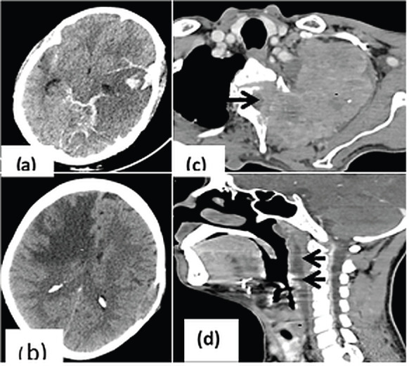
Figure 3. CrC in head and neck region. (a): CT head showing parenchymal as well as subarachnoid bleed in a patient with acute myeloid leukemia; (b): Unenhanced CT showing significant white matter edema (asterisk) in right fronto-parietal region causing mass effect on ipsilateral frontal horn of lateral ventricle; (c): Axial CT image showing pancoast tumour (asterisk) in upper lobe of left lung causing cervical vertebral erosion and intraspinal extension (arrow); (d): Post radiation edema in patient with glottic carcinoma (asterisk) with fluid collection in the retropharyngeal space (arrows) indicating immediate intervention for the same before commencing further treatment.

