Visual Abstract
Keywords: HER2 heterogeneity, esophageal adenocarcinoma, gastric adenocarcinoma, trastuzumab, HER2 PET
Abstract
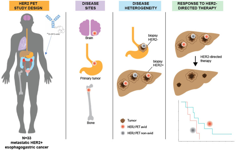
Variations in human epidermal growth factor receptor 2 (HER2) expression between the primary tumor and metastases may contribute to drug resistance in HER2-positive (HER2+) metastatic esophagogastric cancer (mEGC). 89Zr-trastuzumab PET (HER2 PET) holds promise for noninvasive assessment of variations in HER2 expression and target engagement. The aim of this study was to describe HER2 PET findings in patients with mEGC. Methods: Patients with HER2+ mEGC were imaged with HER2 PET, 18F-FDG PET, and CT. Lesions were annotated using measurements (on CT) and maximum SUVs (on HER2 PET). Correlation of visualized disease burden among imaging modalities with clinical and pathologic characteristics was performed. Results: Thirty-three patients with HER2+ mEGC were imaged with HER2 PET and CT (12% esophageal, 64% gastroesophageal junction, and 24% gastric adenocarcinoma), 26 of whom were also imaged with 18F-FDG PET. More lesions were identified on 18F-FDG PET (median, 7 [range, 1–14]) than HER2 PET (median, 4 [range, 0–11]). Of the 8 lesions identified on HER2 but not on 18F-FDG PET, 3 (38%) were in bone and 1 was in the brain. Of the 68 lesions identified on 18F-FDG but not on HER2 PET, 4 (6%) were in bone and the remainder were in the lymph nodes (35, 51%) and liver (16, 24%). Of the 33 total patients, 23 (70%) were HER2 imaging–positive (≥50% of tumor load positive). Only 10 patients had 100% of the tumor load positive; 2 had 0% positive. When only patients receiving HER2-directed therapy as first-line treatment were considered (n = 13), median progression-free survival (PFS) therapy was not significantly different between HER2 imaging–positive and –negative patients. Median PFS for patients with at least 1 intense or very intense lesion (SUV ≥ 10) was 16 (95% CI: 11-not reached) mo (n = 7), compared with 12 (95% CI: 6.3-not reached) mo for patients without an intense or very intense lesion (n = 6) (P = 0.35). Conclusion: HER2 PET may identify heterogeneity of HER2 expression and allow assessment of lesions throughout the entire body. A potential application of HER2 PET is noninvasive evaluation of HER2 status including assessment of intrapatient disease heterogeneity not captured by standard imaging or single-site biopsies.
Esophagogastric cancer (EGC) is the third most common cause of cancer-related death worldwide, and 20%–30% of patients with metastatic EGC (mEGC) have human epidermal growth factor receptor 2 (HER2)–positive disease (1–4). On the basis of data from 2 trials—the phase III randomized controlled ToGA (5), which demonstrated improved response rate and survival when trastuzumab was added to chemotherapy, and the phase III Keynote-811 (6,7), which demonstrated a better response rate and survival when trastuzumab was added to chemotherapy in combination with PD-1 blockade—HER2 is a validated treatment target in mEGC. Although HER2 immunohistochemistry, HER2-to-CEP17 ratio, and ERBB2 gene copy number can be used to predict response to trastuzumab-based chemotherapy (8), many patients with HER2-positive (HER2+) EGC develop resistance to HER2-directed therapies (3). Heterogeneity of HER2 expression between the primary tumor and metastases and loss of HER2 expression during trastuzumab therapy contribute to therapeutic resistance in HER2+ mEGC (9). Whole-body imaging with 89Zr-trastuzumab PET (HER2 PET) has a potential advantage over single-site biopsy as it can noninvasively assess variations in HER2 expression and target engagement.
We previously published the pharmacokinetics, biodistribution, and dosimetry of 89Zr-trastuzumab in HER2+ mEGC (10). HER2 PET images showed optimal tumor visualization 5–8 d after injection, and no clinically significant toxicities were observed. Here, we expand the cohort from 10 to 33 patients to further evaluate the baseline biodistribution of 89Zr-trastuzumab and the association between imaging results and response to treatment. The distribution of 89Zr-trastuzumab uptake, compared with standard imaging with 18F-FDG PET and CT, in HER2+ mEGC and the ability of this metric to predict response to HER2-directed therapy have not been described. We sought to investigate HER2 PET as a noninvasive tool to evaluate disease heterogeneity and predict response to treatment. We hypothesized that the intensity of 89Zr-trastuzumab uptake, as measured by maximum SUV, and HER2 imaging positivity (≥50% of active lesions with 89Zr-trastuzumab uptake) would be associated with response to HER2-directed therapy.
MATERIALS AND METHODS
Patients and Study Design
Eligible patients had HER2+ (immunohistochemistry 3+, immunohistochemistry 2+ and fluorescence in situ hybridization [FISH] > 2.0) mEGC, measurable or evaluable disease, Karnofsky performance ≥ 60%, and adequate organ function. This was a single-site, prospective open-label pilot imaging protocol approved by the institutional review board and ethics committee at Memorial Sloan Kettering Cancer Center (ClinicalTrials.gov identifier NCT02023996). The study included 2 groups of patients who were imaged with HER2 PET, 18F-FDG PET, and CT. The purpose of imaging in the first group of patients (group 1) was to find the optimal time for imaging after injection of the radiotracer and define its pharmacokinetics. Patients in group 2 underwent imaging to increase the study sample size and accomplish the secondary objectives of the study including correlation with tumor molecular analysis and response to treatment, reported here. All patients gave informed consent for participation in the study. All visualized lesions (maximum 5/organ) were annotated in detail using individual-lesion measurements on CT and SUV on HER2 and 18F-FDG PET by Memorial Sloan Kettering radiologists. Clinical characteristics, including baseline demographic data and previous treatments, were manually extracted from the medical record and managed using REDCap electronic data capture tools (11,12). Visualized disease burden on each imaging modality and pathologic tumor characteristics were annotated for each patient.
89Zr-Trastuzumab Drug Product
The details of the drug product, imaging protocol, and biodistribution have been published previously (10). The 89Zr-trastuzumab was manufactured by the Memorial Sloan Kettering Radiochemistry and Molecular Imaging Probes Core Facility in compliance with a Food and Drug Administration investigational new drug application. Clinical-grade trastuzumab (Herceptin; Genentech) was conjugated with p-SCN-Bn-deferoxamine (Macrocyclics) chelator, followed by radiolabeling with 89Zr, a positron emitter with a 78.4-h half-life. Patient unit doses of 185 MBq/3 mg of 89Zr-trastuzumab were mixed with nonradiolabeled trastuzumab to achieve a total mass of 50 mg.
Imaging
Each patient underwent whole-body PET/CT from mid skull to proximal thigh in 3-dimensional mode with attenuation, scatter, and other standard corrections applied and using iterative reconstruction. PET images were acquired 5 d after injection, based on the optimal imaging time of 5–8 d defined previously (10). Patients receiving therapies directed at HER2 were offered repeat imaging 2- to 6-wk after treatment, at the discretion of the treating physician and the study primary investigator, to evaluate changes in tumor uptake.
Patients underwent CT imaging of the chest, abdomen, and pelvis at a median of 7 d from the date of HER2 PET (range, 1–43 d). Localization in the tumor was defined as focal accumulation greater than adjacent background in areas in which physiologic activity was not expected. SUVs normalized to lean body mass were determined. We subclassified each lesion as negative (SUV < 3), low positive (SUV 3–5), moderate (SUV 5–10), intense (SUV 10–15), or very intense (SUV > 15).
Definition of HER2 Imaging Positivity
Any lesion that was identified by one of the above imaging methods and was clinically determined to represent a tumor was categorized as an active lesion. A patient with a HER2+ tumor was considered to be HER2 imaging–positive if ≥50% of the active lesions were detectable by HER2 PET. The total number of active lesions identified on CT, 18F-FDG PET, or HER2 PET was used as the denominator for the tumor load. To determine HER2 imaging positivity, we divided the total number of lesions identified on HER2 PET by the tumor load.
Definition of HER2 Heterogeneity
Heterogeneity of HER2 status on biopsy was defined on the basis of variation in HER2 overexpression in multiple disease sites biopsied (median 3 samples per patient; range, 1–8). For example, a case with 1 lesion that was HER2 immunohistochemistry 3+ or 2+ and amplified by FISH and a second lesion that was either negative (immunohistochemistry 1+ or 0+) or equivocal by FISH would be classified as having heterogeneous disease. Genomic assessment of ERBB2 amplification was not used to establish heterogeneity, as not all patients underwent somatic mutation analysis. Heterogeneity of HER2 expression by HER2 PET was defined in the protocol, on the basis of previously published data (13), by the percentage of tumor load that showed tracer uptake. Group stratification was as follows: group A, the entire tumor load showed tracer uptake (100%); group B, the dominant part of the tumor load showed tracer uptake (≥50%); group C, only a minor part of the tumor load showed tracer uptake (<50%); and group D, the entire tumor load lacked tracer uptake (0%). Groups B and C were considered to have heterogeneous uptake (>0% and <100% of tumor load positive).
Statistical Analysis
The primary objectives of the protocol were to evaluate the feasibility of detecting tumors using HER2 PET in the first 10 patients with HER2+ EGC and to evaluate the safety, biodistribution, and pharmacokinetics of 89Zr-trastuzumab, all of which were reported previously (10). HER2 PET imaging was considered feasible on the basis of antibody imaging positivity in 7 or more of the 10 patients in the first cohort. Secondary objectives, reported here, were to describe tumor molecular analysis with imaging results and to evaluate imaging results in the context of response to treatment. HER2 imaging positivity was estimated on the basis of the 33 patients with the 1-sided 90% confidence limit. Patients with ≥50% of the total tumor load with 89Zr-trastuzumab uptake were considered HER2 imaging–positive, and patients with <50% were considered HER2 imaging–negative.
Overall survival (OS) and progression-free survival (PFS) were calculated from the date of treatment until time of death (for OS) or until the date of progression or death, whichever came first (for PFS). Patients who did not experience the event of interest by the end of the study were censored at the time of last available follow-up (for OS) or last available CT (for PFS). Because the study population was heterogeneous and included both patients receiving first-line treatment for metastatic disease and those receiving treatment for refractory disease, we restricted the OS and PFS analyses to the homogenous group of patients who were receiving first-line therapy at the time of the HER2 PET (n = 13). OS and PFS were estimated using Kaplan–Meier methods and compared between subgroups (A/B vs. C/D; SUV intensity) using the permutated log-rank test. All P values were based on 2-tailed statistical analysis, and a P value of less than 0.05 was considered to indicate statistical significance. All analyses were conducted in R version 4.0.4 (R Development Core Team, 2022) (14).
RESULTS
Summary of Patients
Thirty-three patients with metastatic HER2+ gastric (24%), gastroesophageal junction (GEJ) (64%), or esophageal (12%) adenocarcinoma were imaged with HER2 PET and CT, and 26 of these patients were also imaged with 18F-FDG PET (Table 1). HER2 status was assessed using biopsy or resection specimens of the primary tumor (21/33, 64%) or metastasis (12/33, 36%) and was confirmed by immunohistochemistry 3+ (26/33, 79%), immunohistochemistry 2+ and amplification by FISH (6/33, 18%), or ERBB2 amplification by next-generation sequencing with MSK-IMPACT (Memorial Sloan Kettering-Integrated Mutation Profiling of Actionable Cancer Targets) (1/33, 3%) (15). All patients had metastatic disease at the time of enrollment; most patients had metastases to the lymph nodes (23/33, 70%) or liver (19/33, 58%), followed by lung (11/33, 33%), peritoneum (8/33, 24%), bone (3/33, 9%), or other tissues. Most patients underwent prior treatment with HER2-directed therapy (20/33, 61%, had received at least 1 line of HER2-directed therapy); the median time from diagnosis to HER2 PET was 13 mo. The median number of lines of therapy received at the time of HER2 PET was 2 (range, 1–6), and the median number of total lines of therapy received throughout the course of illness was 3 (range, 1–9). Thirty patients (91%) were receiving HER2-directed therapy, and 13 (39%) were receiving first-line HER2-directed therapy at the time of HER2 PET (Supplemental Table 1; supplemental materials are available at http://jnm.snmjournals.org). Among patients receiving HER2-directed therapy at the time of HER2 PET, the median time on treatment was 4 mo (range, 0–47 mo) for all patients and 14 mo (range, 4–47 mo) for those receiving first-line therapy (Supplemental Table 2).
TABLE 1.
Patient and Treatment Characteristics
| Characteristic | Prior trastuzumab (n = 19) | No prior trastuzumab (n = 14) | Total (n = 33) |
|---|---|---|---|
| Median age at diagnosis (y) | 59 (range, 40–76) | 59 (range, 34–79) | 59 (range, 34–79) |
| Patients with metastasis at diagnosis (n) | 17 (89) | 10 (71) | 27 (82) |
| Primary tumor site (n) | |||
| Esophageal | 2 (11) | 2 (14) | 4 (12) |
| GEJ (Siewert I-II) | 14 (74) | 7 (50) | 21 (64) |
| Gastric | 3 (16) | 5 (36) | 8 (24) |
| Method used to confirm sample is HER2+ (n) | |||
| FISH | 3 (16) | 3 (21) | 6 (18) |
| IHC | 15 (79) | 11 (79) | 26 (79) |
| NGS | 1 (5) | 0 (0) | 1 (3) |
| Patients with HER2 heterogeneity across samples (n) | 3 (16) | 6 (43) | 9 (27) |
| Patients receiving HER2-directed therapy at the time of scan (n) | 16 (84) | 14 (100) | 30 (91) |
| Median no. of lines of treatment at the time of HER2 PET | 3 (range, 2–6) | 1 (range, 1–2) | 2 (range, 1–6) |
| Median total lines of treatment received | 3 (range, 2–7) | 3 (range, 1–9) | 3 (range, 1–9) |
| Median time on treatment at the time of HER2 PET (d) | 93 (range, 0–212) | 394 (range, 7–1,410) | 126 (range, 0–1,410) |
| Total lesions detected on imaging (all patients) | |||
| Median, CT | 5 (range, 1–11) | 5.5 (range, 1–15) | 5 (range, 1–15) |
| Median, HER2 | 4 (range, 1–7) | 3.5 (range, 0–11) | 4 (range, 0–11) |
| Median, 18F-FDG PET | 6.5 (range, 1–13) | 7 (range, 1–14) | 7 (range, 1–14) |
| Patients with ≥ 1 lesion intense or very intense on HER2 PET (n) | 8 (42) | 7 (50) | 15 (45) |
| Median SUVmax per patient on HER2 PET | 7.8 (range, 3.20–23.8) | 9.8 (range, 0–22.2) | 9.2 (range, 0–23.8) |
| Median SUVmean per lesion on HER2 PET | 6.5 (range, 2.8–14.2) | 7.8 (range, 0–15.9) | 7.0 (range, 0–15.9) |
Data are number, with percentages in parentheses, or median, with the minimum to maximum in parentheses.
IHC = immunohistochemistry; NGS = next-generation sequencing.
The total number of lesions identified on each imaging modality is summarized in Table 1. The median number of lesions identified by each modality is as follows: baseline CT, 5 (range, 1–15); HER2 PET, 4 (range, 0–11); and 18F-FDG PET, 7 (range, 1–14) (Supplemental Table 3).
89Zr-Trastuzumab PET Captures Nonstandard Disease Sites
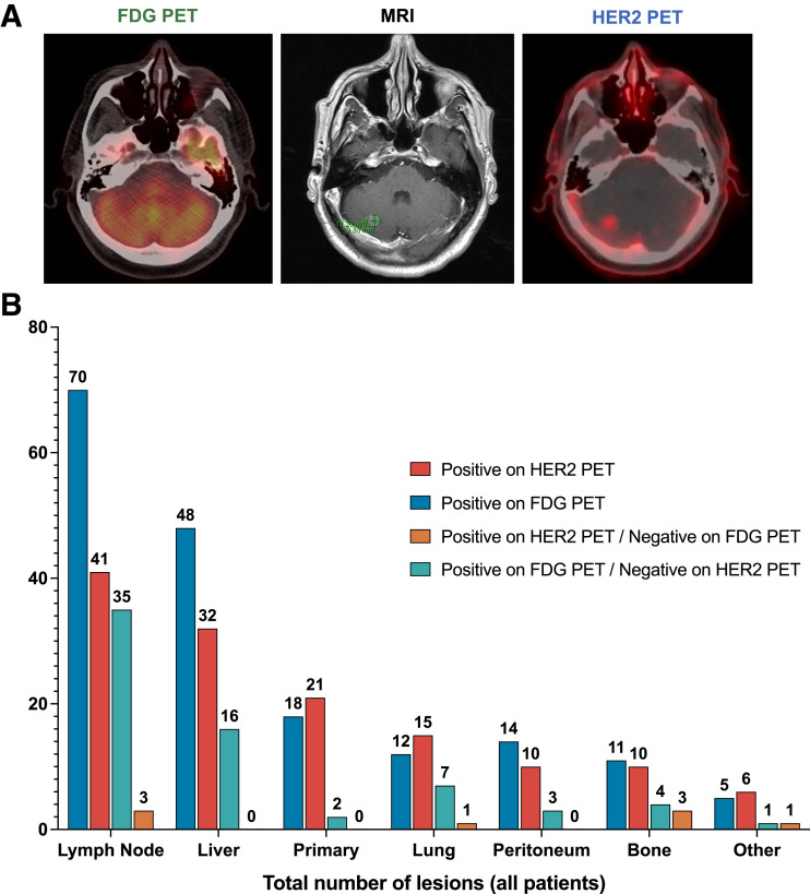
The potential clinical applications of HER2 PET imaging include identification of disease sites not captured by standard imaging, establishment of sites of HER2 heterogeneity not captured by biopsy, and early assessment of response to HER2-directed therapy. We included specific case examples to illustrate these points. The first is a case of metastatic HER2+ GEJ poorly differentiated carcinoma in which HER2 PET identified a right cerebellar metastasis (SUV 2.6) that had not been detected on 18F-FDG PET (Fig. 1A). The finding was confirmed on brain MRI, and the patient underwent stereotactic radiosurgery to treat this lesion.
FIGURE 1.
Disease sites captured by HER2 and 18F-FDG PET. (A) 18F-FDG PET, MRI, and HER2 PET images from a patient with cerebellar metastasis. The images shown are from a patient with de novo metastatic HER2+ GEJ poorly differentiated carcinoma with mixed adeno and squamous differentiation. HER2 PET (right) demonstrated a right cerebellar metastasis (SUV 2.6) without corresponding uptake on 18F-FDG PET (left) and confirmed on brain MRI (middle). (B) Number of lesions identified by HER2 PET and 18F-FDG PET among all patients. Total number of lesions avid on HER2 PET (red) and 18F-FDG PET (blue) is shown. Total numbers of lesions better identified only on HER2 (orange) or 18F-FDG PET (green) are also shown.
Among all patients, more total lesions were visualized on 18F-FDG PET (n = 178) than on HER2 PET (n = 135) (Fig. 1B). Of the total lesions positive on HER2 PET, most were in lymph nodes (41/135 [30%]) or the liver (32/135 [24%]). For 18F-FDG PET, most were also in lymph nodes (70/178 [39%]) or the liver (48/178 [27%]). However, primary tumor and bone lesions were detected at a higher frequency on HER2 PET (primary tumor: 21/135 [16%]; bone: 10/135 [7%]) than on 18F-FDG PET (primary tumor: 18/178 [10%]; bone: 11/178 [6%]).
Five patients had at least 1 lesion positive on HER2 PET and negative on 18F-FDG PET (range, 0–4); 18 patients had at least 1 lesion positive on 18F-FDG PET and negative on HER2 PET (range, 0–8). Of the 8 lesions that were positive on HER2 PET and negative on 18F-FDG PET, 3 were in the bone (38%) and 3 were in lymph nodes (38%) (Fig. 1B). In contrast, most lesions that were positive on 18F-FDG PET and negative on HER2 PET were in the lymph nodes (35/68 [51%]) and the liver (16/68 [24%]); only 4 of 68 (6%) were in the bone.
HER2 PET Illustrates HER2 Heterogeneity
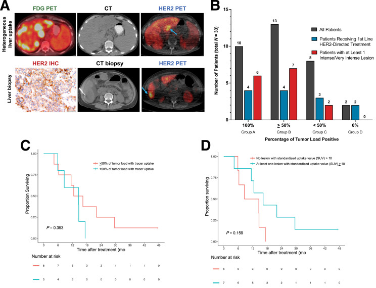
The next case example illustrates heterogeneous liver uptake of the radiotracer on HER2 PET, suggesting that HER2 overexpression is heterogeneous (Fig. 2A). However, liver biopsy for this patient, obtained from a single site of 89Zr-trastuzumab avidity, showed HER2 immunohistochemistry 3+ and, as expected, did not capture the intrapatient heterogeneity seen on imaging.
FIGURE 2.
HER2 disease heterogeneity illustrated by 89Zr-trastuzumab PET (HER2 PET). (A) 18F-FDG PET and HER2 PET images from a patient with metastatic HER2+ gastric adenocarcinoma with heterogeneous HER2 expression in the liver. Heterogeneous 89Zr-trastuzumab uptake on imaging is shown (blue arrows demonstrate positive lesions, upper figure). Liver biopsy at a site of 89Zr-trastuzumab uptake demonstrates HER2 positivity with immunohistochemistry 3+ in 60% of cells (lower). (B) The percentage of tumor load with 89Zr-trastuzumab uptake. Patients were stratified into 4 groups by percentage of tumor load showing tracer uptake. Total patients in groups A–D are shown in gray. Number of patients receiving first-line HER2-directed therapy in each group is represented in blue. Patients with at least 1 intense or very intense lesion on HER2 PET (SUV ≥ 10) are represented in red. Of the 15 patients with at least 1 intense or very intense lesion (15/33 [45%]), 6 were in group A (6/33 [18%]) and 7 were in group B (7/33 [21%]). (C) PFS stratified by percentage of tumor load positive in patients receiving first-line HER2-directed therapy (P = 0.353, using permutated log-rank test comparing the 2 groups). (D) PFS stratified by presence of at least 1 lesion with intense or very intense 89Zr-trastuzumab uptake (SUV ≥ 10) in patients receiving first-line HER2-directed therapy (P = 0.159, using permutated log-rank test comparing the 2 groups). IHC = immunohistochemistry.
As defined in our prespecified analysis, patients with ≥50% of the total tumor load with 89Zr-trastuzumab uptake were considered HER2 imaging–positive, and patients with <50% were considered HER2 imaging–negative. As described in the “Materials and Methods” section, we stratified patients into 4 groups by percentage of tumor load that showed tracer uptake (13). Of the 33 patients, 23 (70%) were HER2 imaging–positive (group A or B) (1-sided 90% confidence limit, 57% for feasibility) (Fig. 2B). Only 10 patients had 100% of the tumor load positive; 2 had 0% positive. Of the 30 patients who were receiving HER2-directed therapy at the time of the scan, 20 (66%) had ≥50% active lesions (group A or B). Of the 13 patients who were receiving first-line HER2-directed therapy at the time of the scan, 8 (62% of the group, 24% of the total cohort) had ≥50% active lesions (group A or B). Although all the patients without any tracer uptake (6% of the cohort) were receiving second-line or later treatment, most patients receiving advanced lines of therapy were HER2 imaging–positive, supporting the notion that HER2 remains a relevant biomarker beyond the first-line setting.
We next describe the proportion of patients in group A or B and group C or D with at least 1 intense or very intense lesion on HER2 PET. Among those with ≥50% of tumor load positive (group A or B), 57% of patients had at least 1 intense or very intense lesion, whereas only 20% in those with <50% of tumor load positive (group C or D) had at least 1 intense or very intense lesion. Biopsy-proven HER2 heterogeneity was present in 30% of patients in group A or B and in 20% of patients in group C or D. A slightly higher proportion of patients in group A or B (61%) had received trastuzumab therapy at the time of the scan, relative to those in group C or D (50%).
In addition to looking at individual-lesion positivity by HER2 PET, we subclassified each lesion as negative (SUV < 3), low positive (SUV 3–5), moderate (SUV 5–10), intense (SUV 10–15), or very intense (SUV > 15). In the case example shown in Figure 2A in which all biopsies were HER2 immunohistochemistry 3+, the tumor load positivity for 89Zr-trastuzumab uptake was <50% (group C). Although the patient had at least 1 intense or very intense lesion, the patient’s PFS on second-line HER2-directed therapy (3 mo) was less than the median among all patients with at least 1 intense or very intense lesion (5 mo).
Survival of Patients Receiving First-Line Treatment at the Time of the Scan
Survival was evaluated only among patients receiving first-line HER2-directed therapy at the time of the HER2 PET (n = 13). The baseline characteristics of this group are summarized in Supplemental Table 4. Among surviving patients (n = 2), the median follow-up time was 50.0 mo (range, 45.8–54.3 mo). At the time of the data lock in July 2021, 11 total deaths and 12 progression events had been observed. When only patients receiving HER2-directed therapy in the first-line setting were considered, the median PFS was 15 mo (95% CI: 8.6–not reached).
The median PFS among patients in group A or B (n = 8) was 14 mo (95% CI: 11.0–not reached), compared with 15 mo (95% CI: 8.6–not reached) among patients in group C or D (n = 5) (Fig. 2C). Among patients receiving HER2-directed therapy in the second-line setting, most patients in both groups (A/B n = 7, C/D n = 2) progressed or died before 3 mo. Given the small number of patients in this subgroup, PFS should be interpreted with caution.
HER2 PET and Response to HER2-Directed Therapy
We next stratified patients by the presence or absence of at least 1 intense or very intense lesion on baseline HER2 PET and compared PFS among patients receiving first-line HER2-directed therapy at the time of the scan (n = 13). The median PFS was 16 mo (95% CI: 11–not reached) and 12 mo (95% CI: 6.3–not reached), respectively (Fig. 2D). Given the small number of patients in this subgroup, PFS should be interpreted with caution.
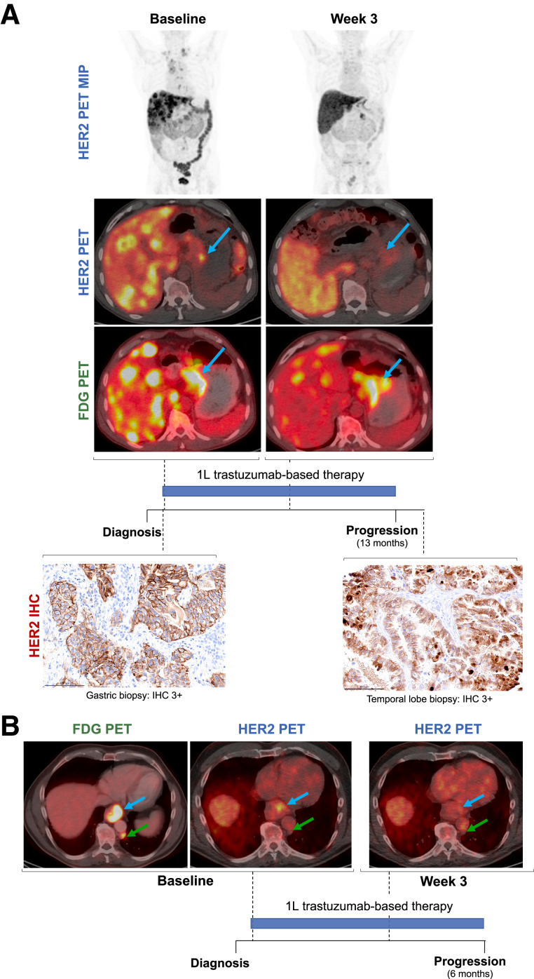
The final 2 case examples (Fig. 3) illustrate the potential role of HER2 PET in predicting response to HER2-directed therapy. In Figure 3A, a patient with HER2+ mEGC had ≥50% tumor load uptake of 89Zr-trastuzumab on baseline imaging (group B) and at least 1 intense or very intense lesion. Both HER2 and 18F-FDG PET showed primary tumor avidity; less than 3 wk after initiation of trastuzumab-based treatment, the primary tumor remained 18F-FDG PET–avid but was no longer avid on HER2 PET, indicating HER2 receptor saturation by trastuzumab. This patient had a PFS of 13 mo on first-line HER2-directed therapy, and the disease remained HER2+ on postprogression biopsy, with subsequent response to second-line HER2-directed therapy. In contrast, the patient in Figure 3B, who had ≥50% tumor load uptake of 89Zr-trastuzumab on baseline imaging (group B) but no intense or very intense lesions, had no change in primary tumor 89Zr-trastuzumab uptake after initiation of first-line HER2-directed treatment and had a relatively short PFS of 6 mo on treatment.
FIGURE 3.
89Zr-trastuzumab PET (HER2 PET) and early assessment of response to HER2-directed therapy. (A) 18F-FDG PET, HER2 PET, and HER2 immunohistochemistry (IHC) in a patient with metastatic HER2+ esophagogastric cancer with a long response to first-line HER2-directed therapy. Patient had > 50% of tumor load with 89Zr-trastuzumab uptake on baseline imaging (group B) and at least 1 lesion with SUV ≥ 10. Although primary tumor was avid on baseline HER2 and 18F-FDG PET, less than 3 wk after initiation of trastuzumab-based treatment, primary tumor remained 18F-FDG PET avid but was no longer avid on HER2 PET, likely indicating HER2 saturation by trastuzumab. This patient had a PFS of 13 mo on first-line HER2-directed therapy. (B) 18F-FDG PET and HER2 PET in a patient metastatic HER2+ esophageal adenocarcinoma with a short response to first-line HER2-directed therapy. Patient had > 50% of the tumor load with 89Zr-trastuzumab uptake on baseline imaging (group B) but no lesions with SUV ≥ 10; patient had no change in primary tumor 89Zr-trastuzumab uptake (SUV 8.1 from 6.6, blue arrows) or in posterior left paraaortic node 89Zr-trastuzumab uptake (green arrows) after initiation of first-line HER2-directed treatment. Patient had a relatively short PFS of 6 mo on treatment. 1 L = first-line.
Patients Not Receiving HER2-Directed Therapy
Of the 3 patients who were not receiving HER2-directed therapy at the time of the scan, 1 underwent repeat liver biopsy that demonstrated equivocal HER2 status by both immunohistochemistry and FISH (the initial specimen, obtained at an outside institution, was HER2 immunohistochemistry 3+). The second patient recently had disease progression on HER2-directed therapy, and biopsy of a splenic lesion demonstrated HER2 immunohistochemistry 1–2+ and nonamplification on FISH. The third patient underwent repeat biopsy of the primary GEJ mass, which showed HER2 immunohistochemistry 1+ (negative); therefore, this patient did not receive additional HER2-directed therapy.
DISCUSSION
Our data suggest that antibody imaging with HER2 PET is feasible and allows noninvasive assessment of global variations in HER2 expression and target enhancement. HER2 PET identified bone lesions more so than soft-tissue lesions. Compared with HER2 PET, 18F-FDG PET identified more lymph node lesions and it is unclear whether these findings represent true disease or inflammation.
HER2 PET may help visualize heterogeneity of HER2 expression and allow assessment of lesions throughout the entire body. The percentage of tumor load positive for 89Zr-trastuzumab varied among patients: approximately two thirds of the patients in our study had ≥50% tumor load positivity and one third had <50% positivity. The percentage of tumor load with tracer uptake was not significantly associated with PFS in the small subgroup analysis of patients receiving first-line HER2-directed therapy. The case example shown in Figure 1 highlights the potential for HER2 PET to identify lesions in the brain before symptomatic presentation, without a biopsy. This has not been previously demonstrated in the literature. In addition, the case example shown in Figure 2 illustrates the potential for biomarker-directed imaging to identify sites of disease heterogeneity that are not captured by standard imaging and biopsy. There was a trend toward improved PFS among patients with at least 1 lesion on HER2 PET with SUV greater than or equal to 10 who were receiving first-line HER2-directed therapy, though this difference was not significant. A larger study would be needed to associate HER2 PET findings with PFS.
Although the clinical application of evaluating disease heterogeneity by HER2 PET has not been clearly established for EGC, HER2 PET has been used to help guide clinical decision making for other HER2+ tumor types. In a study of 20 patients with breast cancer, including 7 patients with metastases that were inaccessible for biopsy, HER2 PET was used to support clinical decision making and changed management in 8 of 20 patients (40%) (16). Similarly, in a cohort of 12 patients with HER2-mutant lung cancer, pretreatment HER2 PET identified 89Zr-trastuzumab–avid lesions in 4 patients, all of whom responded to HER2-directed therapy with ado-trastuzumab emtansine (T-DM1); in contrast, among the 8 patients without uptake of 89Zr-trastuzumab, only 3 (37%) responded to T-DM1 treatment (17).
Our study demonstrates that intensity of 89Zr-trastuzumab uptake varies between and within patients and can be used to stratify patients, although the clinical application of this has not yet been determined. At least 1 lesion with an SUV ≥ 10 on HER2 PET may be associated with response to HER2-directed therapy, though this remains to be validated in future studies. Although the percentage of tumor load positive was used to establish feasibility in this study, it remains unclear whether this is a marker of likelihood to respond to HER2-directed therapy.
HER2 PET is limited by the high background tracer uptake in several key organs, including the liver, making it challenging to identify discrete tumors using this technique. The current study is limited by its descriptive design. In addition, the study is limited by patient exposure to trastuzumab before HER2 PET due to partial target saturation. Further investigation specifically including previously untreated patients is required to determine whether HER2 PET can be used as a clinical predictive tool in patients with HER2+ mEGC.
CONCLUSION
HER2 PET may identify heterogeneity of HER2 expression and allows assessment of lesions throughout the entire body. HER2 PET has a potential advantage over single-site biopsy in assessment of HER2 heterogeneity. Bone lesions were better identified than soft-tissue lesions on HER2 PET. Until further studies validate the preliminary clinical findings presented, we anticipate that HER2 PET will remain a valuable research tool.
DISCLOSURE
Financial support for this study was received from Department of Defense Congressionally Directed Medical Research Program (CA 150646, Yelena Y. Janjigian and Jason S. Lewis), NIH/NCI Cancer Center Support Grant P30 CA008748, 2013 Conquer Cancer Foundation ASCO Career Development Award (Yelena Y. Janjigian), 2014 and 2016 Mr. William H. Goodwin and Mrs. Alice Goodwin and the Commonweath Foundation for Cancer Research and The Center for Experimental Therapeutics at Memorial Sloan Kettering Cancer Center (Yelena Y. Janjigian and Jason S. Lewis), and 2015 Cycle for Survival Award (Yelena Y. Janjigian). Yelena Y. Janjigian has received research funding from Bayer, Bristol Myers Squibb, Cycle for Survival, Department of Defense, Eli Lilly, Fred’s Team, Genentech/Roche, Merck, the National Cancer Institute, and RGENIX and has served on advisory boards or in a consulting role for Amerisource Bergen, Arcus Biosciences, Astra Zeneca, Basilea Pharmaceutica, Bayer, Bristol Myers Squibb, Daiichi Sankyo, Eli Lilly, Geneos Therapeutics, GlaxoSmithKline, Imedex, Imugene, Lynx Health, Merck, Merck Serono, Michael J. Hennessy Associates, Paradigm Medical Communications, PeerView Institute, Pfizer, Research to Practice, RGENIX, Seagen, Silverback Therapeutics, and Zymeworks Inc. Neeta Pandit-Taskar has received research funding from Bayer Health, Bristol Myers Squibb, Clarity Pharmaceuticals, Imaginab, Janssen, and Regeneron; has served in consulting or advisory roles for Illumina and Progenics; and has received honoraria from Actinium Pharmaceuticals and AstraZeneca/MedImmune. Steven B. Maron has received research funding from Guardant Health (Inst) and Roche/Genentech (Inst); has served on advisory boards or in consulting roles for Basilea, Health Advances and Natera; and has stock in Calithera Biosciences. Joseph A. O’Donoghue has served in a consulting or advisory role for Janssen Research & Development. No other potential conflict of interest relevant to this article was reported.
KEY POINTS
QUESTION: Is HER2 PET an effective tool for noninvasive assessment of variations in HER2 expression and target engagement in patients with HER2+ mEGC?
PERTINENT FINDINGS: In a pilot study of HER2 PET in 33 patients with HER2+ mEGC, 70% of patients were HER2 imaging–positive (≥50% of tumor load positive) and most patients had variable HER2 uptake across disease sites. Among patients receiving HER2-directed therapy as first-line treatment, median PFS was longer for those with at least 1 intense or very intense lesion on HER2 PET.
IMPLICATIONS FOR PATIENT CARE: A potential application of HER2 PET is noninvasive evaluation of intrapatient heterogeneity of HER2 status not captured by single-site biopsies in patients with HER2+ mEGC.
REFERENCES
- 1. Bray F, Ferlay J, Soerjomataram I, Siegel RL, Torre LA, Jemal A. Global cancer statistics 2018: GLOBOCAN estimates of incidence and mortality worldwide for 36 cancers in 185 countries. CA Cancer J Clin. 2018;68:394–424. [DOI] [PubMed] [Google Scholar]
- 2. Reichelt U, Duesedau P, Tsourlakis MC, et al. Frequent homogeneous HER-2 amplification in primary and metastatic adenocarcinoma of the esophagus. Mod Pathol. 2007;20:120–129. [DOI] [PubMed] [Google Scholar]
- 3. Janjigian YY, Werner D, Pauligk C, et al. Prognosis of metastatic gastric and gastroesophageal junction cancer by HER2 status: a European and USA International collaborative analysis. Ann Oncol. 2012;23:2656–2662. [DOI] [PubMed] [Google Scholar]
- 4. Schoppmann SF, Jesch B, Friedrich J, et al. Expression of Her-2 in carcinomas of the esophagus. Am J Surg Pathol. 2010;34:1868–1873. [DOI] [PubMed] [Google Scholar]
- 5. Bang YJ, Van Cutsem E, Feyereislova A, et al. Trastuzumab in combination with chemotherapy versus chemotherapy alone for treatment of HER2-positive advanced gastric or gastro-oesophageal junction cancer (ToGA): A phase 3, open-label, randomised controlled trial. Lancet. 2010;376:687–697. [DOI] [PubMed] [Google Scholar]
- 6. Janjigian YY, Maron SB, Chatila WK, et al. First-line pembrolizumab and trastuzumab in HER2-positive oesophageal, gastric, or gastro-oesophageal junction cancer: an open-label, single-arm, phase 2 trial. Lancet Oncol. 2020;21:821–831. [DOI] [PMC free article] [PubMed] [Google Scholar]
- 7. Janjigian YY, Kawazoe A, Yañez P, et al. The KEYNOTE-811 trial of dual PD-1 and HER2 blockade in HER2-positive gastric cancer. Nature. 2021;600:727–730. [DOI] [PMC free article] [PubMed] [Google Scholar]
- 8. Ock CY, Lee KW, Kim JW, et al. Optimal patient selection for trastuzumab treatment in HER2-Positive advanced gastric cancer. Clin Cancer Res. 2015;21:2520–2529. [DOI] [PubMed] [Google Scholar]
- 9. Gajria D, Chandarlapaty S. HER2-amplified breast cancer: mechanisms of trastuzumab resistance and novel targeted therapies. Expert Rev Anticancer Ther. 2011;11:263–275. [DOI] [PMC free article] [PubMed] [Google Scholar]
- 10. O’Donoghue JA, Lewis JS, Pandit-Taskar N, et al. Pharmacokinetics, biodistribution, and radiation dosimetry for 89Zr-trastuzumab in patients with esophagogastric cancer. J Nucl Med. 2018;59:161–166. [DOI] [PMC free article] [PubMed] [Google Scholar]
- 11. Harris PA, Taylor R, Thielke R, Payne J, Gonzalez N, Conde JG. Research electronic data capture (REDCap): a metadata-driven methodology and workflow process for providing translational research informatics support. J Biomed Inform. 2009;42:377–381. [DOI] [PMC free article] [PubMed] [Google Scholar]
- 12. Harris PA, Taylor R, Minor BL, et al. The REDCap consortium: building an international community of software platform partners. J Biomed Inform. 2019;95:103208. [DOI] [PMC free article] [PubMed] [Google Scholar]
- 13. Gebhart G, Lamberts LE, Wimana Z, et al. Molecular imaging as a tool to investigate heterogeneity of advanced HER2-positive breast cancer and to predict patient outcome under trastuzumab emtansine (T-DM1): The ZEPHIR trial. Ann Oncol. 2016;27:619–624. [DOI] [PubMed] [Google Scholar]
- 14.R Core Team (2022). R: A language and environment for statistical computing. R Foundation for Statistical Computing, Vienna, Austria. R Project website. https://www.R-project.org/. Accessed March 24, 2023.
- 15. Cheng DT, Mitchell TN, Zehir A, et al. Memorial Sloan Kettering-Integrated Mutation Profiling of Actionable Cancer Targets (MSK-IMPACT): A hybridization capture-based next-generation sequencing clinical assay for solid tumor molecular oncology. J Mol Diagn. 2015;17:251–264. [DOI] [PMC free article] [PubMed] [Google Scholar]
- 16. Bensch F, Brouwers AH, Hooge MNL, et al. 89Zr-trastuzumab PET supports clinical decision making in breast cancer patients, when HER2 status cannot be determined by standard work up. Eur J Nucl Med Mol Imaging. 2018;45:2300–2306. [DOI] [PMC free article] [PubMed] [Google Scholar]
- 17. Ulaner GA, Hyman DM, Lyashchenko SK, Lewis JS, Carrasquillo JA. 89Zr-trastuzumab PET/CT for detection of human epidermal growth factor receptor 2-positive metastases in patients with human epidermal growth factor receptor 2-negative primary breast cancer. Clin Nucl Med. 2017;42:912–917. [DOI] [PMC free article] [PubMed] [Google Scholar]


