



Abstract
Epigenetics, the inheritance of genomic information independent of DNA sequence, controls the interpretation of extracellular and intracellular signals in cell homeostasis, proliferation and differentiation. On the chromatin level, signal transduction leads to changes in epigenetic marks, such as histone post-translational modifications (PTMs), DNA methylation and chromatin accessibility to regulate gene expression. Crosstalk between different epigenetic mechanisms, such as that between histone PTMs and DNA methylation, leads to an intricate network of chromatin-binding proteins where pre-existing epigenetic marks promote or inhibit the writing of new marks. The recent technical advances in mass spectrometry (MS) -based proteomic methods and in genome-wide DNA sequencing approaches have broadened our understanding of epigenetic networks greatly. However, further development and wider application of these methods is vital in developing treatments for disorders and pathologies that are driven by epigenetic dysregulation.
Introduction
In the eukaryotic nucleus, genomic DNA is packaged into chromatin with proteins to condense the DNA into a highly compacted form and to regulate DNA-dependent processes such as replication, repair and transcription. These events are regulated by cells to maintain homeostasis, to adapt to the environment and to differentiate. Cells decipher external and internal signals to changes in epigenetic marks, such as DNA-methylation, histone post-translational modifications (PTMs) and modifications in non-coding RNAs, that do not involve changes in DNA sequence. These epigenetic patterns provide a means by which even transient alterations in a cellular state can have a lasting impact in a cell but also throughout cell generations when they are inherited to progenitor cells as genomic information.
Epigenetic marks regulate gene activity by influencing chromatin compaction and thus access of DNA for the transcription initiation complex and other chromatin-binding proteins. After being activated by extracellular signals, such as hormones, activated transcription factors (TFs) bind to DNA and recruit histone PTM modifiers and chromatin remodelers to shape the chromatin landscape to activate or repress target genes. Deciphering the effects of signals to epigenetic changes and ultimately to changes in gene expression is a challenging task due to the multiple layers in which these events are regulated. In addition to regulation of the activity and expression levels of cytosolic signaling pathway proteins, the chromatin-associated enzymes that are responsible for reading and writing/erasing chromatin marks are under tight regulation. Chromatin-associated enzymes are directed to specific sites on chromatin by reading chromatin marks, such as their own product to stimulate their own activity [1]. However, when these enzymes read other marks than their own, their function leads to multilayered crosstalk between different chromatin marks. Moreover, by associating with other reader proteins or TFs in multiprotein complexes, their capacity to recognize different chromatin marks increases even more. Modifications on the chromatin readers themselves may also alter their ability to read [2].
Epigenetic dysregulation due to mutations and/or deregulated expression of chromatin-associated enzymes, their protein complex subunits, TFs, ATP-dependent chromatin remodelers, and histones contribute to many human disorders including neurodevelopmental disorders [3,4], psychiatric disorders [5], cardiovascular disease [6], and cancer [7–10]. Understanding epigenetic regulation, especially epigenetic crosstalk, in pathophysiology is vital in developing treatments for these conditions. Here, we will summarize the most-studied histone PTMs, combinatorial PTMs, and their crosstalk with DNA methylation. We will also discuss epigenetic dysregulation in diseases and the therapeutic targeting of epigenetics, followed by a description of genomic and mass-spectrometry-based proteomic approaches in characterizing epigenetic crosstalk. We will collectively refer to all histone and DNA modifications as ‘epigenetic modifications’ even though it is still unknown whether all histone PTMs are inherited during mitosis.
Histone post-translational modifications
Chromatin is decorated with evenly spaced nucleosomes, in which ~147 base pairs of double-stranded DNA is wrapped around histone proteins. Each nucleosome core particle includes eight core histones, canonically, two H3/H4 heterodimers that form the central tetramer, which is capped on each end by a H2A/H2B heterodimer [11,12]. Histone globular domains (also termed folds) form the center of the nucleosome core particle from which the more flexible and unstructured N-terminal tails of all core histones and the C-terminal tail of H2A protrude from [13]. The H1 linker histones bind DNA between nucleosome core particles (linker DNA) both at the entry and exit sites of the nucleosome core particle [14]. The nucleosome core particle together with variable lengths of linker DNA form the nucleosome. The nucleosome together with a bound linker histone is termed the chromatosome core particle [15–17]. Nucleosomes are regularly spaced with ~200 bp distance from each other to form the so called 10-nm chromatin fibers [18,19]. The fibers are assembled to larger functional domains, such as the topologically associated domains (TADs) [20] and the A- and B-compartments [21].
Histone post-translational modifications (PTMs) (i.e. the covalent addition of chemical moieties or small proteins) regulate DNA-dependent processes such as replication, repair and transcription by altering nucleosome dynamics [22,23]. These reversible modifications directly alter histone-DNA and histone-histone interactions to regulate nucleosome sliding, wrapping and assembly, but also recruit hundreds of different chromatin-binding proteins to regulate these events [13,23,24]. Histone tails, especially that of H3, are rich in PTM sites compared to folds (Figure 1.), and they are more readily accessible for protein interactions [13]. However, in addition to histone tails, a combination of different surfaces on the nucleosome, such as segments of DNA, the acidic patch on the H2A/H2B dimer surface and other surfaces on the histone folds, are known to be important for protein interactions on chromatin [12]. Linker histones are also targeted by PTMs, but they have remained far less studied than core histones [25]. Histone PTM levels are affected by signaling cascades triggered by external stimuli [26], histone PTM crosstalk [27], and availability of metabolite precursors for PTMs [28]. Acetylation (ac), methylation (me), phosphorylation (ph) and ubiquitination (ub) are among the most-studied histone PTMs.
Figure 1.
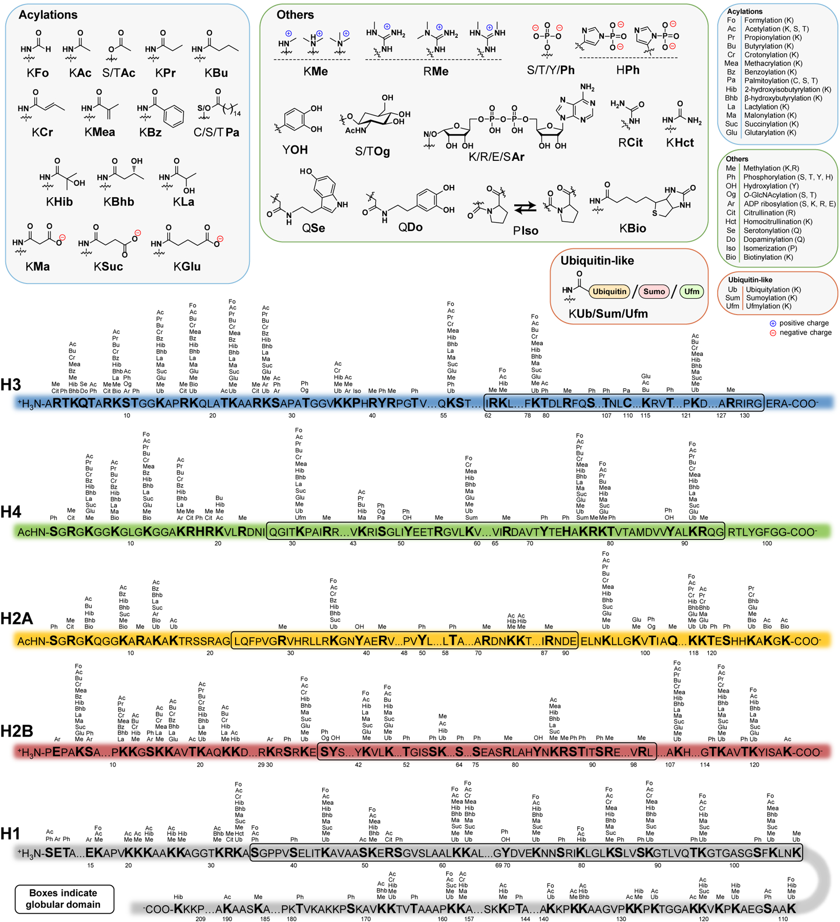
Illustration summarizing the reported human, mouse, and rat histone PTMs. Modified with permission from [386]. Updated modifications are from [28,104,131,387,388].
Acetylation
Acetylation of histone lysine residues is known to associate with active transcription. By neutralizing the positive charge at lysines, histone acetylation weakens histone-DNA and histone-histone interactions that leads to destabilization of nucleosomes and, subsequently, to the exposure of DNA to the transcription machinery [22]. Histone acetylation also recruits proteins with domains that recognize acetylation, such as bromodomain-containing chromatin remodelers that facilitate further unwrapping of the nucleosomes [22,29].
In vitro experiments with nucleosome arrays showed that substituting lysine residues with glutamines to mimic acetylation at the H4, H3, H2A and H2B tails synergistically increase DNA accessibility about 3-fold, possibly by disrupting nucleosome-nucleosome interactions [30,31]. Interestingly, the substitutions at a single nucleosome led to accessibility changes only at linker DNA in the immediate vicinity to the recombinant nucleosome, leaving rest of the array intact, suggesting that the decondensing effect of histone acetylation at a single nucleosome is extremely localized on chromatin [30]. However, the primary function of histone tail acetylation may still lie in the recruitment of chromatin remodelers to evict histones, because simply the presence of H1 linker histones decreased the linker DNA accessibility ~50-fold, to similar inaccessibility levels of DNA bound by core histones. This effect of H1 was completely abolished at nucleosome-free regions [31].Histone acetyltransferases (HATs) and deacetylases (HDACs) control histone acetylation. HATs are enriched at active genes where they facilitate transcription. HDACs at inactive promoters remove acetyl groups added by transient binding of HATs to keep the promoters inactive. However, HDACs are also present in high levels at active genes, suggesting that they protect genes from excessive acetylation or promote the reassembly of chromatin after transcription [32].
Acetylation of H3K27 (H3K27ac) is known to mark active enhancers and to be opposed by trimethylation of H3K27 (H3K27me3) [33]. The acetylation of enhancers is dynamic. For instance, during the differentiation of human embryonic stem cells, some fate-determining inactive enhancers lose their H3K27me3 and acquire H3K27ac to activate [34]. However, H3K27ac may not be necessary to maintain the activity of the enhancers, since active enhancers with H3K27ac were shown to stay primarily in an active state in mouse embryonic stem cells even when H3K27 is mutated and thus acetylation removed [35]. These findings suggest that enhancer activity is determined and maintained by a combination of factors, such as chromatin accessibility, H3K4me1 and histone acetylation at other residues rather than H3K27ac alone.
Methylation
Histones are mono-, di- or trimethylated on lysines and mono- or dimethylated on arginines. Current work has mainly focused on lysine methylations. The positive charge on lysines is not influenced by methylation, suggesting that methylation does not affect nucleosome dynamics as directly as acetylation. Instead, the main mechanism of function by methylation is probably in the recruitment of proteins that contain methylation-recognizing domains such as the plant homeodomain (PHD), chromodomain (CHD) and PWWP domain [22,36]. Histone methylation may also directly compete with acetylation on the same residue, such as methylation at H3K27.
Methylation at histones can either promote or repress transcription, depending on the degree of methylation, the position of the methylated lysine on the histone, and the location of the nucleosome on the genome [37]. In general, actively transcribed genes are associated with high levels of mono-, di- or tri- H3K4 methylations (H3K4me1, H3K4me2 and H3K4me3), di- and trimethylations of H3K36 (H3K36me2 and H3K36me3) and H3K79 (H3K79me2 and H3K79me3) and monomethylations of H2BK5 (H2BK5me1), H3K9 (H3K9me1), H3K27 (H3K27me1), H3K79 (H3K79me1) and H4K20 (H4K20me1), whereas repressed genes tend to be marked with di- and trimethylated H3K9 (H3K9me2 and H3K9me3) and H3K27 (H3K27me2 and H3K27me3) [38–44]. H3K4me1 is primarily associated with enhancers, H3K4me2 with both enhancers and promoters, and H3K4me3 with promoters [22,38,45,46]. H3K36me1 is broadly distributed in the genome [47] and has not been found to have a functional role in transcription (Zaghi et al. 2019). However, H3K36me2 and H3K36me3 have critical roles in maintaining chromatin integrity and regulating gene expression and they display specific genomic distributions, where H3K36me2 is primarily found at active intergenic and regulatory regions [48–50] and H3K36me3 at gene bodies [49,51].
Histone lysine methyltransferases (KMTs) and demethylases (KDMs) regulate the methylation state of histones at specific genomic regions [52–54]. For instance, methyltransferases of the mixed-lineage leukemia (MLL) family catalyze the activating H3K4 methylations [55,56], whereas the repressive H3K27me3 is generated by type 2 polycomb repressive complexes (PRC2) [57]. Other proteins are then recruited to these methylations, such as the ISWI chromatin remodeling complex that is directed to H3K4me3 at developmentally important HOX genes via its PHD finger domain to increase gene expression [58], and the type 1 polycomb complexes (PRC1) that are recruited to H3K27me3 via their CHD-domain containing subunits to further repress genes [59]. H3K36me1 and H3K36me2 are generated by NSD proteins (NSD1–3) [60], ASH1L, SYMD2, SETMAR and SETD3, and H3K36me3 by only one enzyme, SETD2 [61]. Similarly to KMTs, the effect of KDMs on gene transcription also depends on the position of the target lysine and on the location of the nucleosome on chromatin. For instance, demethylases function as transcriptional repressors when they demethylate H3K4 but promote transcription if they demethylate H3K27 [54]. Linker histones can also control core histone methylation. For instance, at repetitive gene elements, linker histones promote H3K9me3 to further silence and compact chromatin [62], and they also inhibit H3K4 methylation [63]. However, it is unclear whether these effects on core histone methylations are direct or a consequence of increased DNA methylation, because the linker histones can also interact with DNA methyltransferases [63].
Phosphorylation
Histone phosphorylations have been less studied than acetylations and methylations [64]. Histone phosphorylation promotes chromatin decondensation at DNA repair sites and at actively transcribed genes [65,66]. Phosphorylation may destabilize nucleosomes by charge repulsion by introducing a negative charge between the histone and negatively charged DNA backbone [67,68], or decondense chromatin by promoting or antagonizing the recruitment of chromatin-binding proteins. For instance, H3 phosphorylation at serine 10 (H3S10ph) was shown to antagonize the binding of HP1 to chromatin [69]. This leads to chromatin decondensing because HP1 forms heterochromatin by creating bridges between nucleosomes with H3K9me3 [70]. Histone phosphorylations may also function as recruitment sites for DNA repair machinery, when for instance the variant histone H2AX is phosphorylated at DNA damage sites. To allow DNA damage checkpoint recovery, phosphatases remove the modification, or the phosphorylated H2AX is completely removed [66].
Especially H3 phosphorylations have been associated with cell cycle progression and active transcription [71–73]. For instance, during mitosis of mammalian cells, H3 is phosphorylated at several sites, such as serines 10 and 28 (H3S10ph and H3S28ph) and threonines 3 and 11 (H3T3ph and H3T11ph) [74–76]. H3S10ph and H3S28ph may play a role in chromosome condensation in mitosis [77,78]. Interestingly, the lysines K9 and K27 adjacent to these phosphorylated serines (S10 and S28) can exist in a methylated state even in the presence of adjacent phosphorylation [74,75], suggesting that there may be effector proteins that can bind the dual modification of methylation and phosphorylation [79]. Cyclin-dependent kinases (CDKs) regulate H1 phosphorylation in a cell-cycle dependent manner with H1 phosphorylation increasing when the cell cycle progresses from G1, S, G2 to the mitotic phase [80]. H1 phosphorylation reaches its peak at the mitotic phase and is thought to play a role in chromosome condensation. Interestingly, the effect of H1 phosphorylation in chromatin condensation seems to be largely dependent on the degree of phosphorylation, where partial phosphorylation leads to chromatin relaxation and hyperphosphorylation to chromatin condensation [81–84].
Histone phosphorylations also regulate gene expression in response to external stimuli, such as growth factors or stress. Growth factors stimulate the Ras mitogen-activated protein kinase (MAPK) signaling pathway, which leads to a rapid increase in H3S10ph and upregulation of the immediate-early genes c-jun, c-fos and c-myc [85,86]. In reporter gene assays, targeting the H3 kinase MSK1 to the c-fos promoter is sufficient to activate gene expression without the need for upstream signaling [87]. The H3S10 specific kinase RSK2 did not induce expression in these experiments, suggesting that it is H3S28ph or the combinatorial phosphorylation at H3S10 and H3S28 (H3S10phS28ph) that promotes the expression since these two phosphorylations are generated by MSK1. Interestingly, at the polycomb-silenced α-globin gene, MSK1 recruitment leads to reduction of H3K27me3 and to the increase of the combinatorial modification H3K27acS28ph, suggesting that H3 phosphorylation contributes to counteracting polycomb-mediated silencing as well [87]. In addition to H3, phosphorylation of other histones, such as H2A and H2B and different phosphatases contribute to growth factor signaling [87,88].
MSK1 and MSK2 -mediated H3S28ph also contributes to expression of stress-responsive genes [89]. In mouse fibroblasts, activation of the MAPK signaling pathway by the stress inducer anisomycin led to an increase in H3S28ph at the promoters of roughly half of all stress-responsive genes. At these promoter regions, reduced binding of HDACs and increased histone acetylation were observed, suggesting that H3 phosphorylation induces gene expression by promoting histone acetylation [89]. In androgen signaling, phosphorylation of H3T11 and H3T6 increases the expression of androgen-regulated genes by promoting removal of the repressive H3K9 methylation but also by preventing demethylation of the activating H3K4 [90,91]. H3T11ph may also control H3K9ac levels at DNA damage sites to regulate gene repression [92].
Ubiquitination
The mechanism of how histone monoubiquitination regulates nucleosome dynamics is not as well understood as it is for histone acetylation, methylation and phosphorylation [22]. Ubiquitin is a small (76 amino-acids) globular protein but still far larger than the other histone PTMs that consist of small chemical moieties. In contrast to polyubiquitination that directs target proteins to proteasomal degradation, histone monoubiquitination recruits reader proteins to regulate DNA repair and transcription. The function of histone monoubiquitinations is thought to be largely context-dependent, because they can either promote or repress gene transcription [22,93–96]. Histone monoubiquitinations are generated by E3 ubiquitin ligases and removed by deubiquitinases (DUBs) [57]. Best-characterized histone monoubiquitinations are those of histone H2AK119 (H2AK119ub1) and histone H2BK120 (H2BK120ub1).
H2AK119ub1 is catalyzed by type 1 polycomb repressor complexes (PRC1) to silence genes regulating development and cell differentiation [94,97,98]. H2AK119ub -mediated gene repression was shown to depend on the remodeling and spacing factor 1 (RSF1) and linker histone (H1) but the exact mechanism of repression remains to be elucidated [99]. In contrast, H2B120ub1 is catalyzed by the RNF20/RNF40 E3 ubiquitin ligase complex [100] and is associated with actively transcribed genes [95,101–103]. The RBBP5 subunit of MLL complexes was shown to recruit these complexes to H2BK120ub1 sites to induce methylation of H3K4, that then recruits chromatin remodelers to increase chromatin accessibility and promote transcription [55].
Other histone PTMs
To date, 13 different acylation marks have been identified in addition to the common acetylation. Most of the recently found novel histone PTMs belong to these non-acetyl lysine acylations, such as methacrylation [104], benzoylation [105] and lactylation [106]. Acylations are classified to three groups based on their chemical structure: 1) hydrophobic (formylation, acetylation, propionylation, butyrylation, crotonylation, methacrylation, benzoylation, palmitoylation), 2) polar (2-hydroxyisobutyrylation, β-hydroxybutyrylation and lactylation), and 3) negatively charged (malonylation, succinylation and glutarylation). Hydrophobic residues increase the hydrophobicity and bulkiness (except formylation) of the lysine residue, compromising its capability in forming hydrogen bonds and electrostatic interactions with negatively charged residues. The increased lysine hydrophobicity promotes van der Waals interactions with hydrophobic molecules. Polar acylations enable the modified lysine to still form hydrogen bonds, whereas the negatively charged acylations drastically change the charge of the lysine residue from +1 to −1 in neutral pH [28].
Some of the acetyl writers, readers and erasers can also target non-acetyl acylations, but the specificity is highly dependent on the acylation and enzyme in question. For instance, the p300 is the most promiscuous acyltransferase that in addition to the common acetylations also writes lysine propionylations [107,108], butyrylations [107], crotonylations [109], β-hydroxybutyrylations [110] and glutarylations [111]. However, acyltransferases belonging to the other two transferase families, GNATs (Gcn5-related N-acetyltransferases) and MYSTs (named for founding members: MOZ, Ybf2 (Sas3), Sas2 and Tip60), are more restricted in their activity and can catalyze only propionylations or butyrylations and with slower rates than acetylatios [112–114]. Likewise, reader domains show specificity, like bromodomains that bind acetylated and propionylated lysines but not butyrylated, crotonylated or acidic acyl groups [115–117]. HDAC3 was shown to target crotonylation [118], but otherwise the role of HDACs in erasing non-acetyl acylations has remained unknown. However, there is increasing evidence that sirtuins, that form the second major family of deacetylases, have a significant role in erasing different acylations. For instance, SIRT5 was shown to remove succinylations, malonylations and glutarylations better than acetylations [111,119–121].
Levels of intracellular acetyl-CoA have been shown to control histone acetylation, thus linking metabolism to epigenetic regulation [122,123]. All three families of HATs can use different acyl-CoA forms as substrates, and availability of different acyl-CoA forms in the nucleus also regulate the levels of histone non-acetyl acylations [28]. The different acyl-CoA forms compete for acetyl-transferase activity. For instance, knocking down ATP Citrate Lyase (ACL), the major source of acetyl-CoA [123], leads to decreased histone acetylation, but increased crotonylation [109]. This effect can be reversed by recovering acetyl-CoA pools with acetate [109]. Growing cells in high-glucose conditions may drive them towards a state of artificially high histone acetylation and underrepresented non-acetyl acylation because the citrate used by ACL is derived from glucose [28]. Levels of the different acyl-CoAs and their respective histone modifications are also controlled by the availability of short-chain fatty acids such as β-hydroxybutyrate and crotonate [109,124]. Interestingly, increased levels of β-hydroxybutyrate during starvation leads to an increase in histone β-hydroxybutyrylation at genes that are induced during starvation [124]. Similarly, histone crotonylation was shown to increase gene expression signal-dependently. Inducing an inflammatory response with ipopolysaccharide (LPS) leads to p300 recruitment to chromatin and increased histone acetylation and crotonylation at inflammatory-response genes [109].
Histone ADP-ribosylation is induced during DNA replication, repair, and transcription [125,126]. Histone ADP-ribosylation leads to chromatin relaxation and unwinding [127,128] and destabilizes histone-DNA interactions increasing DNA accessibility after LPS-treatment [129]. Poly(ADP-ribose) transferases (PARPs) are the primary enzymes that transfer ADP-ribosyl from positively charged nicotinamide adenine dinucleotide (NAD+) to acceptor molecules [130]. ADP-ribosylation of chromatin-bound proteins is especially important in DNA damage responses [126,131,132]. Histones are also targets of modification with O-linked N-acetylglucosamine (O-GlcNAc) by O-GlcNAc transferase (OGT). O-GlcNAcylation of H2B at S112 enhanced H2BK120 monoubiquitination, suggesting that it promotes transcriptional activation [133]. Interestingly, depleting extracellular glucose reduced histone O-GlcNAcylation, suggesting that the modification is regulated by the metabolic state of the cell [133].
Histone PTM crosstalk
In histone PTM crosstalk, pre-existing PTM(s) promote or inhibit the deposition of new PTMs. This crosstalk can occur between two or more PTMs on the same histone tail (cis-histone) or between separate histone tails (trans-histone). Furthermore, crosstalk between the two separate histone tail PTMs could also occur on histones that are on the same nucleosomes (intranucleosome) or different nucleosomes (internucleosome) [134] (Figure 2.).
Figure 2.
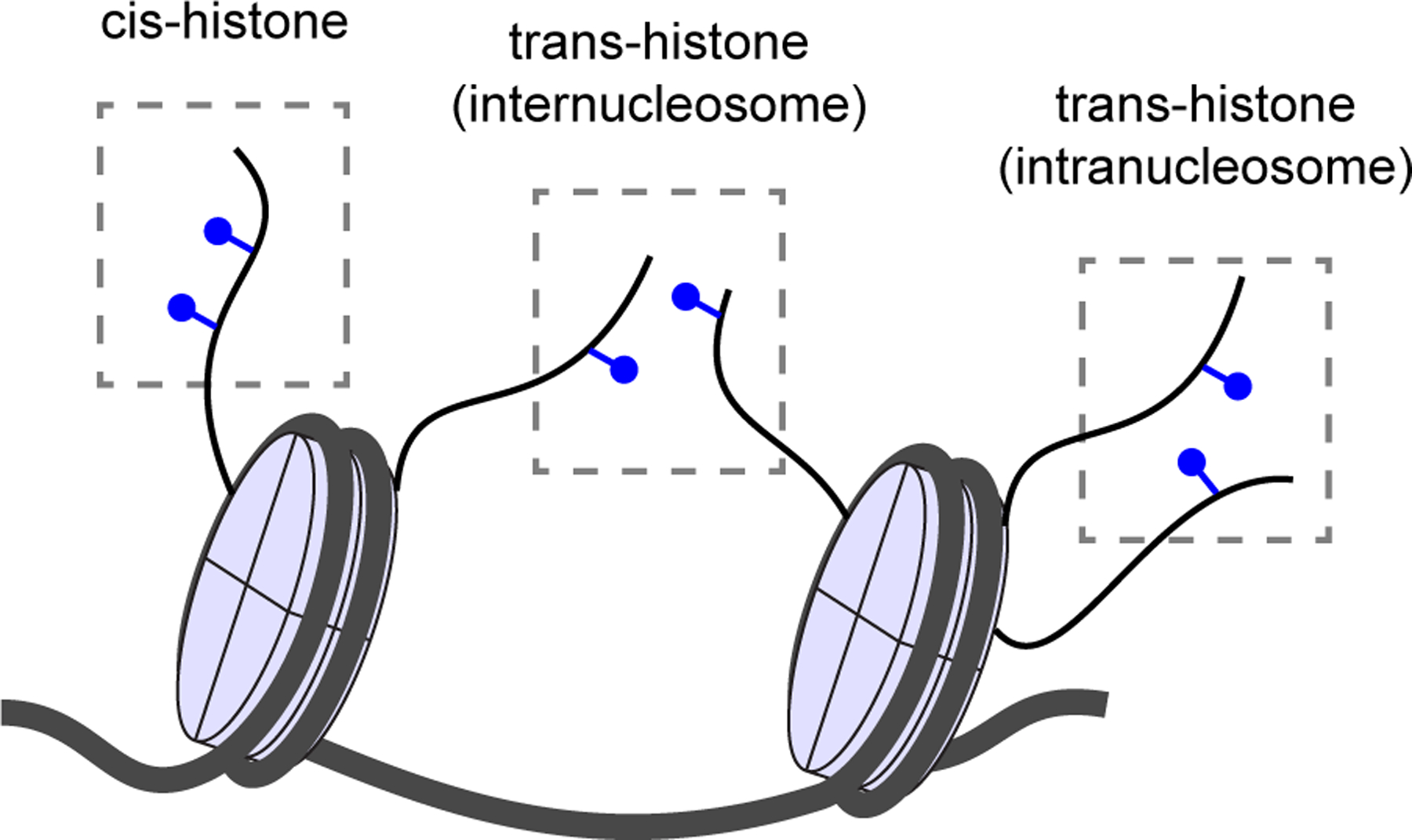
Combinatorial histone PTM patterns. In the cis-histone pattern, PTMs are found on the same histone molecule. In trans-histone (internucleosome) the PTMs occur on different nucleosomes, and in the trans-histone (intranucleosome) on the same nucleosome. Adapted from [134].
Histone PTMs have been shown to use different mechanisms to occlude PTMs that drive transcription to the opposing direction (e.g. activating vs repressing). Some histone PTMs are rarely found together at the same genomic regions, such as the repressive H3K27me3 with PTMs that are associated with active transcription such as H3K27ac [135], H3K4me3 [136,137], H3K36me2 and H3K36me3 [138]. It is worth noting that there are exceptions to these rules, such as at “bivalent” genomic regions in embryonic stem cells, where H3K27me3 can be found together with H3K4me3 and H3K36me3 on the same nucleosomes, albeit at different H3 tails [139]. Pre-existing histone PTMs on the same histone tail may directly compete with a modification on the same residue, such as the different methylation states of H3K27 (H3K27me1, H3K27me2 and H3K27me3) that are mutually exclusive with H3K27ac. However, histone PTMs may also inhibit writers for other PTMs. H3K4me3, H3K36me2 and H3K36me3 were shown to inhibit PRC2 activity to antagonize the deposition of H3K27me3 [137,140]. C-terminal domain of the PRC2 component SUZ12 was shown to mediate the allosteric inhibition by H3K4me3 [137], while the unmodified H3K36 was shown to occupy a critical position at EZH2-DNA interface, suggesting that H3K36 methylation allosterically inhibits H3K27 from interacting with the active site of EZH2 [141].
The inhibitory mechanisms listed above are likely to prevent abrupt repression of transcription since H3K4me3, H3K36me2 and H3K36me3 are deposited at actively transcribed genes. Interestingly, H3K4me3 also interferes with the repressive H3K9 methylation by inhibiting binding of H3K9 methyltransferases [142,143]. Histone PTMs may also promote their own deposition or the deposition of PTMs that drive gene expression to the same direction. For instance, activity of PRC2 is enhanced when it binds its own product H3K27me3, leading to the spread of H3K27me3 on the genome [1,27,144]. H3K27me3 also recruits PRC1 complexes that monoubiquitinate H2AK119 (H2AK119ub1) to repress the genes further [57]. Similarly, H3K4 methylation has been shown to facilitate the deposition of the activating H3K27ac [32], but also H3K27ac to promote deposition of H3K4me3 [145], possibly forming a positive feedback loop of activating histone PTMs.
In general, the degree of H3K27 methylation (H3K27me1/me2/me3) correlates with the activity state of the genomic region, where me3 is found mainly at repressed sites and me1 and me2 also at active sites together with other PTMs associated with activation [1]. It is unclear whether the low degrees of methylations, such as H3K27me1 and me2, H3K36me1 and H3K4me1 and me2 have roles with biological consequences or whether they solely function as intermediates to the di- or trimethylated state. The differential genomic distribution of the different degrees of methylations, such as the localization of H3K4me1 primarily at enhancers and H3K4me3 at promoters, suggests that at least some of these low degree methylation states could have specific biological roles. There may be unknown reader proteins that specifically recognize these modifications. Since these intermediates can exist simultaneously with other PTMs on the same nucleosomes or even on the same histone tail, they could mediate interactions of chromatin-binding proteins or complexes that can read two or more modifications at once. On the other hand, their presence may inhibit the activity of writers for other PTMs if they happen to occupy a critical position on an enzyme, such as the H3K36me2 or me3 that inhibit H3K27me3 deposition by interfering with the DNA-EZH2 interface [137].
The Important role of combinatorial histone PTMs in biology has only very recently begun to be understood. For instance, the DNA methyltransferase DNMT1 was shown to bind to the combinatorial modification of H3K9me3 and ubiquitylated H3 (H3Ub) to maintain DNA methylation [146]. Likewise, H3K4me3 combined with H3 serotonylation at position 5 (H3K4me3Q5ser) was shown to be crucial in promoting transcription of genes that control differentiation [147]. With histone PTM crosstalk it is difficult to determine the sequence of events and usually only negative or positive correlations can be observed. For instance, phosphorylation of H3S10 (H3S10ph) is often found together with H3K9me2 and me3 on the same histone tail, suggesting that crosstalk between these two modifications exists [148,149]. The crosstalk may play a role in the inheritance of histone PTMs to nascent chromatin during mitosis because inhibition of H3S10ph by Aurora B kinase also led to reduced deposition of H3K9me2 on freshly synthetized chromatin [149], but the proteins that read or are inhibited by the dual modification are not known and the biological consequences of the crosstalk remain to be elucidated. The above examples have focused on crosstalk of PTMs on the same histone tail, but crosstalk between PTMs on different histones in the same or different nucleosomes is also likely to have consequences. These possibilities increase the complexity of histone PTM crosstalk even further.
Crosstalk between histone PTMs and DNA methylation
In 1975, DNA methylation at cytosine residues was for the first time suggested to function as an epigenetic mark that could be inherited through somatic cell divisions to silence genes [150,151]. Most of the DNA methylation in mammals is found as 5-methylcytosine (5mC) at CpG dinucleotides, while methylation of other sequences is abundant in plants and fungi [152,153]. Methylated cytosines are frequently mutated to thymine by deamination, which has been thought to lead to a general loss of CpGs in the vertebrate genome through evolution [154]. Methylation is the default state of most CpG dinucleotides in the mammalian genome [155], with the exception of short (approximately 1kb) CpG-rich regions known as CpG islands (CGIs) half of which are found at transcription start sites (TSSs) in gene promoters [156]. CGIs are thought to be protected from deamination by staying primarily in an unmethylated state. Seventy-five percent of all vertebrate promoters are within CGIs and in an unmethylated state [155,157–159]. Current work on DNA methylation has mainly focused on CGIs at TSSs, whereas methylation at gene bodies and intergenic regions such as enhancers has remained less studied [160].
In somatic cells, some CGIs remain in a continuously methylated state for long-term repression of genomic elements such as imprinted genes (one of the two copies of a gene is permanently silenced), X chromosome inactivation (in females, one of the two X chromosomes is silenced to adjust gene dosage), transposons and to prevent expression of germ cell exclusive genes. The repression at these elements is relayed to progenitor cells in mitosis and is maintained through the lifetime of the organism. Failure to maintain DNA methylation at these elements leads to serious pathologies such as the ICF syndrome type 1 that is characterized by chromosome instability, severe immunodeficiency and facial anomalies [161]. ICF syndrome 1 is caused by homozygous loss-of-function mutations in the DNA methyltransferase DNMT3B leading to loss of DNA methylation at multiple regions [162]. Similarly, Tatton-Brown-Rahman overgrowth syndrome is caused by heterozygous germ line mutations in DNMT3A, a paralogue of DNMT3B, and is characterized by tall stature, characteristic facial anomalies and intellectual disabilities [163]. Interestingly, the syndrome shows strong phenotypic similarities with Weaver syndrome (heterozygous missense mutation in the H3 K27 methyltransferase EZH2) [164], Sotos syndrome (heterozygous mutation in the H3 K36 methyltransferase NSD1) [165,166], and the genomic imprinting Beckwith-Wiedemann syndrome [167], suggesting that they all involve disruption of imprinted gene expression [168].
DNA methylation is correlated with histone PTM patterns, indicating that crosstalk between these two epigenetic mechanisms exists. DNA methyltransferases have the ability to read histone PTMs, while DNA methylation has been shown to influence the binding of other chromatin-associated enzymes [169]. In cancer, CGI TSSs that are repressed by Polycomb proteins in polycomb repressive complexes (PRC) 1 and 2 are more likely to become methylated than TSSs of other genes, suggesting that histone PTM mediated silencing precedes DNA methylation [170–172]. DNA methylation holds inactive TSSs in a repressed state by recruiting proteins with methyl-CpG-binding domains (MBD) that specifically recognize methylated DNA [173,174]. MBD proteins then recruit histone deacetylases to maintain repression [175–177]. One of the roles of DNA methylation in epigenetic regulation may be to stabilize or ‘lock in’ silencing initiated by histone PTMs. This view is supported by work showing that the de novo DNA methyltransferases DNMT3A and DNMT3B methylate primarily nucleosomal DNA. The catalytically inactive DNA methyltransferase DNMT3L that is exclusively expressed in germ cells, was shown to direct DNMT3A and DNMT3B to methylate DNA at nucleosomes especially in the absence of H3K4 methylation [178]. Unmethylated H3K4 is associated with methylated DNA and inactive promoters [178]. DNA methylation at active TSSs probably does not happen because they are devoid of nucleosomes and the nucleosomes flanking the active TSS often contain the histone mark H3K4me3 and histone variant H2A.Z that seem to repel DNA methylation [179,180]. H3K4me3 was shown to bind to the ADD domain in DNMT3A/B to inhibit the activity of these enzymes [181].
DNA methylation established at the germ cell stage is faithfully maintained by the methyl transferase DNMT1 through mitotic cell divisions for the whole lifetime of the organism. DNMT1 is active only at hemimethylated DNA (one of the two DNA strands is methylated) which prevents it from performing de novo methylation. The CXXC domain on DNMT1 binds specifically unmethylated CpG nucleotides, but in doing so autoinhibits the methyl transferase activity of the protein, preventing activity at unmethylated sites [182]. In addition, interaction with UHRF1 (ubiquitin-like with PHD and ring finger domains 1) directs DNMT1 to hemimethylated regions because UHRF1 contains an SRA domain that binds hemimethylated DNA [183]. Null alleles of Uhrf1 phenocopy null alleles of Dnmt1 in mice, indicating that UHRF1 is vital for methylation maintenance by DNMT1 [183]. Recently, the replication foci targeting sequence (RFTS) domain of DNMT1 was shown to bind directly to a combinatorial modification of H3K9me3 and ubiquitylated H3 (H3Ub). Disruption of this interaction led to impairment of global DNA methylation and genomic stability, showing that H3Ub together with H3K9me3 reinforces maintenance of DNA methylation [146]. UHRF1 generates the mono-ubiquitination of H3 for DNMT1 binding [184] and was later shown to also mono-ubiquitinate PCNA-associated factor 15 (PAF15) that also recruits DNMT1 [185]. The two bromo-adjacent homology (BAH) domains on DNMT1 possibly mediate direct interactions with histone PTMs as well, but these interactions have not been identified yet [168].
The CXXC domain that binds specifically unmethylated CpG dinucleotides is, in addition to DNMT1, found on 13 other nuclear proteins, some of which are histone PTM modifiers [182]. One of these proteins is the PRC1 component and H3K36 and H3K4 demethylase KDM2B (also known as FBXL10, JHDM1B, and CXXC2) that was found to protect a specific subclass of CGIs from de novo methylation. Removal of KDM2B led to hypermethylation and repression of CGI promoters only at CGIs that were bound by PRC complexes, suggesting that KDM2B specifically protects polycomb-regulated genes from DNA methylation [186]. However, catalytically inactive KDM2B was able to revert the hypermethylation, suggesting that histone demethylase activity is not important for the protective function of KDM2B in this case. The detailed mechanism of how KDM2B protects these promoters from methylation remains to be elucidated.
Conversely, KDM1A, a paralogue of KDM2B, was shown to play an active role in removing H3K36me2 from CGIs, because its knockdown resulted in an increase in H3K36me2 at these regions [187]. H3K36me2 has been shown to inhibit transcription, suggesting that the demethylase activity of KDM2A could promote transcription from CGIs [188]. CXXC1 (CFP1) has been suggested to also contribute to the unmethylated state of CGIs by recruiting the H3K4 methyltransferases SETD1A and SETD1B that place H3K4 di- and trimethylation (H3K4me2 and H3K4me3) [189–192]. The H3K4 methyltransferases MLL1 and MLL2 also contain CXXC domains and may contribute to this process [193–195]. Ten-eleven translocation (TET) proteins TET1 and TET3 also contain the CXXC domain and may protect CGIs from methylation by catalyzing the conversion of 5mC to 5-hydroxymethylcytosine. Interestingly, TET1 has been shown to play a dual role by recruiting also the SIN3A histone deacetylase complex to CGIs to repress a subset of target genes in embryonic stem cells [196].
At gene bodies, DNA methylation does not prevent elongation of the primary transcript but can block transcription from downstream TSSs. This has been suggested to be a mechanism to allow silencing of repeated elements at gene bodies while allowing the primary gene to be transcribed [197]. However, gene body methylation may also have an important role in controlling alternative promoter usage to regulate expression of alternative transcripts in a tissue- and cell type-specific manner [198]. It may also regulate splicing since exons contain higher levels of DNA methylation than introns [199]. This may be influenced by the higher occupancy levels of nucleosomes at exons [200] since nucleosomes are the preferential substrate for DNA methyltrasferases [201]. CTCF possibly functions as a link between DNA methylation and splicing because it seems to prefer binding to unmethylated DNA and its binding leads to RNA polymerase II pausing that affects splicing [202,203]. H3K36me3 is associated with transcription elongation and DNA methylation at gene bodies [204–206]. The PWWP domain in DNMT3A and DNMT3B has been shown to mediate their recruitment to intragenic H3K36me3 [204,207]. The DNMT3B PWWP domain mutant (S270P) expressed in ICF syndrome 1 showed impaired recruitment to H3K36me3 [204,207,208] and a decrease in DNA methylation at pericentromeric satellite repeat II that is observed in ICF syndrome 1 [209–211]. Interestingly, the linker histone H1 can directly interact with DNMT1A and DNMT3B, but not with DNMT3A, to promote gene silencing. The DNMTs seem to prefer some H1 subtypes more than others [63].
At intergenic regions, enhancer activity and DNA methylation levels have been shown to correlate. For instance, enhancer activation by glucocorticoid receptor binding was shown to decrease DNA methylation at these sites [212]. DNA methylation may also affect insulators that regulate the interaction between an enhancer and a promoter. The preferential binding of CTCF to unmethylated DNA may mediate these functions [202]. H3K36me3 is largely enriched at gene bodies, whereas H3K36me2 is more diffusely found both at gene bodies and intergenic regions. DNMT3A was shown to prefer H3K36me2 over H3K36me3, whereas DNMT3B seems to prefer H3K36me3. These differences in preference may partially explain the genomic distributions of DNMT3A and DNMT3B [213]. NSD1-mediated H3K36me2 was shown to recruit DNMT3A to intergenic regions to maintain DNA methylation. Removal of the H3K36me2 methyltransferase NSD1 redistributed DNMT3A to H3K36me3 at gene bodies and reduced intergenic DNA methylation. The DNMT3A PWWP domain mutants (W297del, I310N, Y365C) found in Tatton-Brown-Rahman overgrowth syndrome impaired DNMT3A recruitment to H3K36me2 and reduced DNA methylation at these sites [213]. DNA methylome in Tatton-Brown-Rahman overgrowth syndrome was similar to that of the Sotos syndrome (NSD1 mutations), suggesting that they have the same underlying mechanism [213]. Interestingly, H2AK119ub1 was recently shown to recruit DNMT3A to CGIs, suggesting that there are alternative histone PTM-mediated ways for DNMT3A recruitment [214].
Epigenetic dysregulation in cancer
Genetic mutations of tumor suppressor proteins, such as those of the tumor protein p53, are a major driving force of cancer. However, cancer cells also display a drastically distorted epigenetic landscape that plays an important role in cancer development and progression [215]. Genetic mutations can lead to epigenetic dysregulation when mutations occur at epigenetic writer, eraser or reader proteins. However, epigenetic dysregulation itself can also lead to accumulation of further genetic mutations, for instance, by causing chromosome instability. DNA methylation, histone acetylation and histone methylation are the most-studied epigenetic mechanisms in cancer.
Cancer cells tend to show 20–60% decreased genome-wide 5mC methylation compared to non-cancerous cells [216]. This global loss of DNA methylation is thought to reactivate silenced repetitive elements, leading to chromatin instability and translocations, potentially causing further mutations (e.g. fusions) that can promote cancer progression. Additionally, hypomethylation at the promoters of oncogenes and imprinted genes can upregulate their expression and thus facilitate cancer growth [217]. Hypomethylated and overexpressed genes in cancer include genes such as CYP1B1 in prostate cancer, BRCA2 in sporadic ovarian cancer, MASPIN in colorectal cancer, thyroid cancer and pancreatic cancer and HOX11 in T-cell acute lymphoblastic leukemia [218–224], among others. The expression level of some of these genes, such as BRCA2, was also found to correlate with tumor stage [218]. Interestingly, even though DNMTs play such a central role in establishing and maintaining DNA methylation, inactivating mutations in DNMTs have been only found in AML [225] and not in other cancers. This is why dysregulated DNMT recruitment by other factors such as histone PTMs has been proposed to be the major cause for hypomethylation [217].
In contrast to hypomethylation, hypermethylation can promote cancer progression when it occurs at CpG islands at promoters of tumor suppressor genes that would normally remain in an unmethylated and thus active state. Hypermethylation and subsequent downregulation has been observed at genes regulating important cellular processes such as DNA repair (hMLH1, MGMT, WRN, BRCA1), Ras signaling (RASSFIA, NOREIA), cell cycle (P16INK4a, P15INK4B, RB), p53 network (P14ARF, TP73, HIC-1), Wnt signaling (APC, DKK-1, IGFBP-3) and apoptosis (TMS1, DAPK1, WIF-1, SFRP1) [226]. Aberrant hypermethylation in cancer has also been observed at CpG island shores, that are defined as 2 kb -long regions that lie on both sides of CpG islands. CpG island shore hypermethylation is observed specifically at genes that regulate differentiation, such as HOXA2, GATA2, TGFB1 and PAX5. [227–229]. Additionally, cancer cells tend to show global downregulation of miRNA expression, caused by hypermethylation at miRNA promoters [230]. Hypermethylation patterns are highly tumor and gene-specific and there does not seem to be a simple common mechanism for hypermethylation. It has been proposed that fusion proteins could recruit DNMTs to wrong genomic sites to induce hypermethylation [231], or that methylation simply spreads from highly methylated regions to surrounding sequences [232]. However, DNMT overexpression may also drive hypermethylation since DNMTs are overexpressed in various cancer types [233]. Inactivating mutations in TET proteins that erase DNA methylation may also function as a major driver of hypermethylation. Indeed, inactivating TET mutations are known to play a key role in the progression of various hematological cancers and solid tumors [234]. In addition, in gliomas and AML, gain-of-function (GOF) mutations in the tricarboxylic acid (TCA) cycle enzymes isocitrate dehydrogenase 1 and 2 (IDH1 and IDH2) drive disease phenotype by producing a metabolite that inhibits DNA and histone demethylation, thus causing DNA and histone hypermethylation [235,236].
In cancer, DNA hypomethylation at specific genes was shown to correlate with loss of H4K16 acetylation (H4K16ac). Indeed, global reduction in H4K16ac is the most common histone PTM dysregulation observed in cancer [237]. Imbalance in histone acetylation is thought to be driven by overexpressed or mutated HDACs often together with mutated HATs [237–240]. HAT fusions are common in several cancer types [239,240]. For instance, HDAC1 inactivation was recently shown to promote lymphomagenesis in mice [241]. The HAT KAT7 was shown to act as an oncogene in leukemia where it induces H3K14ac to upregulate expression of HOXA9 and HOX10 oncogenes [242].
Histone methylation is also often dysregulated in cancer, and it is driven by aberrant expression of KMTs and KDMs [243]. In general, cancer cells are characterized by a global loss in the activating mark H3K4me3 and the repressive mark H4K20me3 and gain repressive marks H3K9me3 and H3K27me3 [237,244–246]. However, the miswritten epigenetic landscape is highly tumor and gene specific, and these generalized rules do not always apply. For instance, the catalytic PRC2 subunit EZH2, that deposits H3K27me3, has both oncogenic and tumor-suppressive functions depending on the context [247,248]. GOF mutations in EZH2 promote pathogenesis of lymphoma and melanoma by enhancing EZH2 ability to methylate H3K27me1 and H3K27me2 to higher methylation states thus leading to increased H3K27me3 levels [247,249–251]. In lymphoma, the increased H3K27me3 levels facilitate cell cycle progression by repressing transcription of cell cycle inhibitors (such as CDKN2A and CDKN1A), among other genes [251–253]. In this context, EZH2 also represses expression of chemokines and major histocompatibility complex class 1 antigen processing genes leading to suppression of inflammatory responses and tumor immune evasion [254–256]. However, loss-of-function mutations of EZH2 or EED and SUZ12 (two other core PRC2 components) are also prevalent in cancers such as myeloid neoplasms, T-cell acute lymphoblastic leukemia and malignant peripheral nerve sheath tumor (MPNST) [257–259]. In mouse models, knockout of PRC2 subunits promoted progression of leukemia and MPNST, further validating the tumor suppressive role of PRC2 in these cancers [260,261]. Interestingly, loss of PRC2 in MPNST is associated with an increase in DNA methylation, suggesting that DNA methylation tries to compensate for the lack of repression by H3K27me3. These PRC2-deficient cells become highly sensitive for DNMT inhibitors, providing an opportunity for therapeutic intervention [262].
Mutations in the MLL family of methyltransferases that catalyze H3K4 methylation are common especially in leukemias. MLL1 fusions in acute myelogenous leukemia (AML) or acute lymphoblastic leukemia (ALL) are known to be a classic example of how dysregulated histone methylation can facilitate cancer progression [263–265]. More than 100 different MLL1 fusions have been identified in leukemias with elongation factors being the most common fusion partners [266]. These GOF fusions lead to abnormal accumulation of H3K4me3 and activation of oncogenes that then promote cancer progression. Other MLL family members MLL3 and MLL4 are often mutated in lung cancer, melanoma and B-cell lymphomas, but the mutations are loss-of-function mutations [267–271]. In healthy cells, MLL3 and MLL4 establish H3K4me1 at enhancers to upregulate expression of tumor suppressors, differentiation genes and suppressors of cytokine signaling [268,270,272].
H3K36 methyltransferases can be oncogenic or tumor suppressive depending on the context. SETD2 that catalyzes H3K36me3 is a tumor suppressor in acute leukemia, T cell lymphoma, lung cancer and renal cancer [50]. Likewise, NSD1 that catalyzes H3K36me2, functions as a tumor suppressor in epithelial cell cancer [273,274]. However, NSD2 that catalyzes H3K36me2, is an oncogene in multiple myeloma and pediatric ALLs where it is often rearranged or harbors GOF mutations to maintain elevated H3K36me2 levels [50,275–277]. In a similar manner, NSD3 overexpression and GOF mutations significantly contribute to development and progression of lung squamous cell carcinoma by upregulating H3K36me2 to reprogram oncogenic gene expression signatures [278]. Since DNMT3A and DNMT3B can bind to H3K36me2/3, dysregulation of the histone methyltransferases is also reflected in DNA methylation levels and often also changes the H3K27me3 profile [51,276,279]. Finally, H3K79 methylation functions as an activating mark in breast cancer where it repels HDAC1 and DNMT1 from epithelial-mesenchymal transition genes and thus promotes cancer progression [280]. H3K79 is catalyzed by DOT1L that is best known for its role in leukemias where it interacts with MLL fusion proteins to promote oncogenesis [266].
Mutations in histones (i.e. oncohistones) and mutations in chromatin remodeling complexes are known to cause a dysregulated epigenetic landscape in cancer and drive cancer progression. Oncohistones have been found in cancers such as glioma, sarcoma, and lymphoma [7,281–283]. They influence the binding of reader and writer proteins to chromatin, thus causing dysregulated epigenetic patterning affecting DNA repair and transcription [7,281,282,284,285]. Mutations in chromatin remodelers affect about 10–20% of all cancers [286]. Chromatin remodeler mutations lead to dysregulated chromatin accessibility and thus altered gene expression. Alterations in histone PTM patterning in cancer may also influence the recruitment of chromatin remodelers since the remodelers are recruited by histone PTMs [287,288].
Epigenetic dysregulation in non-malignant diseases
Epigenetic dysregulation plays a major role in the development and progression of cancer, but also in non-malignant diseases such as neurological diseases, autoimmune diseases and cardiovascular and metabolic diseases [3–10,215,227,289–292]. Mutations in epigenetic writers and readers are known to participate in the development and progression of several neurodevelopmental and neurological disorders. For instance, Rett syndrome is a progressive X-linked neurodevelopmental disorder caused by inactivating mutations in methyl-CpG-binding protein 2 (MECP2) [293,294]. MECP2 binds methylated DNA to repress transcription at methylated promoters. It is expressed especially in mature nerve cells where it possibly maintains neuronal maturation, activity, and plasticity [295]. DNA hypo- and hypermethylation sites have been identified in several neurodevelopmental and neurological disorders. Mutations in DNMT3A lead to hypomethylation in the Tatton-Brown-Rahman overgrowth syndrome [213]. Overexpression of TNFα due to promoter hypomethylation leads to apoptosis of neuronal cells in Parkinson’s disease [296]. In multiple sclerosis, promoter of the PADI2 gene was also found to be hypomethylated [297]. Fragile X syndrome, characterized by mild to severe intellectual disability, is caused by silencing of the FMR1 promoter via hypermethylation [298]. The promoters of genes NEP in Alzheimer’s disease, FXN in Friedreich’s ataxia and SMN2 in spinal muscular atrophy, are also hypermethylated in these disorders [297].
In neurological diseases, histone hypoacetylation is the most frequently observed histone PTM change [227]. For instance, inactivating mutations in the HATs CBP and EP300 have been found in Rubinstein-Taybi syndrome, a neurodevelopmental disorder that is characterized by moderate to severe intellectual disability, distinctive facial features, and short stature [297,299]. In amyotrophic lateral sclerosis (ALS), aggregates of FUS protein bind and inhibit the HAT activity of CBP, leading to hypoacetylation of its target genes [297]. Hypoacetylation has also been found in Parkinson’s and Huntington’s disease [297] and Friedreich’s ataxia [300]. Histone phosphorylation is dysregulated in Coffin-Lowry syndrome, where the RSK2 serine/threonine kinase that targets histones is mutated [301,302]. Histone methylation patterns are also dysregulated in various neurological disorders [227,297]. For instance, Sotos syndrome is caused by haploinsufficiency in the methyltransferase NSD1 that catalyzes H3K36me2 [213]. Interestingly, histone mutations were recently found to be the causative agent of a novel class of neurodevelopmental disorders [4]. This was the first time when histone mutations were associated with a neurodevelopmental disorder and not cancer. These mutated H3.3 histones (variant of H3) cause aberrant local and global histone PTM patterns and transcriptional dysregulation. The patients have not developed malignancies, suggesting that the mutations are distinct from cancer-associated somatic histone mutations [4].
Epigenetic alterations have also been found in autoimmune diseases and cardiovascular and metabolic diseases. DNA methylation is altered in the ICF (immunodeficiency, centromeric instability and facial anomalies) syndrome caused by heterozygous mutations in DNMT3B, that leads to hypomethylation at pericentromeric satellite repeats and other repeat sequences [161,162]. Hypomethylated sites have been also found in Systemic lupus erythematosus (SLE) and rheumatoid arthritis. Rheumatoid arthritis also shows hypermethylation at specific genes [303,304]. In addition, histone acetylation, methylation and phosphorylation are known to regulate inflammatory processes in SLE and rheumatoid arthritis [227]. Interestingly, histone acetylation also regulates cardiac morphology and cardiovascular diseases in animal models. For instance, HDAC2 is known to drive stress-induced increases in heart muscle mass (hypertrophy) and regulate cardiac development together with HDAC1 [6]. Overexpression of HDAC2 increased heart mass and knockdown of both HDAC1 and HDAC2 led to cardiac defects in mice [305,306]. HDACs are also involved in the inflammatory response at infarction injury sites, where they regulate the polarization of macrophages [307]. Together with genetic factors, epigenetic dysregulation also contributes to the development of obesity and type 2 diabetes. For instance, in type 2 diabetes, altered DNA methylation has been discovered at several genes that regulate metabolism, and single nucleotide polymorphism (SNPs) that add or remove CpG sites, may contribute to dysregulation of DNA methylation at these sites [308].
Therapeutic targeting of epigenetics
Epigenetic dysregulation not only activates tumorigenesis but can also be harnessed by tumor cells to escape from chemotherapy and host immune surveillance [309]. Some drugs that target epigenetics have already been effective for the treatment of hematological and solid tumors, but they have taken a long time to become accepted [292]. There are several ongoing clinical trials that aim to develop epigenetic drugs further or combine different therapies [309,310]. The epigenetic drugs that are currently in clinical trials can be divided into two classes: broad reprogrammers that cause large-scale changes in gene expression, and targeted therapies that target a specific mutation [292].
DNMT inhibitors (DNMTi), histone deacetylase inhibitors (HDACi) and bromodomain and extra-terminal motif protein inhibitors (BETi) are broad reprogrammers that generally reverse cancer-specific gene alterations by causing genome-wide changes in the epigenome [311,312]. The DNMTis 5-azacytidine and decitabine reduced malignant cell burden, improved blood cell count and improved survival in 15% of patients with myelodysplastic syndrome or AML [313,314]. They were accepted by the US Food and Drug Administration (FDA) for the treatment of these conditions. DNMTis can induce delayed responses, suggesting that they can reprogram the epigenome in addition to causing direct cytotoxicity [315,316]. However, the activity of DNMTis is limited to solid tumors [309,317] and patients often develop resistance so that they either respond to the drug poorly to begin with (primary resistance) or develop resistance over the course of treatment (secondary resistance) [318,319]. Guadecitabine is a promising new DNMTi with longer half-life and bioavailability than its predecessors and it is currently in clinical trials [292,320].
The HDACis vorinostat, belinostat and romidepsin have been accepted for the treatment of T cell lymphomas, and the HDACi panobinostat in combination with the proteasome inhibitor bortezomib for the treatment of drug-resistant multiple myeloma [292,321]. Specific inhibitors that target certain HDAC isoforms to increase efficiency and decrease side effects are under development [292]. BETis bind to the bromodomain of the chromatin remodeling BET proteins that use the bromodomain to recognize acetylated histones. Originally, BETis were developed to target the chromatin remodeler BRD4 that is translocated in some cancers and promotes expression of the MYC oncogene [322–324]. BETis are in preclinical studies and have entered early-stage clinical trials [292]. For instance, BETis attenuate the growth of castration-resistant prostate cancer cells [325–328].
Targeted therapies can be used to specifically target epigenetic regulators with GOF mutations with the potential of causing less off-target effects than with broad reprogrammers [292,309]. For instance, inhibiting EZH2 induced cell death specifically in lymphoma cells that were expressing the EZH2 GOF mutation [329]. Likewise, IDH inhibitors have been promising in early clinical trials of AML and gliomas that express IDH GOF mutations [330]. These cancers are characterized by DNA and histone hypermethylation [235,236]. However, DNMTis were more effective in inhibiting growth of IDH1 mutant glioma cells, suggesting that the use of broad reprogrammers might be beneficial in these cases [331]. Targeted therapies can also be used to inhibit proteins that act in the same signaling network or directly interact with the mutant protein. For instance, inhibition of the H3K79 methyltransferase DOT1L can be used to selectively kill MLL cells that express MLL1 translocations [332]. Expression of the MLL1 fusion mutant leads to recruitment of DOT1L to aberrant gene locations, where DOT1L mediated H3K79 methylation upregulates gene expression. Inhibition of DOT1L selectively kills cultured cells bearing MLL1 translocations by blocking H3K79 methylation and inhibiting expression of leukemogenic genes [332]. Similarly, inhibition of the histone demethylase 1 (LSD1, KDM1A) seems to specifically kill cancer cells with certain DNA methylation patterns, such as small-cell lung carcinoma cells [333]. Discovering new epigenetic writers, erasers and readers in cancer signaling networks can potentially provide novel targets for cancer therapy [334–337].
Drug combinations that target both DNA methylation and histone PTMs simultaneously are currently in development [292]. Combining inhibitors can increase the efficacy of each of the single agents. For instance, HDACi together with DNMTi upregulates transcription at a larger number of genomic sites than alone [230,338–341]. DNA methylation and EZH2-mediated H3K27me3 can both silence CpG islands, but they are mutually exclusive. H3K27me3 often compensates for loss of DNA methylation to maintain repression at repetitive elements such as endogenous retroviruses (ERVs) after DNMTi treatment [170,342,343]. Combining DNMTis with EZH2 inhibitors or other histone methyltransferase inhibitors can potentially promote the expression of ERVs, releasing antigens that render the cancer cells visible to the host immune system [292,344]. Interestingly, in hematological malignancies, physiological concentrations of vitamin C can boost the effects of DNMTi to inhibit proliferation and induce apoptosis of cancer cells. Since vitamin C is a cofactor of TET proteins that demethylate DNA, it is possibly the increase in DNA demethylase activity synergizing with DNMT inhibition that strengthens the DNA hypomethylation phenotype [292,345,346].
The literature on therapeutic targeting of epigenetics in non-malignant diseases such as neurological diseases is scarce, possibly because epigenetic dysregulation in these disorders tend to be caused by inactivating mutations that are difficult to target. However, at least HDACis have been promising in the treatment of cardiovascular and metabolic diseases [6]. For instance, HDACis improve healing at myocardial infarction injury sites by promoting macrophage polarization to reparative type in mouse models [307] and improve insulin secretion in pancreatic islets from human donors with type 2 diabetes [308]. It is worth noting that HDACs can also deacetylate a plethora of non-histone proteins, meaning that the effect of the HDACis is not necessarily solely histone-mediated, and depends on the specificity of the inhibitor in question [6].
Genomic and proteomic methods to characterize epigenetic crosstalk
Recent technical advances in mass spectrometry (MS), DNA sequencing methods such as chromatin-immunoprecipitation coupled to genome-wide sequencing (ChIP-seq) and in the generation of recombinant histones and reader domains have accelerated the study of epigenetic crosstalk, especially that of combinatorial histone PTMs [134,347,348].
ChIP-seq [349] is a very powerful technique in the study of histone PTMs since it allows the mapping of the PTMs to different genomic regions (e.g. enhancers, promoters or gene bodies) and to specific genes, providing a plethora of information on the behavior of the PTM. When ChIP-seq is combined with other genome-wide methods that provide information on the transcriptional state of the genomic region, such as RNA sequencing (RNA-seq) [350] or global run-on sequencing (GRO-seq) [351], it can provide crucial information of whether the PTM tends to associate with repressed or active regions. When further combined with perturbation assays (e.g. enzyme knock out, knock down or inhibitors) information can be gathered on the importance of a specific PTM for the expression of a given gene or gene sets.
Combining ChIP-seq with assays that interrogate chromatin accessibility, such as DNAse I hypersensitive site sequencing (DNAse-seq) [352], assay for transposase-accessible chromatin (ATAC-seq) [353] or micrococcal nuclease sequencing (MNase-seq) [354,355], can provide information on whether the histone PTM associates with accessible or inaccessible chromatin regions. These chromatin accessibility methods are based on quantifying the susceptibility of chromatin to enzymatic cleavage. Chromatin-bound proteins, such as histones, protect DNA from cleavage and the protective effect is dependent on the density and residence time of the chromatin-bound proteins. DNAse-seq utilizes DNAse I to release DNA fragments from accessible regions, after which the fragments are used in library preparation and sequenced. ATAC-seq utilizes the Tn5 transposase that directly ligates sequencing adaptors to DNA fragments from accessible regions, simplifying the sample preparation workflow, and it is also suitable for lower cell numbers. In MNase-seq, in contrast, all accessible linker DNA is degraded, and the DNA protected by core histones is sequenced. MNase-seq can be used to assay chromatin accessibility at the single nucleosome level [356,357].
ChIP-seq canbe used to determine whether histone PTMs reside on the same regions, which would suggest possible crosstalk between these PTMs. In traditional ChIP-seq, the resolution limit is about 150–300bp due to the size of sonication-derived chromatin fragments, but this has been improved to near-single-bp scale with lambda exonuclease in ChIP-exo [358] and more recently with micrococcal endo/exo nuclease in high-resolution X-ChIP [359]. Moreover, recent alternative methods, CUT&Run [360] and CUT&Tag [361] offer high resolution, easier workflows and are suitable for lower cell numbers. However, even with improved resolution, the histone PTMs are pulled down one at a time and analyzed from a population of cells or single cells, meaning that the overlapping signal from two or more PTMs never originates from the same nucleosome in the same cell. In re-ChIP experiments (or sequential ChIP) histone PTM pulldown is followed by a pulldown of the second modification, resulting in a pool of chromatin fragments that are enriched for both modifications [139,362]. The downside of the sequential pulldown is that there may not be enough material at the end for sequencing, limiting its use to abundant PTMs that have good antibodies available. These problems could be avoided with the use of antibodies that directly recognize combinatorial modifications, but there are only few antibodies available. Antibodies may also nonspecifically recognize wrong PTMs, or their binding may be blocked by PTMs in the vicinity of the binding site [363], leading to unpredictable bias in ChIP-seq data.
Mass spectrometry (MS) provides a powerful tool for identifying combinatorial histone PTMs on the histone tail and complete histone level [134,347,348,364]. Histone MS can be classified into bottom-up, middle-down and top-down, based on the size of the input peptide or protein (Figure 3.) [365–367]. In bottom-up approach, histones are digested with proteases (e.g. trypsin), to acquire small peptide fragments (5–20 amino acids) for MS analysis. Bottom-up is suitable for the detection of combinatorial histone PTMs that are close to each other, such as those at H3K27 combined with H3K36 [368–370]. Middle-down and top-down approaches are used to detect PTM combinations at a longer distance, because they use larger molecules as input. In middle-down, specific proteases, usually Glu-C for H3 and Asp-N for H4, are used to generate longer peptide fragments (20–50 amino acids) to cover the most prevalently studied PTM combinations [365,371]. Middle-down MS has been used to identify combinatorial PTMs such as H3K4 methylation with H3K27 acetylation [372]. In top-down, intact histones (more than 100 amino acids) without proteolytic digestion are analyzed with MS, allowing the detection of combinatorial PTMs also at longer distances. For example, top-down approach was applied in multiple myeloma cells to detect the combinatorial hypermethylation at H3K9, H3K27 and H3K36 driven by elevated expression of the MMSET methyltransferase [373]. Bottom-up approach failed to detect this relationship. The benefit of the bottom-up approach is that it is well optimized and the most accurate and robust method for detecting histone PTMs [374], whereas the middle-down and top-down methods are less sensitive, technically more challenging and require specialized data-analysis. However, middle-down is the preferred method for combinatorial histone PTM analysis because it falls between the bottom-up and top-down in difficulty and suitability for combinatorial analysis [371].
Figure 3.
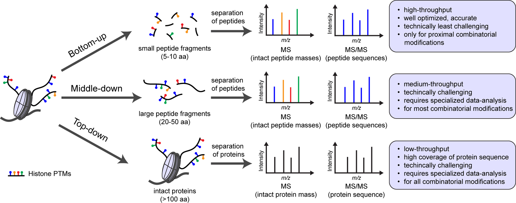
Histone MS approaches based on the size of input. In bottom-up and middle-down methods, peptides are first fragmented to 5–10 aa and 20–50 aa fragments, respectively. In top-down method, intact proteins are used as input. Benefits and limitations of each method are listed.
The MS-based methods can be combined with genome-wide methods to understand histone PTM crosstalk in a more detailed manner. However, the limitation of all the MS-based methods is that the genomic location of the combinatorial PTMs cannot be inferred from the data. Combinatorial histone PTM analysis can be potentially performed on genomic regions that are enriched by specific chromatin-binding proteins (e.g. TF of interest) by antibody or tag-based pulldowns, but this yields to a global histone PTM profile on all binding sites of the protein of interest. Similar approach could be used by engineering a protein or oligonucleotide that only binds specific chromatin regions, as has been done with transcription activator-like proteins to identify telomere-binding proteins by MS [375] or with DNA oligomers that hybridize with target telomeric regions [376]. CasID utilizes a catalytically inactive Cas9 to direct a biotin ligase to specific gene sequences [377]. It could also be potentially used to biotinylate and purify histones for combinatorial PTM analysis at selected enhancers or promoters of interest, but the guide RNAs would have to be individually designed for each target sequence. CasID also suffers from high signal to noise ratio because it is limited to two alleles. However, the signal to noise ratio has been improved with proximal biotinylation by episomal recruitment (PROBER) that uses episomes to amplify the sequence of interest [378].
With metabolically labeled histones, it is possible to assign histone PTMs to accessible or inaccessible chromatin regions by MS [379]. It is based on the principle that at accessible (transcriptionally active) chromatin regions histone turnover is higher than at inaccessible (transcriptionally inactive) regions. This leads to higher incorporation of metabolically labeled histones at accessible regions compared to inaccessible ones, and the metabolically labeled histones with their PTMs can be distinguished from the non-labeled ones with MS. This way, PTM combinations can be assigned to active or repressed regions providing information on their possible biological function. The findings correlate well with genome-wide methods [379].
Crosslinking MS has been applied to identify proteins that interact with RNA [380,381] and DNA [382,383]. With crosslinking MS, peptides together with the crosslinked single nucleotide adducts can be identified [381]. However, longer oligonucleotide sequences together with crosslinked peptides still cannot be identified using MS. It is tempting to speculate that if the technical hurdles of sequencing oligonucleotides together with peptides would be overcome, crosslinking MS could have the potential to map peptides with their PTMs to genomic locations. This would require the sequencing of long DNA fragments of 25–150 base pairs for unambiguous mapping, depending on the genomic region [384].
Concluding remarks
Crosstalk between cellular signaling pathways and epigenetic networks is a complicated process that can be regulated at multiple levels, such as the presence or absence of extracellular signaling molecules and the expression and activity of cytosolic signaling cascade proteins and epigenetic regulators. Deregulated expression or mutations in epigenetic reader and writer proteins leads to misinterpretation of cellular signals at the chromatin level and to the onset and progression of a variety of disorders and pathologies. Discovering new mechanisms of epigenetic regulation, such as histone PTM crosstalk, in the onset and progression of diseases could aid in the development of targeted therapeutics.
In histone PTM crosstalk, pre-existing PTMs promote or inhibit the deposition of new histone PTMs. Especially histone PTMs that are known to primarily associate with a specific transcriptional state of a genomic region (i.e. active or repressed) seem to antagonize writers that deposit PTMs with the opposite function. These mechanisms possibly exist to maintain the transcriptional state of the genomic region until a signal is received that shifts the balance (e.g. signal-activated TF binds and recruits activators). Higher protein copy number of writers for repressive histone PTMs over activating ones may also create a repressive “pressure” in the nucleus to ensure only a limited number of genes are expressed at a given time [385] and contribute to turning off transcription.
In the past two decades, the exciting new technological developments in both genome-wide sequencing and MS-based proteomics methods have opened new avenues to interrogate mechanisms of epigenetic crosstalk in an unprecedented manner. Combinatorial histone PTMs are now begun to be understood as key in biological processes such as regulation of DNA methylation [146] and transcription [147]. Further development and wider application of MS-based approaches such as top-down proteomics in different biological contexts is likely to increase our understanding and respect for the complexity of epigenetic crosstalk yet to another level.
Acknowledgements
J.K.L was supported by the Sigrid Jusélius Foundation and the Maud Kuistila Foundation. This work was supported by the National Institutes of Health (NIH) grants HD106051, CA196539 and AI118891.
Abbreviations
- 5mC
5-methylcytosine
- ACL
ATAC-seq, assay for transposase-accessible chromatin
- ATP
citrate lyase
- CGI
CpG islands
- CHD
chromodomain
- ChIP-seq
chromatin immunoprecipitation genome-wide sequencing
- DNAse-seq
DNAse I hypersensitive site sequencing
- ERV
endogenous retrovirus
- GOF
gain-of-function
- HAT
histone acetyltransferase
- HDAC
histone deacetylase
- KDM
histone lysine demethylase
- KMT
histone lysine methyltransferase
- MAPK
mitogen-activated protein kinase
- MBD
methyl-CpG-binding domain
- MLL
mixed-lineage leukemia
- MNase-seq
micrococcal nuclease sequencing
- MPNST
malignant peripheral nerve sheath tumor
- MS
mass spectrometry
- PHD
plant homeodomain
- PRC1
polycomb repressive complex 1
- PRC2
polycomb repressive complex 2
- PTM
post-translational modification
- TF
transcription factor
- TSS
transcription start site
Footnotes
Competing interests
The authors declare that there are no competing interests associated with the manuscript.
CrediT Author Contribution
J.K.L: Conceptualization, Investigation, Visualization, Writing – original draft, Writing – review and editing. B.A.G: Supervision, Conceptualization.
References
- 1.Laugesen A, Højfeldt JW and Helin K (2019) Molecular Mechanisms Directing PRC2 Recruitment and H3K27 Methylation. Mol Cell 74, 8–18. 10.1016/j.molcel.2019.03.011. [DOI] [PMC free article] [PubMed] [Google Scholar]
- 2.Murakami Y (2019) Phosphorylation of repressive histone code readers by casein kinase 2 plays diverse roles in heterochromatin regulation. J Biochem 166, 3–6. 10.1093/jb/mvz045. [DOI] [PubMed] [Google Scholar]
- 3.Kim J-H, Lee JH, Lee I-S, Lee SB and Cho KS (2017) Histone Lysine Methylation and Neurodevelopmental Disorders. Int J Mol Sci 18, E1404. 10.3390/ijms18071404. [DOI] [PMC free article] [PubMed] [Google Scholar]
- 4.Bryant L, Li D, Cox SG, Marchione D, Joiner EF, Wilson K, Janssen K, Lee P, March ME, Nair D, et al. (2020) Histone H3.3 beyond cancer: Germline mutations in Histone 3 Family 3A and 3B cause a previously unidentified neurodegenerative disorder in 46 patients. Sci Adv 6, eabc9207. 10.1126/sciadv.abc9207. [DOI] [PMC free article] [PubMed] [Google Scholar]
- 5.Peter CJ and Akbarian S (2011) Balancing histone methylation activities in psychiatric disorders. Trends Mol Med 17, 372–379. 10.1016/j.molmed.2011.02.003. [DOI] [PMC free article] [PubMed] [Google Scholar]
- 6.Bagchi RA and Weeks KL (2019) Histone deacetylases in cardiovascular and metabolic diseases. J Mol Cell Cardiol 130, 151–159. 10.1016/j.yjmcc.2019.04.003. [DOI] [PubMed] [Google Scholar]
- 7.Nacev BA, Feng L, Bagert JD, Lemiesz AE, Gao J, Soshnev AA, Kundra R, Schultz N, Muir TW and Allis CD (2019) The expanding landscape of “oncohistone” mutations in human cancers. Nature 567, 473–478. 10.1038/s41586-019-1038-1. [DOI] [PMC free article] [PubMed] [Google Scholar]
- 8.Mio C, Bulotta S, Russo D and Damante G (2019) Reading Cancer: Chromatin Readers as Druggable Targets for Cancer Treatment. Cancers (Basel) 11, E61. 10.3390/cancers11010061. [DOI] [PMC free article] [PubMed] [Google Scholar]
- 9.Nishiyama A and Nakanishi M (2021) Navigating the DNA methylation landscape of cancer. Trends Genet 37, 1012–1027. 10.1016/j.tig.2021.05.002. [DOI] [PubMed] [Google Scholar]
- 10.Zhao Z and Shilatifard A (2019) Epigenetic modifications of histones in cancer. Genome Biol 20, 245. 10.1186/s13059-019-1870-5. [DOI] [PMC free article] [PubMed] [Google Scholar]
- 11.Luger K, Mäder AW, Richmond RK, Sargent DF and Richmond TJ (1997) Crystal structure of the nucleosome core particle at 2.8 A resolution. Nature 389, 251–260. 10.1038/38444. [DOI] [PubMed] [Google Scholar]
- 12.Zhou K, Gaullier G and Luger K (2019) Nucleosome structure and dynamics are coming of age. Nat Struct Mol Biol 26, 3–13. 10.1038/s41594-018-0166-x. [DOI] [PMC free article] [PubMed] [Google Scholar]
- 13.Iwasaki W, Miya Y, Horikoshi N, Osakabe A, Taguchi H, Tachiwana H, Shibata T, Kagawa W and Kurumizaka H (2013) Contribution of histone N-terminal tails to the structure and stability of nucleosomes. FEBS Open Bio 3, 363–369. 10.1016/j.fob.2013.08.007. [DOI] [PMC free article] [PubMed] [Google Scholar]
- 14.Zhou B-R, Jiang J, Feng H, Ghirlando R, Xiao TS and Bai Y (2015) Structural Mechanisms of Nucleosome Recognition by Linker Histones. Mol Cell 59, 628–638. 10.1016/j.molcel.2015.06.025. [DOI] [PMC free article] [PubMed] [Google Scholar]
- 15.Simpson RT (1978) Structure of the chromatosome, a chromatin particle containing 160 base pairs of DNA and all the histones. Biochemistry 17, 5524–5531. 10.1021/bi00618a030. [DOI] [PubMed] [Google Scholar]
- 16.Olins AL and Olins DE (1974) Spheroid chromatin units (v bodies). Science 183, 330–332. 10.1126/science.183.4122.330. [DOI] [PubMed] [Google Scholar]
- 17.Kornberg RD (1974) Chromatin structure: a repeating unit of histones and DNA. Science 184, 868–871. 10.1126/science.184.4139.868. [DOI] [PubMed] [Google Scholar]
- 18.Li G, Margueron R, Hu G, Stokes D, Wang Y-H and Reinberg D (2010) Highly compacted chromatin formed in vitro reflects the dynamics of transcription activation in vivo. Mol Cell 38, 41–53. 10.1016/j.molcel.2010.01.042. [DOI] [PMC free article] [PubMed] [Google Scholar]
- 19.Maeshima K, Ide S and Babokhov M (2019) Dynamic chromatin organization without the 30-nm fiber. Curr Opin Cell Biol 58, 95–104. 10.1016/j.ceb.2019.02.003. [DOI] [PubMed] [Google Scholar]
- 20.Dixon JR, Selvaraj S, Yue F, Kim A, Li Y, Shen Y, Hu M, Liu JS and Ren B (2012) Topological domains in mammalian genomes identified by analysis of chromatin interactions. Nature 485, 376–380. 10.1038/nature11082. [DOI] [PMC free article] [PubMed] [Google Scholar]
- 21.Lieberman-Aiden E, van Berkum NL, Williams L, Imakaev M, Ragoczy T, Telling A, Amit I, Lajoie BR, Sabo PJ, Dorschner MO, et al. (2009) Comprehensive mapping of long-range interactions reveals folding principles of the human genome. Science 326, 289–293. 10.1126/science.1181369. [DOI] [PMC free article] [PubMed] [Google Scholar]
- 22.Zentner GE and Henikoff S (2013) Regulation of nucleosome dynamics by histone modifications. Nat Struct Mol Biol 20, 259–266. 10.1038/nsmb.2470. [DOI] [PubMed] [Google Scholar]
- 23.Bowman GD and Poirier MG (2015) Post-translational modifications of histones that influence nucleosome dynamics. Chem Rev 115, 2274–2295. 10.1021/cr500350x. [DOI] [PMC free article] [PubMed] [Google Scholar]
- 24.Zhao Y and Garcia BA (2015) Comprehensive Catalog of Currently Documented Histone Modifications. Cold Spring Harb Perspect Biol 7, a025064. 10.1101/cshperspect.a025064. [DOI] [PMC free article] [PubMed] [Google Scholar]
- 25.Fyodorov DV, Zhou B-R, Skoultchi AI and Bai Y (2018) Emerging roles of linker histones in regulating chromatin structure and function. Nat Rev Mol Cell Biol 19, 192–206. 10.1038/nrm.2017.94. [DOI] [PMC free article] [PubMed] [Google Scholar]
- 26.Grunt TW (2018) Interacting Cancer Machineries: Cell Signaling, Lipid Metabolism, and Epigenetics. Trends Endocrinol Metab 29, 86–98. 10.1016/j.tem.2017.11.003. [DOI] [PubMed] [Google Scholar]
- 27.Zhang T, Cooper S and Brockdorff N (2015) The interplay of histone modifications - writers that read. EMBO Rep 16, 1467–1481. 10.15252/embr.201540945. [DOI] [PMC free article] [PubMed] [Google Scholar]
- 28.Sabari BR, Zhang D, Allis CD and Zhao Y (2017) Metabolic regulation of gene expression through histone acylations. Nat Rev Mol Cell Biol 18, 90–101. 10.1038/nrm.2016.140. [DOI] [PMC free article] [PubMed] [Google Scholar]
- 29.Zeng L and Zhou MM (2002) Bromodomain: an acetyl-lysine binding domain. FEBS Lett 513, 124–128. 10.1016/s0014-5793(01)03309-9. [DOI] [PubMed] [Google Scholar]
- 30.Mishra LN, Pepenella S, Rogge R, Hansen JC and Hayes JJ (2016) Acetylation Mimics Within a Single Nucleosome Alter Local DNA Accessibility In Compacted Nucleosome Arrays. Sci Rep 6, 34808. 10.1038/srep34808. [DOI] [PMC free article] [PubMed] [Google Scholar]
- 31.Mishra LN and Hayes JJ (2018) A nucleosome-free region locally abrogates histone H1-dependent restriction of linker DNA accessibility in chromatin. J Biol Chem 293, 19191–19200. 10.1074/jbc.RA118.005721. [DOI] [PMC free article] [PubMed] [Google Scholar]
- 32.Wang Z, Zang C, Cui K, Schones DE, Barski A, Peng W and Zhao K (2009) Genome-wide mapping of HATs and HDACs reveals distinct functions in active and inactive genes. Cell 138, 1019–1031. 10.1016/j.cell.2009.06.049. [DOI] [PMC free article] [PubMed] [Google Scholar]
- 33.Zentner GE, Tesar PJ and Scacheri PC (2011) Epigenetic signatures distinguish multiple classes of enhancers with distinct cellular functions. Genome Res 21, 1273–1283. 10.1101/gr.122382.111. [DOI] [PMC free article] [PubMed] [Google Scholar]
- 34.Rada-Iglesias A, Bajpai R, Swigut T, Brugmann SA, Flynn RA and Wysocka J (2011) A unique chromatin signature uncovers early developmental enhancers in humans. Nature 470, 279–283. 10.1038/nature09692. [DOI] [PMC free article] [PubMed] [Google Scholar]
- 35.Zhang T, Zhang Z, Dong Q, Xiong J and Zhu B (2020) Histone H3K27 acetylation is dispensable for enhancer activity in mouse embryonic stem cells. Genome Biol 21, 45. 10.1186/s13059-020-01957-w. [DOI] [PMC free article] [PubMed] [Google Scholar]
- 36.Musselman CA, Khorasanizadeh S and Kutateladze TG (2014) Towards understanding methyllysine readout. Biochim Biophys Acta 1839, 686–693. 10.1016/j.bbagrm.2014.04.001. [DOI] [PMC free article] [PubMed] [Google Scholar]
- 37.Zaghi M, Broccoli V and Sessa A (2019) H3K36 Methylation in Neural Development and Associated Diseases. Front Genet 10, 1291. 10.3389/fgene.2019.01291. [DOI] [PMC free article] [PubMed] [Google Scholar]
- 38.Barski A, Cuddapah S, Cui K, Roh T-Y, Schones DE, Wang Z, Wei G, Chepelev I and Zhao K (2007) High-resolution profiling of histone methylations in the human genome. Cell 129, 823–837. 10.1016/j.cell.2007.05.009. [DOI] [PubMed] [Google Scholar]
- 39.Huang C and Zhu B (2018) Roles of H3K36-specific histone methyltransferases in transcription: antagonizing silencing and safeguarding transcription fidelity. Biophys Rep 4, 170–177. 10.1007/s41048-018-0063-1. [DOI] [PMC free article] [PubMed] [Google Scholar]
- 40.Godfrey L, Crump NT, O’Byrne S, Lau I-J, Rice S, Harman JR, Jackson T, Elliott N, Buck G, Connor C, et al. (2021) H3K79me2/3 controls enhancer-promoter interactions and activation of the pan-cancer stem cell marker PROM1/CD133 in MLL-AF4 leukemia cells. Leukemia 35, 90–106. 10.1038/s41375-020-0808-y. [DOI] [PMC free article] [PubMed] [Google Scholar]
- 41.Im H, Park C, Feng Q, Johnson KD, Kiekhaefer CM, Choi K, Zhang Y and Bresnick EH (2003) Dynamic regulation of histone H3 methylated at lysine 79 within a tissue-specific chromatin domain. J Biol Chem 278, 18346–18352. 10.1074/jbc.M300890200. [DOI] [PubMed] [Google Scholar]
- 42.Wood K, Tellier M and Murphy S (2018) DOT1L and H3K79 Methylation in Transcription and Genomic Stability. Biomolecules 8, 11. 10.3390/biom8010011. [DOI] [PMC free article] [PubMed] [Google Scholar]
- 43.Nguyen AT and Zhang Y (2011) The diverse functions of Dot1 and H3K79 methylation. Genes Dev 25, 1345–1358. 10.1101/gad.2057811. [DOI] [PMC free article] [PubMed] [Google Scholar]
- 44.Li Y, Wen H, Xi Y, Tanaka K, Wang H, Peng D, Ren Y, Jin Q, Dent SYR, Li W, et al. (2014) AF9 YEATS domain links histone acetylation to DOT1L-mediated H3K79 methylation. Cell 159, 558–571. 10.1016/j.cell.2014.09.049. [DOI] [PMC free article] [PubMed] [Google Scholar]
- 45.Heintzman ND, Stuart RK, Hon G, Fu Y, Ching CW, Hawkins RD, Barrera LO, Van Calcar S, Qu C, Ching KA, et al. (2007) Distinct and predictive chromatin signatures of transcriptional promoters and enhancers in the human genome. Nat Genet 39, 311–318. 10.1038/ng1966. [DOI] [PubMed] [Google Scholar]
- 46.Heintzman ND, Hon GC, Hawkins RD, Kheradpour P, Stark A, Harp LF, Ye Z, Lee LK, Stuart RK, Ching CW, et al. (2009) Histone modifications at human enhancers reflect global cell-type-specific gene expression. Nature 459, 108–112. 10.1038/nature07829. [DOI] [PMC free article] [PubMed] [Google Scholar]
- 47.Kim A, Kiefer CM and Dean A (2007) Distinctive signatures of histone methylation in transcribed coding and noncoding human beta-globin sequences. Mol Cell Biol 27, 1271–1279. 10.1128/MCB.01684-06. [DOI] [PMC free article] [PubMed] [Google Scholar]
- 48.Bannister AJ, Schneider R, Myers FA, Thorne AW, Crane-Robinson C and Kouzarides T (2005) Spatial distribution of di- and tri-methyl lysine 36 of histone H3 at active genes. J Biol Chem 280, 17732–17736. 10.1074/jbc.M500796200. [DOI] [PubMed] [Google Scholar]
- 49.Fang D, Gan H, Lee J-H, Han J, Wang Z, Riester SM, Jin L, Chen J, Zhou H, Wang J, et al. (2016) The histone H3.3K36M mutation reprograms the epigenome of chondroblastomas. Science 352, 1344–1348. 10.1126/science.aae0065. [DOI] [PMC free article] [PubMed] [Google Scholar]
- 50.Li J, Ahn JH and Wang GG (2019) Understanding histone H3 lysine 36 methylation and its deregulation in disease. Cell Mol Life Sci 76, 2899–2916. 10.1007/s00018-019-03144-y. [DOI] [PMC free article] [PubMed] [Google Scholar]
- 51.Popovic R, Martinez-Garcia E, Giannopoulou EG, Zhang Q, Zhang Q, Ezponda T, Shah MY, Zheng Y, Will CM, Small EC, et al. (2014) Histone methyltransferase MMSET/NSD2 alters EZH2 binding and reprograms the myeloma epigenome through global and focal changes in H3K36 and H3K27 methylation. PLoS Genet 10, e1004566. 10.1371/journal.pgen.1004566. [DOI] [PMC free article] [PubMed] [Google Scholar]
- 52.Shi Y and Whetstine JR (2007) Dynamic regulation of histone lysine methylation by demethylases. Mol Cell 25, 1–14. 10.1016/j.molcel.2006.12.010. [DOI] [PubMed] [Google Scholar]
- 53.Nicholson TB and Chen T (2009) LSD1 demethylates histone and non-histone proteins. Epigenetics 4, 129–132. 10.4161/epi.4.3.8443. [DOI] [PubMed] [Google Scholar]
- 54.Hyun K, Jeon J, Park K and Kim J (2017) Writing, erasing and reading histone lysine methylations. Exp Mol Med 49, e324. 10.1038/emm.2017.11. [DOI] [PMC free article] [PubMed] [Google Scholar]
- 55.Li Y, Han J, Zhang Y, Cao F, Liu Z, Li S, Wu J, Hu C, Wang Y, Shuai J, et al. (2016) Structural basis for activity regulation of MLL family methyltransferases. Nature 530, 447–452. 10.1038/nature16952. [DOI] [PMC free article] [PubMed] [Google Scholar]
- 56.Xue H, Yao T, Cao M, Zhu G, Li Y, Yuan G, Chen Y, Lei M and Huang J (2019) Structural basis of nucleosome recognition and modification by MLL methyltransferases. Nature 573, 445–449. 10.1038/s41586-019-1528-1. [DOI] [PubMed] [Google Scholar]
- 57.Schuettengruber B, Bourbon H-M, Di Croce L and Cavalli G (2017) Genome Regulation by Polycomb and Trithorax: 70 Years and Counting. Cell 171, 34–57. 10.1016/j.cell.2017.08.002. [DOI] [PubMed] [Google Scholar]
- 58.Wysocka J, Swigut T, Xiao H, Milne TA, Kwon SY, Landry J, Kauer M, Tackett AJ, Chait BT, Badenhorst P, et al. (2006) A PHD finger of NURF couples histone H3 lysine 4 trimethylation with chromatin remodelling. Nature 442, 86–90. 10.1038/nature04815. [DOI] [PubMed] [Google Scholar]
- 59.Fischle W, Wang Y, Jacobs SA, Kim Y, Allis CD and Khorasanizadeh S (2003) Molecular basis for the discrimination of repressive methyl-lysine marks in histone H3 by Polycomb and HP1 chromodomains. Genes Dev 17, 1870–1881. 10.1101/gad.1110503. [DOI] [PMC free article] [PubMed] [Google Scholar]
- 60.Topchu I, Pangeni RP, Bychkov I, Miller SA, Izumchenko E, Yu J, Golemis E, Karanicolas J and Boumber Y (2022) The role of NSD1, NSD2, and NSD3 histone methyltransferases in solid tumors. Cell Mol Life Sci 79, 285. 10.1007/s00018-022-04321-2. [DOI] [PMC free article] [PubMed] [Google Scholar]
- 61.Husmann D and Gozani O (2019) Histone lysine methyltransferases in biology and disease. Nat Struct Mol Biol 26, 880–889. 10.1038/s41594-019-0298-7. [DOI] [PMC free article] [PubMed] [Google Scholar]
- 62.Healton SE, Pinto HD, Mishra LN, Hamilton GA, Wheat JC, Swist-Rosowska K, Shukeir N, Dou Y, Steidl U, Jenuwein T, et al. (2020) H1 linker histones silence repetitive elements by promoting both histone H3K9 methylation and chromatin compaction. Proc Natl Acad Sci U S A 117, 14251–14258. 10.1073/pnas.1920725117. [DOI] [PMC free article] [PubMed] [Google Scholar]
- 63.Yang S-M, Kim BJ, Norwood Toro L and Skoultchi AI (2013) H1 linker histone promotes epigenetic silencing by regulating both DNA methylation and histone H3 methylation. Proc Natl Acad Sci U S A 110, 1708–1713. 10.1073/pnas.1213266110. [DOI] [PMC free article] [PubMed] [Google Scholar]
- 64.Cerutti H and Casas-Mollano JA (2009) Histone H3 phosphorylation: universal code or lineage specific dialects? Epigenetics 4, 71–75. 10.4161/epi.4.2.7781. [DOI] [PubMed] [Google Scholar]
- 65.Rossetto D, Avvakumov N and Côté J (2012) Histone phosphorylation: a chromatin modification involved in diverse nuclear events. Epigenetics 7, 1098–1108. 10.4161/epi.21975. [DOI] [PMC free article] [PubMed] [Google Scholar]
- 66.Gil RS and Vagnarelli P (2019) Protein phosphatases in chromatin structure and function. Biochim Biophys Acta Mol Cell Res 1866, 90–101. 10.1016/j.bbamcr.2018.07.016. [DOI] [PMC free article] [PubMed] [Google Scholar]
- 67.Banerjee T and Chakravarti D (2011) A peek into the complex realm of histone phosphorylation. Mol Cell Biol 31, 4858–4873. 10.1128/MCB.05631-11. [DOI] [PMC free article] [PubMed] [Google Scholar]
- 68.North JA, Javaid S, Ferdinand MB, Chatterjee N, Picking JW, Shoffner M, Nakkula RJ, Bartholomew B, Ottesen JJ, Fishel R, et al. (2011) Phosphorylation of histone H3(T118) alters nucleosome dynamics and remodeling. Nucleic Acids Res 39, 6465–6474. 10.1093/nar/gkr304. [DOI] [PMC free article] [PubMed] [Google Scholar]
- 69.Hirota T, Lipp JJ, Toh B-H and Peters J-M (2005) Histone H3 serine 10 phosphorylation by Aurora B causes HP1 dissociation from heterochromatin. Nature 438, 1176–1180. 10.1038/nature04254. [DOI] [PubMed] [Google Scholar]
- 70.Machida S, Takizawa Y, Ishimaru M, Sugita Y, Sekine S, Nakayama J-I, Wolf M and Kurumizaka H (2018) Structural Basis of Heterochromatin Formation by Human HP1. Mol Cell 69, 385–397.e8. 10.1016/j.molcel.2017.12.011. [DOI] [PubMed] [Google Scholar]
- 71.Strahl BD and Allis CD (2000) The language of covalent histone modifications. Nature 403, 41–45. 10.1038/47412. [DOI] [PubMed] [Google Scholar]
- 72.Johansen KM and Johansen J (2006) Regulation of chromatin structure by histone H3S10 phosphorylation. Chromosome Res 14, 393–404. 10.1007/s10577-006-1063-4. [DOI] [PubMed] [Google Scholar]
- 73.Kouzarides T (2007) Chromatin modifications and their function. Cell 128, 693–705. 10.1016/j.cell.2007.02.005. [DOI] [PubMed] [Google Scholar]
- 74.Garcia BA, Barber CM, Hake SB, Ptak C, Turner FB, Busby SA, Shabanowitz J, Moran RG, Allis CD and Hunt DF (2005) Modifications of human histone H3 variants during mitosis. Biochemistry 44, 13202–13213. 10.1021/bi050906n. [DOI] [PubMed] [Google Scholar]
- 75.Bonenfant D, Towbin H, Coulot M, Schindler P, Mueller DR and van Oostrum J (2007) Analysis of dynamic changes in post-translational modifications of human histones during cell cycle by mass spectrometry. Mol Cell Proteomics 6, 1917–1932. 10.1074/mcp.M700070-MCP200. [DOI] [PubMed] [Google Scholar]
- 76.Zhou H, Li D, Song L, Liu R, Chen J and Huang X (2008) Thr11 phosphorylated H3 is associated with centromere DNA during mitosis in MCF-7 cells. Mol Cell Biochem 311, 45–50. 10.1007/s11010-007-9692-2. [DOI] [PubMed] [Google Scholar]
- 77.Van Hooser A, Goodrich DW, Allis CD, Brinkley BR and Mancini MA (1998) Histone H3 phosphorylation is required for the initiation, but not maintenance, of mammalian chromosome condensation. J Cell Sci 111 (Pt 23), 3497–3506. 10.1242/jcs.111.23.3497. [DOI] [PubMed] [Google Scholar]
- 78.Kang T-H, Park D-Y, Choi YH, Kim K-J, Yoon HS and Kim K-T (2007) Mitotic histone H3 phosphorylation by vaccinia-related kinase 1 in mammalian cells. Mol Cell Biol 27, 8533–8546. 10.1128/MCB.00018-07. [DOI] [PMC free article] [PubMed] [Google Scholar]
- 79.Fischle W, Wang Y and Allis CD (2003) Binary switches and modification cassettes in histone biology and beyond. Nature 425, 475–479. 10.1038/nature02017. [DOI] [PubMed] [Google Scholar]
- 80.Roth SY and Allis CD (1992) Chromatin condensation: does histone H1 dephosphorylation play a role? Trends Biochem Sci 17, 93–98. 10.1016/0968-0004(92)90243-3. [DOI] [PubMed] [Google Scholar]
- 81.Contreras A, Hale TK, Stenoien DL, Rosen JM, Mancini MA and Herrera RE (2003) The dynamic mobility of histone H1 is regulated by cyclin/CDK phosphorylation. Mol Cell Biol 23, 8626–8636. 10.1128/MCB.23.23.8626-8636.2003. [DOI] [PMC free article] [PubMed] [Google Scholar]
- 82.Hao F, Mishra LN, Jaya P, Jones R and Hayes JJ (2022) Identification and Analysis of Six Phosphorylation Sites Within the Xenopus laevis Linker Histone H1.0 C-Terminal Domain Indicate Distinct Effects on Nucleosome Structure. Mol Cell Proteomics 21, 100250. 10.1016/j.mcpro.2022.100250. [DOI] [PMC free article] [PubMed] [Google Scholar]
- 83.Roque A, Ponte I, Arrondo JLR and Suau P (2008) Phosphorylation of the carboxy-terminal domain of histone H1: effects on secondary structure and DNA condensation. Nucleic Acids Res 36, 4719–4726. 10.1093/nar/gkn440. [DOI] [PMC free article] [PubMed] [Google Scholar]
- 84.Lopez R, Sarg B, Lindner H, Bartolomé S, Ponte I, Suau P and Roque A (2015) Linker histone partial phosphorylation: effects on secondary structure and chromatin condensation. Nucleic Acids Res 43, 4463–4476. 10.1093/nar/gkv304. [DOI] [PMC free article] [PubMed] [Google Scholar]
- 85.Chadee DN, Hendzel MJ, Tylipski CP, Allis CD, Bazett-Jones DP, Wright JA and Davie JR (1999) Increased Ser-10 phosphorylation of histone H3 in mitogen-stimulated and oncogene-transformed mouse fibroblasts. J Biol Chem 274, 24914–24920. 10.1074/jbc.274.35.24914. [DOI] [PubMed] [Google Scholar]
- 86.Choi HS, Choi BY, Cho Y-Y, Mizuno H, Kang BS, Bode AM and Dong Z (2005) Phosphorylation of histone H3 at serine 10 is indispensable for neoplastic cell transformation. Cancer Res 65, 5818–5827. 10.1158/0008-5472.CAN-05-0197. [DOI] [PMC free article] [PubMed] [Google Scholar]
- 87.Lau PNI and Cheung P (2011) Histone code pathway involving H3 S28 phosphorylation and K27 acetylation activates transcription and antagonizes polycomb silencing. Proc Natl Acad Sci U S A 108, 2801–2806. 10.1073/pnas.1012798108. [DOI] [PMC free article] [PubMed] [Google Scholar]
- 88.Reyes G, Niederst M, Cohen-Katsenelson K, Stender JD, Kunkel MT, Chen M, Brognard J, Sierecki E, Gao T, Nowak DG, et al. (2014) Pleckstrin homology domain leucine-rich repeat protein phosphatases set the amplitude of receptor tyrosine kinase output. Proc Natl Acad Sci U S A 111, E3957–3965. 10.1073/pnas.1404221111. [DOI] [PMC free article] [PubMed] [Google Scholar]
- 89.Sawicka A, Hartl D, Goiser M, Pusch O, Stocsits RR, Tamir IM, Mechtler K and Seiser C (2014) H3S28 phosphorylation is a hallmark of the transcriptional response to cellular stress. Genome Res 24, 1808–1820. 10.1101/gr.176255.114. [DOI] [PMC free article] [PubMed] [Google Scholar]
- 90.Metzger E, Yin N, Wissmann M, Kunowska N, Fischer K, Friedrichs N, Patnaik D, Higgins JMG, Potier N, Scheidtmann K-H, et al. (2008) Phosphorylation of histone H3 at threonine 11 establishes a novel chromatin mark for transcriptional regulation. Nat Cell Biol 10, 53–60. 10.1038/ncb1668. [DOI] [PMC free article] [PubMed] [Google Scholar]
- 91.Metzger E, Imhof A, Patel D, Kahl P, Hoffmeyer K, Friedrichs N, Müller JM, Greschik H, Kirfel J, Ji S, et al. (2010) Phosphorylation of histone H3T6 by PKCbeta(I) controls demethylation at histone H3K4. Nature 464, 792–796. 10.1038/nature08839. [DOI] [PubMed] [Google Scholar]
- 92.Shimada M, Niida H, Zineldeen DH, Tagami H, Tanaka M, Saito H and Nakanishi M (2008) Chk1 is a histone H3 threonine 11 kinase that regulates DNA damage-induced transcriptional repression. Cell 132, 221–232. 10.1016/j.cell.2007.12.013. [DOI] [PubMed] [Google Scholar]
- 93.Fleming AB, Kao C-F, Hillyer C, Pikaart M and Osley MA (2008) H2B ubiquitylation plays a role in nucleosome dynamics during transcription elongation. Mol Cell 31, 57–66. 10.1016/j.molcel.2008.04.025. [DOI] [PubMed] [Google Scholar]
- 94.Zhou W, Zhu P, Wang J, Pascual G, Ohgi KA, Lozach J, Glass CK and Rosenfeld MG (2008) Histone H2A monoubiquitination represses transcription by inhibiting RNA polymerase II transcriptional elongation. Mol Cell 29, 69–80. 10.1016/j.molcel.2007.11.002. [DOI] [PMC free article] [PubMed] [Google Scholar]
- 95.Minsky N, Shema E, Field Y, Schuster M, Segal E and Oren M (2008) Monoubiquitinated H2B is associated with the transcribed region of highly expressed genes in human cells. Nat Cell Biol 10, 483–488. 10.1038/ncb1712. [DOI] [PubMed] [Google Scholar]
- 96.Lee J-S, Garrett AS, Yen K, Takahashi Y-H, Hu D, Jackson J, Seidel C, Pugh BF and Shilatifard A (2012) Codependency of H2B monoubiquitination and nucleosome reassembly on Chd1. Genes Dev 26, 914–919. 10.1101/gad.186841.112. [DOI] [PMC free article] [PubMed] [Google Scholar]
- 97.Wang H, Wang L, Erdjument-Bromage H, Vidal M, Tempst P, Jones RS and Zhang Y (2004) Role of histone H2A ubiquitination in Polycomb silencing. Nature 431, 873–878. 10.1038/nature02985. [DOI] [PubMed] [Google Scholar]
- 98.Wang Z, Gearhart MD, Lee Y-W, Kumar I, Ramazanov B, Zhang Y, Hernandez C, Lu AY, Neuenkirchen N, Deng J, et al. (2018) A Non-canonical BCOR-PRC1.1 Complex Represses Differentiation Programs in Human ESCs. Cell Stem Cell 22, 235–251.e9. 10.1016/j.stem.2017.12.002. [DOI] [PMC free article] [PubMed] [Google Scholar]
- 99.Zhang Z, Jones AE, Wu W, Kim J, Kang Y, Bi X, Gu Y, Popov IK, Renfrow MB, Vassylyeva MN, et al. (2017) Role of remodeling and spacing factor 1 in histone H2A ubiquitination-mediated gene silencing. Proc Natl Acad Sci U S A 114, E7949–E7958. 10.1073/pnas.1711158114. [DOI] [PMC free article] [PubMed] [Google Scholar]
- 100.Kim J, Hake SB and Roeder RG (2005) The human homolog of yeast BRE1 functions as a transcriptional coactivator through direct activator interactions. Mol Cell 20, 759–770. 10.1016/j.molcel.2005.11.012. [DOI] [PubMed] [Google Scholar]
- 101.Sun Z-W and Allis CD (2002) Ubiquitination of histone H2B regulates H3 methylation and gene silencing in yeast. Nature 418, 104–108. 10.1038/nature00883. [DOI] [PubMed] [Google Scholar]
- 102.Fierz B, Chatterjee C, McGinty RK, Bar-Dagan M, Raleigh DP and Muir TW (2011) Histone H2B ubiquitylation disrupts local and higher-order chromatin compaction. Nat Chem Biol 7, 113–119. 10.1038/nchembio.501. [DOI] [PMC free article] [PubMed] [Google Scholar]
- 103.Fierz B, Kilic S, Hieb AR, Luger K and Muir TW (2012) Stability of nucleosomes containing homogenously ubiquitylated H2A and H2B prepared using semisynthesis. J Am Chem Soc 134, 19548–19551. 10.1021/ja308908p. [DOI] [PMC free article] [PubMed] [Google Scholar]
- 104.Delaney K, Tan M, Zhu Z, Gao J, Dai L, Kim S, Ding J, He M, Halabelian L, Yang L, et al. (2021) Histone lysine methacrylation is a dynamic post-translational modification regulated by HAT1 and SIRT2. Cell Discov 7, 122. 10.1038/s41421-021-00344-4. [DOI] [PMC free article] [PubMed] [Google Scholar]
- 105.Huang H, Zhang D, Wang Y, Perez-Neut M, Han Z, Zheng YG, Hao Q and Zhao Y (2018) Lysine benzoylation is a histone mark regulated by SIRT2. Nat Commun 9, 3374. 10.1038/s41467-018-05567-w. [DOI] [PMC free article] [PubMed] [Google Scholar]
- 106.Zhang D, Tang Z, Huang H, Zhou G, Cui C, Weng Y, Liu W, Kim S, Lee S, Perez-Neut M, et al. (2019) Metabolic regulation of gene expression by histone lactylation. Nature 574, 575–580. 10.1038/s41586-019-1678-1. [DOI] [PMC free article] [PubMed] [Google Scholar]
- 107.Chen Y, Sprung R, Tang Y, Ball H, Sangras B, Kim SC, Falck JR, Peng J, Gu W and Zhao Y (2007) Lysine propionylation and butyrylation are novel post-translational modifications in histones. Mol Cell Proteomics 6, 812–819. 10.1074/mcp.M700021-MCP200. [DOI] [PMC free article] [PubMed] [Google Scholar]
- 108.Cheng Z, Tang Y, Chen Y, Kim S, Liu H, Li SSC, Gu W and Zhao Y (2009) Molecular characterization of propionyllysines in non-histone proteins. Mol Cell Proteomics 8, 45–52. 10.1074/mcp.M800224-MCP200. [DOI] [PMC free article] [PubMed] [Google Scholar]
- 109.Sabari BR, Tang Z, Huang H, Yong-Gonzalez V, Molina H, Kong HE, Dai L, Shimada M, Cross JR, Zhao Y, et al. (2015) Intracellular crotonyl-CoA stimulates transcription through p300-catalyzed histone crotonylation. Mol Cell 58, 203–215. 10.1016/j.molcel.2015.02.029. [DOI] [PMC free article] [PubMed] [Google Scholar]
- 110.Kaczmarska Z, Ortega E, Goudarzi A, Huang H, Kim S, Márquez JA, Zhao Y, Khochbin S and Panne D (2017) Structure of p300 in complex with acyl-CoA variants. Nat Chem Biol 13, 21–29. 10.1038/nchembio.2217. [DOI] [PMC free article] [PubMed] [Google Scholar]
- 111.Tan M, Peng C, Anderson KA, Chhoy P, Xie Z, Dai L, Park J, Chen Y, Huang H, Zhang Y, et al. (2014) Lysine glutarylation is a protein posttranslational modification regulated by SIRT5. Cell Metab 19, 605–617. 10.1016/j.cmet.2014.03.014. [DOI] [PMC free article] [PubMed] [Google Scholar]
- 112.Berndsen CE, Albaugh BN, Tan S and Denu JM (2007) Catalytic mechanism of a MYST family histone acetyltransferase. Biochemistry 46, 623–629. 10.1021/bi602513x. [DOI] [PMC free article] [PubMed] [Google Scholar]
- 113.Leemhuis H, Packman LC, Nightingale KP and Hollfelder F (2008) The human histone acetyltransferase P/CAF is a promiscuous histone propionyltransferase. Chembiochem 9, 499–503. 10.1002/cbic.200700556. [DOI] [PubMed] [Google Scholar]
- 114.Ringel AE and Wolberger C (2016) Structural basis for acyl-group discrimination by human Gcn5L2. Acta Crystallogr D Struct Biol 72, 841–848. 10.1107/S2059798316007907. [DOI] [PMC free article] [PubMed] [Google Scholar]
- 115.Vollmuth F and Geyer M (2010) Interaction of propionylated and butyrylated histone H3 lysine marks with Brd4 bromodomains. Angew Chem Int Ed Engl 49, 6768–6772. 10.1002/anie.201002724. [DOI] [PubMed] [Google Scholar]
- 116.Flynn EM, Huang OW, Poy F, Oppikofer M, Bellon SF, Tang Y and Cochran AG (2015) A Subset of Human Bromodomains Recognizes Butyryllysine and Crotonyllysine Histone Peptide Modifications. Structure 23, 1801–1814. 10.1016/j.str.2015.08.004. [DOI] [PubMed] [Google Scholar]
- 117.Goudarzi A, Zhang D, Huang H, Barral S, Kwon OK, Qi S, Tang Z, Buchou T, Vitte A-L, He T, et al. (2016) Dynamic Competing Histone H4 K5K8 Acetylation and Butyrylation Are Hallmarks of Highly Active Gene Promoters. Mol Cell 62, 169–180. 10.1016/j.molcel.2016.03.014. [DOI] [PMC free article] [PubMed] [Google Scholar]
- 118.Madsen AS and Olsen CA (2012) Profiling of substrates for zinc-dependent lysine deacylase enzymes: HDAC3 exhibits decrotonylase activity in vitro. Angew Chem Int Ed Engl 51, 9083–9087. 10.1002/anie.201203754. [DOI] [PubMed] [Google Scholar]
- 119.Peng C, Lu Z, Xie Z, Cheng Z, Chen Y, Tan M, Luo H, Zhang Y, He W, Yang K, et al. (2011) The first identification of lysine malonylation substrates and its regulatory enzyme. Mol Cell Proteomics 10, M111.012658. 10.1074/mcp.M111.012658. [DOI] [PMC free article] [PubMed] [Google Scholar]
- 120.Du J, Zhou Y, Su X, Yu JJ, Khan S, Jiang H, Kim J, Woo J, Kim JH, Choi BH, et al. (2011) Sirt5 is a NAD-dependent protein lysine demalonylase and desuccinylase. Science 334, 806–809. 10.1126/science.1207861. [DOI] [PMC free article] [PubMed] [Google Scholar]
- 121.Park J, Chen Y, Tishkoff DX, Peng C, Tan M, Dai L, Xie Z, Zhang Y, Zwaans BMM, Skinner ME, et al. (2013) SIRT5-mediated lysine desuccinylation impacts diverse metabolic pathways. Mol Cell 50, 919–930. 10.1016/j.molcel.2013.06.001. [DOI] [PMC free article] [PubMed] [Google Scholar]
- 122.Cai L, Sutter BM, Li B and Tu BP (2011) Acetyl-CoA induces cell growth and proliferation by promoting the acetylation of histones at growth genes. Mol Cell 42, 426–437. 10.1016/j.molcel.2011.05.004. [DOI] [PMC free article] [PubMed] [Google Scholar]
- 123.Wellen KE, Hatzivassiliou G, Sachdeva UM, Bui TV, Cross JR and Thompson CB (2009) ATP-citrate lyase links cellular metabolism to histone acetylation. Science 324, 1076–1080. 10.1126/science.1164097. [DOI] [PMC free article] [PubMed] [Google Scholar]
- 124.Xie Z, Zhang D, Chung D, Tang Z, Huang H, Dai L, Qi S, Li J, Colak G, Chen Y, et al. (2016) Metabolic Regulation of Gene Expression by Histone Lysine β-Hydroxybutyrylation. Mol Cell 62, 194–206. 10.1016/j.molcel.2016.03.036. [DOI] [PMC free article] [PubMed] [Google Scholar]
- 125.Messner S and Hottiger MO (2011) Histone ADP-ribosylation in DNA repair, replication and transcription. Trends Cell Biol 21, 534–542. 10.1016/j.tcb.2011.06.001. [DOI] [PubMed] [Google Scholar]
- 126.Hottiger MO (2015) Nuclear ADP-Ribosylation and Its Role in Chromatin Plasticity, Cell Differentiation, and Epigenetics. Annu Rev Biochem 84, 227–263. 10.1146/annurev-biochem-060614-034506. [DOI] [PubMed] [Google Scholar]
- 127.Poirier GG, de Murcia G, Jongstra-Bilen J, Niedergang C and Mandel P (1982) Poly(ADP-ribosyl)ation of polynucleosomes causes relaxation of chromatin structure. Proc Natl Acad Sci U S A 79, 3423–3427. 10.1073/pnas.79.11.3423. [DOI] [PMC free article] [PubMed] [Google Scholar]
- 128.Aubin RJ, Fréchette A, de Murcia G, Mandel P, Lord A, Grondin G and Poirier GG (1983) Correlation between endogenous nucleosomal hyper(ADP-ribosyl)ation of histone H1 and the induction of chromatin relaxation. EMBO J 2, 1685–1693. 10.1002/j.1460-2075.1983.tb01643.x. [DOI] [PMC free article] [PubMed] [Google Scholar]
- 129.Martinez-Zamudio R and Ha HC (2012) Histone ADP-ribosylation facilitates gene transcription by directly remodeling nucleosomes. Mol Cell Biol 32, 2490–2502. 10.1128/MCB.06667-11. [DOI] [PMC free article] [PubMed] [Google Scholar]
- 130.Gupte R, Liu Z and Kraus WL (2017) PARPs and ADP-ribosylation: recent advances linking molecular functions to biological outcomes. Genes Dev 31, 101–126. 10.1101/gad.291518.116. [DOI] [PMC free article] [PubMed] [Google Scholar]
- 131.Leidecker O, Bonfiglio JJ, Colby T, Zhang Q, Atanassov I, Zaja R, Palazzo L, Stockum A, Ahel I and Matic I (2016) Serine is a new target residue for endogenous ADP-ribosylation on histones. Nat Chem Biol 12, 998–1000. 10.1038/nchembio.2180. [DOI] [PMC free article] [PubMed] [Google Scholar]
- 132.Bartlett E, Bonfiglio JJ, Prokhorova E, Colby T, Zobel F, Ahel I and Matic I (2018) Interplay of Histone Marks with Serine ADP-Ribosylation. Cell Rep 24, 3488–3502.e5. 10.1016/j.celrep.2018.08.092. [DOI] [PMC free article] [PubMed] [Google Scholar]
- 133.Fujiki R, Hashiba W, Sekine H, Yokoyama A, Chikanishi T, Ito S, Imai Y, Kim J, He HH, Igarashi K, et al. (2011) GlcNAcylation of histone H2B facilitates its monoubiquitination. Nature 480, 557–560. 10.1038/nature10656. [DOI] [PMC free article] [PubMed] [Google Scholar]
- 134.Su Z and Denu JM (2016) Reading the Combinatorial Histone Language. ACS Chem Biol 11, 564–574. 10.1021/acschembio.5b00864. [DOI] [PMC free article] [PubMed] [Google Scholar]
- 135.Bartosovic M, Kabbe M and Castelo-Branco G (2021) Single-cell CUT&Tag profiles histone modifications and transcription factors in complex tissues. Nat Biotechnol 39, 825–835. 10.1038/s41587-021-00869-9. [DOI] [PMC free article] [PubMed] [Google Scholar]
- 136.Liu X, Wang C, Liu W, Li J, Li C, Kou X, Chen J, Zhao Y, Gao H, Wang H, et al. (2016) Distinct features of H3K4me3 and H3K27me3 chromatin domains in pre-implantation embryos. Nature 537, 558–562. 10.1038/nature19362. [DOI] [PubMed] [Google Scholar]
- 137.Schmitges FW, Prusty AB, Faty M, Stützer A, Lingaraju GM, Aiwazian J, Sack R, Hess D, Li L, Zhou S, et al. (2011) Histone methylation by PRC2 is inhibited by active chromatin marks. Mol Cell 42, 330–341. 10.1016/j.molcel.2011.03.025. [DOI] [PubMed] [Google Scholar]
- 138.Chen H, Hu B, Horth C, Bareke E, Rosenbaum P, Kwon SY, Sirois J, Weinberg DN, Robison FM, Garcia BA, et al. (2022) H3K36 dimethylation shapes the epigenetic interaction landscape by directing repressive chromatin modifications in embryonic stem cells. Genome Res 32, 825–837. 10.1101/gr.276383.121. [DOI] [PMC free article] [PubMed] [Google Scholar]
- 139.Voigt P, LeRoy G, Drury WJ, Zee BM, Son J, Beck DB, Young NL, Garcia BA and Reinberg D (2012) Asymmetrically modified nucleosomes. Cell 151, 181–193. 10.1016/j.cell.2012.09.002. [DOI] [PMC free article] [PubMed] [Google Scholar]
- 140.Yuan W, Xu M, Huang C, Liu N, Chen S and Zhu B (2011) H3K36 methylation antagonizes PRC2-mediated H3K27 methylation. J Biol Chem 286, 7983–7989. 10.1074/jbc.M110.194027. [DOI] [PMC free article] [PubMed] [Google Scholar]
- 141.Finogenova K, Bonnet J, Poepsel S, Schäfer IB, Finkl K, Schmid K, Litz C, Strauss M, Benda C and Müller J (2020) Structural basis for PRC2 decoding of active histone methylation marks H3K36me2/3. Elife 9, e61964. 10.7554/eLife.61964. [DOI] [PMC free article] [PubMed] [Google Scholar]
- 142.Nishioka K, Chuikov S, Sarma K, Erdjument-Bromage H, Allis CD, Tempst P and Reinberg D (2002) Set9, a novel histone H3 methyltransferase that facilitates transcription by precluding histone tail modifications required for heterochromatin formation. Genes Dev 16, 479–489. 10.1101/gad.967202. [DOI] [PMC free article] [PubMed] [Google Scholar]
- 143.Zegerman P, Canas B, Pappin D and Kouzarides T (2002) Histone H3 lysine 4 methylation disrupts binding of nucleosome remodeling and deacetylase (NuRD) repressor complex. J Biol Chem 277, 11621–11624. 10.1074/jbc.C200045200. [DOI] [PubMed] [Google Scholar]
- 144.Holoch D and Margueron R (2017) Mechanisms Regulating PRC2 Recruitment and Enzymatic Activity. Trends Biochem Sci 42, 531–542. 10.1016/j.tibs.2017.04.003. [DOI] [PubMed] [Google Scholar]
- 145.Nightingale KP, Gendreizig S, White DA, Bradbury C, Hollfelder F and Turner BM (2007) Cross-talk between histone modifications in response to histone deacetylase inhibitors: MLL4 links histone H3 acetylation and histone H3K4 methylation. J Biol Chem 282, 4408–4416. 10.1074/jbc.M606773200. [DOI] [PubMed] [Google Scholar]
- 146.Ren W, Fan H, Grimm SA, Guo Y, Kim JJ, Yin J, Li L, Petell CJ, Tan X-F, Zhang Z-M, et al. (2020) Direct readout of heterochromatic H3K9me3 regulates DNMT1-mediated maintenance DNA methylation. Proc Natl Acad Sci U S A 117, 18439–18447. 10.1073/pnas.2009316117. [DOI] [PMC free article] [PubMed] [Google Scholar]
- 147.Farrelly LA, Thompson RE, Zhao S, Lepack AE, Lyu Y, Bhanu NV, Zhang B, Loh Y-HE, Ramakrishnan A, Vadodaria KC, et al. (2019) Histone serotonylation is a permissive modification that enhances TFIID binding to H3K4me3. Nature 567, 535–539. 10.1038/s41586-019-1024-7. [DOI] [PMC free article] [PubMed] [Google Scholar]
- 148.Jeong YS, Cho S, Park JS, Ko Y and Kang Y-K (2010) Phosphorylation of serine-10 of histone H3 shields modified lysine-9 selectively during mitosis. Genes Cells 15, 181–192. 10.1111/j.1365-2443.2009.01375.x. [DOI] [PubMed] [Google Scholar]
- 149.Lin S, Yuan Z-F, Han Y, Marchione DM and Garcia BA (2016) Preferential Phosphorylation on Old Histones during Early Mitosis in Human Cells. J Biol Chem 291, 15342–15357. 10.1074/jbc.M116.726067. [DOI] [PMC free article] [PubMed] [Google Scholar]
- 150.Holliday R and Pugh JE (1975) DNA modification mechanisms and gene activity during development. Science 187, 226–232. [PubMed] [Google Scholar]
- 151.Riggs AD (1975) X inactivation, differentiation, and DNA methylation. Cytogenet Cell Genet 14, 9–25. 10.1159/000130315. [DOI] [PubMed] [Google Scholar]
- 152.Cokus SJ, Feng S, Zhang X, Chen Z, Merriman B, Haudenschild CD, Pradhan S, Nelson SF, Pellegrini M and Jacobsen SE (2008) Shotgun bisulphite sequencing of the Arabidopsis genome reveals DNA methylation patterning. Nature 452, 215–219. 10.1038/nature06745. [DOI] [PMC free article] [PubMed] [Google Scholar]
- 153.Rountree MR and Selker EU (1997) DNA methylation inhibits elongation but not initiation of transcription in Neurospora crassa. Genes Dev 11, 2383–2395. 10.1101/gad.11.18.2383. [DOI] [PMC free article] [PubMed] [Google Scholar]
- 154.Bird AP (1980) DNA methylation and the frequency of CpG in animal DNA. Nucleic Acids Res 8, 1499–1504. 10.1093/nar/8.7.1499. [DOI] [PMC free article] [PubMed] [Google Scholar]
- 155.Edwards JR, O’Donnell AH, Rollins RA, Peckham HE, Lee C, Milekic MH, Chanrion B, Fu Y, Su T, Hibshoosh H, et al. (2010) Chromatin and sequence features that define the fine and gross structure of genomic methylation patterns. Genome Res 20, 972–980. 10.1101/gr.101535.109. [DOI] [PMC free article] [PubMed] [Google Scholar]
- 156.Illingworth RS, Gruenewald-Schneider U, Webb S, Kerr ARW, James KD, Turner DJ, Smith C, Harrison DJ, Andrews R and Bird AP (2010) Orphan CpG islands identify numerous conserved promoters in the mammalian genome. PLoS Genet 6, e1001134. 10.1371/journal.pgen.1001134. [DOI] [PMC free article] [PubMed] [Google Scholar]
- 157.Bestor TH, Edwards JR and Boulard M (2015) Notes on the role of dynamic DNA methylation in mammalian development. Proc Natl Acad Sci U S A 112, 6796–6799. 10.1073/pnas.1415301111. [DOI] [PMC free article] [PubMed] [Google Scholar]
- 158.Lister R, Pelizzola M, Dowen RH, Hawkins RD, Hon G, Tonti-Filippini J, Nery JR, Lee L, Ye Z, Ngo Q-M, et al. (2009) Human DNA methylomes at base resolution show widespread epigenomic differences. Nature 462, 315–322. 10.1038/nature08514. [DOI] [PMC free article] [PubMed] [Google Scholar]
- 159.Ioshikhes IP and Zhang MQ (2000) Large-scale human promoter mapping using CpG islands. Nat Genet 26, 61–63. 10.1038/79189. [DOI] [PubMed] [Google Scholar]
- 160.Jones PA (2012) Functions of DNA methylation: islands, start sites, gene bodies and beyond. Nat Rev Genet 13, 484–492. 10.1038/nrg3230. [DOI] [PubMed] [Google Scholar]
- 161.Xu GL, Bestor TH, Bourc’his D, Hsieh CL, Tommerup N, Bugge M, Hulten M, Qu X, Russo JJ and Viegas-Péquignot E (1999) Chromosome instability and immunodeficiency syndrome caused by mutations in a DNA methyltransferase gene. Nature 402, 187–191. 10.1038/46052. [DOI] [PubMed] [Google Scholar]
- 162.Simo-Riudalbas L, Diaz-Lagares A, Gatto S, Gagliardi M, Crujeiras AB, Matarazzo MR, Esteller M and Sandoval J (2015) Genome-Wide DNA Methylation Analysis Identifies Novel Hypomethylated Non-Pericentromeric Genes with Potential Clinical Implications in ICF Syndrome. PLoS One 10, e0132517. 10.1371/journal.pone.0132517. [DOI] [PMC free article] [PubMed] [Google Scholar]
- 163.Tatton-Brown K, Loveday C, Yost S, Clarke M, Ramsay E, Zachariou A, Elliott A, Wylie H, Ardissone A, Rittinger O, et al. (2017) Mutations in Epigenetic Regulation Genes Are a Major Cause of Overgrowth with Intellectual Disability. Am J Hum Genet 100, 725–736. 10.1016/j.ajhg.2017.03.010. [DOI] [PMC free article] [PubMed] [Google Scholar]
- 164.Tatton-Brown K, Murray A, Hanks S, Douglas J, Armstrong R, Banka S, Bird LM, Clericuzio CL, Cormier-Daire V, Cushing T, et al. (2013) Weaver syndrome and EZH2 mutations: Clarifying the clinical phenotype. Am J Med Genet A 161A, 2972–2980. 10.1002/ajmg.a.36229. [DOI] [PubMed] [Google Scholar]
- 165.Kurotaki N, Imaizumi K, Harada N, Masuno M, Kondoh T, Nagai T, Ohashi H, Naritomi K, Tsukahara M, Makita Y, et al. (2002) Haploinsufficiency of NSD1 causes Sotos syndrome. Nat Genet 30, 365–366. 10.1038/ng863. [DOI] [PubMed] [Google Scholar]
- 166.Rayasam GV, Wendling O, Angrand P-O, Mark M, Niederreither K, Song L, Lerouge T, Hager GL, Chambon P and Losson R (2003) NSD1 is essential for early post-implantation development and has a catalytically active SET domain. EMBO J 22, 3153–3163. 10.1093/emboj/cdg288. [DOI] [PMC free article] [PubMed] [Google Scholar]
- 167.Wiedemann HR (1964) [FAMILIAL MALFORMATION COMPLEX WITH UMBILICAL HERNIA AND MACROGLOSSIA--A “NEW SYNDROME”?]. J Genet Hum 13, 223–232. [PubMed] [Google Scholar]
- 168.Edwards JR, Yarychkivska O, Boulard M and Bestor TH (2017) DNA methylation and DNA methyltransferases. Epigenetics Chromatin 10, 23. 10.1186/s13072-017-0130-8. [DOI] [PMC free article] [PubMed] [Google Scholar]
- 169.Soshnev AA, Josefowicz SZ and Allis CD (2016) Greater Than the Sum of Parts: Complexity of the Dynamic Epigenome. Mol Cell 62, 681–694. 10.1016/j.molcel.2016.05.004. [DOI] [PMC free article] [PubMed] [Google Scholar]
- 170.Gal-Yam EN, Egger G, Iniguez L, Holster H, Einarsson S, Zhang X, Lin JC, Liang G, Jones PA and Tanay A (2008) Frequent switching of Polycomb repressive marks and DNA hypermethylation in the PC3 prostate cancer cell line. Proc Natl Acad Sci U S A 105, 12979–12984. 10.1073/pnas.0806437105. [DOI] [PMC free article] [PubMed] [Google Scholar]
- 171.Ohm JE, McGarvey KM, Yu X, Cheng L, Schuebel KE, Cope L, Mohammad HP, Chen W, Daniel VC, Yu W, et al. (2007) A stem cell-like chromatin pattern may predispose tumor suppressor genes to DNA hypermethylation and heritable silencing. Nat Genet 39, 237–242. 10.1038/ng1972. [DOI] [PMC free article] [PubMed] [Google Scholar]
- 172.Schlesinger Y, Straussman R, Keshet I, Farkash S, Hecht M, Zimmerman J, Eden E, Yakhini Z, Ben-Shushan E, Reubinoff BE, et al. (2007) Polycomb-mediated methylation on Lys27 of histone H3 pre-marks genes for de novo methylation in cancer. Nat Genet 39, 232–236. 10.1038/ng1950. [DOI] [PubMed] [Google Scholar]
- 173.Meehan RR, Lewis JD, McKay S, Kleiner EL and Bird AP (1989) Identification of a mammalian protein that binds specifically to DNA containing methylated CpGs. Cell 58, 499–507. 10.1016/0092-8674(89)90430-3. [DOI] [PubMed] [Google Scholar]
- 174.Lewis JD, Meehan RR, Henzel WJ, Maurer-Fogy I, Jeppesen P, Klein F and Bird A (1992) Purification, sequence, and cellular localization of a novel chromosomal protein that binds to methylated DNA. Cell 69, 905–914. 10.1016/0092-8674(92)90610-o. [DOI] [PubMed] [Google Scholar]
- 175.Wade PA and Wolffe AP (2001) ReCoGnizing methylated DNA. Nat Struct Biol 8, 575–577. 10.1038/89593. [DOI] [PubMed] [Google Scholar]
- 176.Klose RJ and Bird AP (2006) Genomic DNA methylation: the mark and its mediators. Trends Biochem Sci 31, 89–97. 10.1016/j.tibs.2005.12.008. [DOI] [PubMed] [Google Scholar]
- 177.Lin JC, Jeong S, Liang G, Takai D, Fatemi M, Tsai YC, Egger G, Gal-Yam EN and Jones PA (2007) Role of nucleosomal occupancy in the epigenetic silencing of the MLH1 CpG island. Cancer Cell 12, 432–444. 10.1016/j.ccr.2007.10.014. [DOI] [PMC free article] [PubMed] [Google Scholar]
- 178.Ooi SKT, Qiu C, Bernstein E, Li K, Jia D, Yang Z, Erdjument-Bromage H, Tempst P, Lin S-P, Allis CD, et al. (2007) DNMT3L connects unmethylated lysine 4 of histone H3 to de novo methylation of DNA. Nature 448, 714–717. 10.1038/nature05987. [DOI] [PMC free article] [PubMed] [Google Scholar]
- 179.Zilberman D, Coleman-Derr D, Ballinger T and Henikoff S (2008) Histone H2A.Z and DNA methylation are mutually antagonistic chromatin marks. Nature 456, 125–129. 10.1038/nature07324. [DOI] [PMC free article] [PubMed] [Google Scholar]
- 180.Conerly ML, Teves SS, Diolaiti D, Ulrich M, Eisenman RN and Henikoff S (2010) Changes in H2A.Z occupancy and DNA methylation during B-cell lymphomagenesis. Genome Res 20, 1383–1390. 10.1101/gr.106542.110. [DOI] [PMC free article] [PubMed] [Google Scholar]
- 181.Noh K-M, Wang H, Kim HR, Wenderski W, Fang F, Li CH, Dewell S, Hughes SH, Melnick AM, Patel DJ, et al. (2015) Engineering of a Histone-Recognition Domain in Dnmt3a Alters the Epigenetic Landscape and Phenotypic Features of Mouse ESCs. Mol Cell 59, 89–103. 10.1016/j.molcel.2015.05.017. [DOI] [PMC free article] [PubMed] [Google Scholar]
- 182.Song J, Rechkoblit O, Bestor TH and Patel DJ (2011) Structure of DNMT1-DNA complex reveals a role for autoinhibition in maintenance DNA methylation. Science 331, 1036–1040. 10.1126/science.1195380. [DOI] [PMC free article] [PubMed] [Google Scholar]
- 183.Bostick M, Kim JK, Estève P-O, Clark A, Pradhan S and Jacobsen SE (2007) UHRF1 plays a role in maintaining DNA methylation in mammalian cells. Science 317, 1760–1764. 10.1126/science.1147939. [DOI] [PubMed] [Google Scholar]
- 184.Ishiyama S, Nishiyama A, Saeki Y, Moritsugu K, Morimoto D, Yamaguchi L, Arai N, Matsumura R, Kawakami T, Mishima Y, et al. (2017) Structure of the Dnmt1 Reader Module Complexed with a Unique Two-Mono-Ubiquitin Mark on Histone H3 Reveals the Basis for DNA Methylation Maintenance. Mol Cell 68, 350–360.e7. 10.1016/j.molcel.2017.09.037. [DOI] [PubMed] [Google Scholar]
- 185.Nishiyama A, Mulholland CB, Bultmann S, Kori S, Endo A, Saeki Y, Qin W, Trummer C, Chiba Y, Yokoyama H, et al. (2020) Two distinct modes of DNMT1 recruitment ensure stable maintenance DNA methylation. Nat Commun 11, 1222. 10.1038/s41467-020-15006-4. [DOI] [PMC free article] [PubMed] [Google Scholar]
- 186.Boulard M, Edwards JR and Bestor TH (2015) FBXL10 protects Polycomb-bound genes from hypermethylation. Nat Genet 47, 479–485. 10.1038/ng.3272. [DOI] [PubMed] [Google Scholar]
- 187.Blackledge NP, Zhou JC, Tolstorukov MY, Farcas AM, Park PJ and Klose RJ (2010) CpG islands recruit a histone H3 lysine 36 demethylase. Mol Cell 38, 179–190. 10.1016/j.molcel.2010.04.009. [DOI] [PMC free article] [PubMed] [Google Scholar]
- 188.Long HK, Blackledge NP and Klose RJ (2013) ZF-CxxC domain-containing proteins, CpG islands and the chromatin connection. Biochem Soc Trans 41, 727–740. 10.1042/BST20130028. [DOI] [PMC free article] [PubMed] [Google Scholar]
- 189.Lee J-H and Skalnik DG (2005) CpG-binding protein (CXXC finger protein 1) is a component of the mammalian Set1 histone H3-Lys4 methyltransferase complex, the analogue of the yeast Set1/COMPASS complex. J Biol Chem 280, 41725–41731. 10.1074/jbc.M508312200. [DOI] [PubMed] [Google Scholar]
- 190.Lee J-H, Tate CM, You J-S and Skalnik DG (2007) Identification and characterization of the human Set1B histone H3-Lys4 methyltransferase complex. J Biol Chem 282, 13419–13428. 10.1074/jbc.M609809200. [DOI] [PubMed] [Google Scholar]
- 191.Thomson JP, Skene PJ, Selfridge J, Clouaire T, Guy J, Webb S, Kerr ARW, Deaton A, Andrews R, James KD, et al. (2010) CpG islands influence chromatin structure via the CpG-binding protein Cfp1. Nature 464, 1082–1086. 10.1038/nature08924. [DOI] [PMC free article] [PubMed] [Google Scholar]
- 192.Ernst P and Vakoc CR (2012) WRAD: enabler of the SET1-family of H3K4 methyltransferases. Brief Funct Genomics 11, 217–226. 10.1093/bfgp/els017. [DOI] [PMC free article] [PubMed] [Google Scholar]
- 193.Birke M, Schreiner S, García-Cuéllar M-P, Mahr K, Titgemeyer F and Slany RK (2002) The MT domain of the proto-oncoprotein MLL binds to CpG-containing DNA and discriminates against methylation. Nucleic Acids Res 30, 958–965. 10.1093/nar/30.4.958. [DOI] [PMC free article] [PubMed] [Google Scholar]
- 194.Ruthenburg AJ, Allis CD and Wysocka J (2007) Methylation of lysine 4 on histone H3: intricacy of writing and reading a single epigenetic mark. Mol Cell 25, 15–30. 10.1016/j.molcel.2006.12.014. [DOI] [PubMed] [Google Scholar]
- 195.Bach C, Mueller D, Buhl S, Garcia-Cuellar MP and Slany RK (2009) Alterations of the CxxC domain preclude oncogenic activation of mixed-lineage leukemia 2. Oncogene 28, 815–823. 10.1038/onc.2008.443. [DOI] [PubMed] [Google Scholar]
- 196.Williams K, Christensen J and Helin K (2011) DNA methylation: TET proteins-guardians of CpG islands? EMBO Rep 13, 28–35. 10.1038/embor.2011.233. [DOI] [PMC free article] [PubMed] [Google Scholar]
- 197.Nguyen CT, Gonzales FA and Jones PA (2001) Altered chromatin structure associated with methylation-induced gene silencing in cancer cells: correlation of accessibility, methylation, MeCP2 binding and acetylation. Nucleic Acids Res 29, 4598–4606. 10.1093/nar/29.22.4598. [DOI] [PMC free article] [PubMed] [Google Scholar]
- 198.Maunakea AK, Nagarajan RP, Bilenky M, Ballinger TJ, D’Souza C, Fouse SD, Johnson BE, Hong C, Nielsen C, Zhao Y, et al. (2010) Conserved role of intragenic DNA methylation in regulating alternative promoters. Nature 466, 253–257. 10.1038/nature09165. [DOI] [PMC free article] [PubMed] [Google Scholar]
- 199.Laurent L, Wong E, Li G, Huynh T, Tsirigos A, Ong CT, Low HM, Kin Sung KW, Rigoutsos I, Loring J, et al. (2010) Dynamic changes in the human methylome during differentiation. Genome Res 20, 320–331. 10.1101/gr.101907.109. [DOI] [PMC free article] [PubMed] [Google Scholar]
- 200.Schwartz S, Meshorer E and Ast G (2009) Chromatin organization marks exon-intron structure. Nat Struct Mol Biol 16, 990–995. 10.1038/nsmb.1659. [DOI] [PubMed] [Google Scholar]
- 201.Chodavarapu RK, Feng S, Bernatavichute YV, Chen P-Y, Stroud H, Yu Y, Hetzel JA, Kuo F, Kim J, Cokus SJ, et al. (2010) Relationship between nucleosome positioning and DNA methylation. Nature 466, 388–392. 10.1038/nature09147. [DOI] [PMC free article] [PubMed] [Google Scholar]
- 202.Bell AC and Felsenfeld G (2000) Methylation of a CTCF-dependent boundary controls imprinted expression of the Igf2 gene. Nature 405, 482–485. 10.1038/35013100. [DOI] [PubMed] [Google Scholar]
- 203.Shukla S, Kavak E, Gregory M, Imashimizu M, Shutinoski B, Kashlev M, Oberdoerffer P, Sandberg R and Oberdoerffer S (2011) CTCF-promoted RNA polymerase II pausing links DNA methylation to splicing. Nature 479, 74–79. 10.1038/nature10442. [DOI] [PMC free article] [PubMed] [Google Scholar]
- 204.Baubec T, Colombo DF, Wirbelauer C, Schmidt J, Burger L, Krebs AR, Akalin A and Schübeler D (2015) Genomic profiling of DNA methyltransferases reveals a role for DNMT3B in genic methylation. Nature 520, 243–247. 10.1038/nature14176. [DOI] [PubMed] [Google Scholar]
- 205.Hahn MA, Wu X, Li AX, Hahn T and Pfeifer GP (2011) Relationship between gene body DNA methylation and intragenic H3K9me3 and H3K36me3 chromatin marks. PLoS One 6, e18844. 10.1371/journal.pone.0018844. [DOI] [PMC free article] [PubMed] [Google Scholar]
- 206.Lee J-S and Shilatifard A (2007) A site to remember: H3K36 methylation a mark for histone deacetylation. Mutat Res 618, 130–134. 10.1016/j.mrfmmm.2006.08.014. [DOI] [PubMed] [Google Scholar]
- 207.Rondelet G, Dal Maso T, Willems L and Wouters J (2016) Structural basis for recognition of histone H3K36me3 nucleosome by human de novo DNA methyltransferases 3A and 3B. J Struct Biol 194, 357–367. 10.1016/j.jsb.2016.03.013. [DOI] [PubMed] [Google Scholar]
- 208.Dhayalan A, Rajavelu A, Rathert P, Tamas R, Jurkowska RZ, Ragozin S and Jeltsch A (2010) The Dnmt3a PWWP domain reads histone 3 lysine 36 trimethylation and guides DNA methylation. J Biol Chem 285, 26114–26120. 10.1074/jbc.M109.089433. [DOI] [PMC free article] [PubMed] [Google Scholar]
- 209.Ge Y-Z, Pu M-T, Gowher H, Wu H-P, Ding J-P, Jeltsch A and Xu G-L (2004) Chromatin targeting of de novo DNA methyltransferases by the PWWP domain. J Biol Chem 279, 25447–25454. 10.1074/jbc.M312296200. [DOI] [PubMed] [Google Scholar]
- 210.Chen T, Tsujimoto N and Li E (2004) The PWWP domain of Dnmt3a and Dnmt3b is required for directing DNA methylation to the major satellite repeats at pericentric heterochromatin. Mol Cell Biol 24, 9048–9058. 10.1128/MCB.24.20.9048-9058.2004. [DOI] [PMC free article] [PubMed] [Google Scholar]
- 211.Shirohzu H, Kubota T, Kumazawa A, Sado T, Chijiwa T, Inagaki K, Suetake I, Tajima S, Wakui K, Miki Y, et al. (2002) Three novel DNMT3B mutations in Japanese patients with ICF syndrome. Am J Med Genet 112, 31–37. 10.1002/ajmg.10658. [DOI] [PubMed] [Google Scholar]
- 212.Wiench M, John S, Baek S, Johnson TA, Sung M-H, Escobar T, Simmons CA, Pearce KH, Biddie SC, Sabo PJ, et al. (2011) DNA methylation status predicts cell type-specific enhancer activity. EMBO J 30, 3028–3039. 10.1038/emboj.2011.210. [DOI] [PMC free article] [PubMed] [Google Scholar]
- 213.Weinberg DN, Papillon-Cavanagh S, Chen H, Yue Y, Chen X, Rajagopalan KN, Horth C, McGuire JT, Xu X, Nikbakht H, et al. (2019) The histone mark H3K36me2 recruits DNMT3A and shapes the intergenic DNA methylation landscape. Nature 573, 281–286. 10.1038/s41586-019-1534-3. [DOI] [PMC free article] [PubMed] [Google Scholar]
- 214.Weinberg DN, Rosenbaum P, Chen X, Barrows D, Horth C, Marunde MR, Popova IK, Gillespie ZB, Keogh M-C, Lu C, et al. (2021) Two competing mechanisms of DNMT3A recruitment regulate the dynamics of de novo DNA methylation at PRC1-targeted CpG islands. Nat Genet 53, 794–800. 10.1038/s41588-021-00856-5. [DOI] [PMC free article] [PubMed] [Google Scholar]
- 215.Zhao S, Allis CD and Wang GG (2021) The language of chromatin modification in human cancers. Nat Rev Cancer 21, 413–430. 10.1038/s41568-021-00357-x. [DOI] [PMC free article] [PubMed] [Google Scholar]
- 216.Goelz SE, Vogelstein B, Hamilton SR and Feinberg AP (1985) Hypomethylation of DNA from benign and malignant human colon neoplasms. Science 228, 187–190. 10.1126/science.2579435. [DOI] [PubMed] [Google Scholar]
- 217.Wilson AS, Power BE and Molloy PL (2007) DNA hypomethylation and human diseases. Biochim Biophys Acta 1775, 138–162. 10.1016/j.bbcan.2006.08.007. [DOI] [PubMed] [Google Scholar]
- 218.Chan KYK, Ozçelik H, Cheung ANY, Ngan HYS and Khoo U-S (2002) Epigenetic factors controlling the BRCA1 and BRCA2 genes in sporadic ovarian cancer. Cancer Res 62, 4151–4156. [PubMed] [Google Scholar]
- 219.Bettstetter M, Woenckhaus M, Wild PJ, Rümmele P, Blaszyk H, Hartmann A, Hofstädter F and Dietmaier W (2005) Elevated nuclear maspin expression is associated with microsatellite instability and high tumour grade in colorectal cancer. J Pathol 205, 606–614. 10.1002/path.1732. [DOI] [PubMed] [Google Scholar]
- 220.Ogasawara S, Maesawa C, Yamamoto M, Akiyama Y, Wada K, Fujisawa K, Higuchi T, Tomisawa Y, Sato N, Endo S, et al. (2004) Disruption of cell-type-specific methylation at the Maspin gene promoter is frequently involved in undifferentiated thyroid cancers. Oncogene 23, 1117–1124. 10.1038/sj.onc.1207211. [DOI] [PubMed] [Google Scholar]
- 221.Sato N, Maitra A, Fukushima N, van Heek NT, Matsubayashi H, Iacobuzio-Donahue CA, Rosty C and Goggins M (2003) Frequent hypomethylation of multiple genes overexpressed in pancreatic ductal adenocarcinoma. Cancer Res 63, 4158–4166. [PubMed] [Google Scholar]
- 222.Sato N, Fukushima N, Matsubayashi H and Goggins M (2004) Identification of maspin and S100P as novel hypomethylation targets in pancreatic cancer using global gene expression profiling. Oncogene 23, 1531–1538. 10.1038/sj.onc.1207269. [DOI] [PubMed] [Google Scholar]
- 223.Watt PM, Kumar R and Kees UR (2000) Promoter demethylation accompanies reactivation of the HOX11 proto-oncogene in leukemia. Genes Chromosomes Cancer 29, 371–377. 10.1002/1098-2264(2000)9999:9999<::aid-gcc1050>3.0.co;2-y. [DOI] [PubMed] [Google Scholar]
- 224.Tokizane T, Shiina H, Igawa M, Enokida H, Urakami S, Kawakami T, Ogishima T, Okino ST, Li L-C, Tanaka Y, et al. (2005) Cytochrome P450 1B1 is overexpressed and regulated by hypomethylation in prostate cancer. Clin Cancer Res 11, 5793–5801. 10.1158/1078-0432.CCR-04-2545. [DOI] [PubMed] [Google Scholar]
- 225.Russler-Germain DA, Spencer DH, Young MA, Lamprecht TL, Miller CA, Fulton R, Meyer MR, Erdmann-Gilmore P, Townsend RR, Wilson RK, et al. (2014) The R882H DNMT3A mutation associated with AML dominantly inhibits wild-type DNMT3A by blocking its ability to form active tetramers. Cancer Cell 25, 442–454. 10.1016/j.ccr.2014.02.010. [DOI] [PMC free article] [PubMed] [Google Scholar]
- 226.Esteller M (2007) Epigenetic gene silencing in cancer: the DNA hypermethylome. Hum Mol Genet 16 Spec No 1, R50–59. 10.1093/hmg/ddm018. [DOI] [PubMed] [Google Scholar]
- 227.Portela A and Esteller M (2010) Epigenetic modifications and human disease. Nat Biotechnol 28, 1057–1068. 10.1038/nbt.1685. [DOI] [PubMed] [Google Scholar]
- 228.Doi A, Park I-H, Wen B, Murakami P, Aryee MJ, Irizarry R, Herb B, Ladd-Acosta C, Rho J, Loewer S, et al. (2009) Differential methylation of tissue- and cancer-specific CpG island shores distinguishes human induced pluripotent stem cells, embryonic stem cells and fibroblasts. Nat Genet 41, 1350–1353. 10.1038/ng.471. [DOI] [PMC free article] [PubMed] [Google Scholar]
- 229.Irizarry RA, Ladd-Acosta C, Wen B, Wu Z, Montano C, Onyango P, Cui H, Gabo K, Rongione M, Webster M, et al. (2009) The human colon cancer methylome shows similar hypo- and hypermethylation at conserved tissue-specific CpG island shores. Nat Genet 41, 178–186. 10.1038/ng.298. [DOI] [PMC free article] [PubMed] [Google Scholar]
- 230.Saito Y, Liang G, Egger G, Friedman JM, Chuang JC, Coetzee GA and Jones PA (2006) Specific activation of microRNA-127 with downregulation of the proto-oncogene BCL6 by chromatin-modifying drugs in human cancer cells. Cancer Cell 9, 435–443. 10.1016/j.ccr.2006.04.020. [DOI] [PubMed] [Google Scholar]
- 231.Di Croce L, Raker VA, Corsaro M, Fazi F, Fanelli M, Faretta M, Fuks F, Lo Coco F, Kouzarides T, Nervi C, et al. (2002) Methyltransferase recruitment and DNA hypermethylation of target promoters by an oncogenic transcription factor. Science 295, 1079–1082. 10.1126/science.1065173. [DOI] [PubMed] [Google Scholar]
- 232.Frigola J, Song J, Stirzaker C, Hinshelwood RA, Peinado MA and Clark SJ (2006) Epigenetic remodeling in colorectal cancer results in coordinate gene suppression across an entire chromosome band. Nat Genet 38, 540–549. 10.1038/ng1781. [DOI] [PubMed] [Google Scholar]
- 233.Miremadi A, Oestergaard MZ, Pharoah PDP and Caldas C (2007) Cancer genetics of epigenetic genes. Hum Mol Genet 16 Spec No 1, R28–49. 10.1093/hmg/ddm021. [DOI] [PubMed] [Google Scholar]
- 234.Huang Y and Rao A (2014) Connections between TET proteins and aberrant DNA modification in cancer. Trends Genet 30, 464–474. 10.1016/j.tig.2014.07.005. [DOI] [PMC free article] [PubMed] [Google Scholar]
- 235.Lu C, Ward PS, Kapoor GS, Rohle D, Turcan S, Abdel-Wahab O, Edwards CR, Khanin R, Figueroa ME, Melnick A, et al. (2012) IDH mutation impairs histone demethylation and results in a block to cell differentiation. Nature 483, 474–478. 10.1038/nature10860. [DOI] [PMC free article] [PubMed] [Google Scholar]
- 236.Turcan S, Rohle D, Goenka A, Walsh LA, Fang F, Yilmaz E, Campos C, Fabius AWM, Lu C, Ward PS, et al. (2012) IDH1 mutation is sufficient to establish the glioma hypermethylator phenotype. Nature 483, 479–483. 10.1038/nature10866. [DOI] [PMC free article] [PubMed] [Google Scholar]
- 237.Fraga MF, Ballestar E, Villar-Garea A, Boix-Chornet M, Espada J, Schotta G, Bonaldi T, Haydon C, Ropero S, Petrie K, et al. (2005) Loss of acetylation at Lys16 and trimethylation at Lys20 of histone H4 is a common hallmark of human cancer. Nat Genet 37, 391–400. 10.1038/ng1531. [DOI] [PubMed] [Google Scholar]
- 238.Zhu P, Martin E, Mengwasser J, Schlag P, Janssen K-P and Göttlicher M (2004) Induction of HDAC2 expression upon loss of APC in colorectal tumorigenesis. Cancer Cell 5, 455–463. 10.1016/s1535-6108(04)00114-x. [DOI] [PubMed] [Google Scholar]
- 239.Moore SDP, Herrick SR, Ince TA, Kleinman MS, Dal Cin P, Morton CC and Quade BJ (2004) Uterine leiomyomata with t(10;17) disrupt the histone acetyltransferase MORF. Cancer Res 64, 5570–5577. 10.1158/0008-5472.CAN-04-0050. [DOI] [PubMed] [Google Scholar]
- 240.Bryan EJ, Jokubaitis VJ, Chamberlain NL, Baxter SW, Dawson E, Choong DYH and Campbell IG (2002) Mutation analysis of EP300 in colon, breast and ovarian carcinomas. Int J Cancer 102, 137–141. 10.1002/ijc.10682. [DOI] [PubMed] [Google Scholar]
- 241.Vlaming H, McLean CM, Korthout T, Alemdehy MF, Hendriks S, Lancini C, Palit S, Klarenbeek S, Kwesi-Maliepaard EM, Molenaar TM, et al. (2019) Conserved crosstalk between histone deacetylation and H3K79 methylation generates DOT1L-dose dependency in HDAC1-deficient thymic lymphoma. EMBO J 38, e101564. 10.15252/embj.2019101564. [DOI] [PMC free article] [PubMed] [Google Scholar]
- 242.MacPherson L, Anokye J, Yeung MM, Lam EYN, Chan Y-C, Weng C-F, Yeh P, Knezevic K, Butler MS, Hoegl A, et al. (2020) HBO1 is required for the maintenance of leukaemia stem cells. Nature 577, 266–270. 10.1038/s41586-019-1835-6. [DOI] [PubMed] [Google Scholar]
- 243.Chi P, Allis CD and Wang GG (2010) Covalent histone modifications--miswritten, misinterpreted and mis-erased in human cancers. Nat Rev Cancer 10, 457–469. 10.1038/nrc2876. [DOI] [PMC free article] [PubMed] [Google Scholar]
- 244.Hamamoto R, Furukawa Y, Morita M, Iimura Y, Silva FP, Li M, Yagyu R and Nakamura Y (2004) SMYD3 encodes a histone methyltransferase involved in the proliferation of cancer cells. Nat Cell Biol 6, 731–740. 10.1038/ncb1151. [DOI] [PubMed] [Google Scholar]
- 245.Kondo Y, Shen L, Suzuki S, Kurokawa T, Masuko K, Tanaka Y, Kato H, Mizuno Y, Yokoe M, Sugauchi F, et al. (2007) Alterations of DNA methylation and histone modifications contribute to gene silencing in hepatocellular carcinomas. Hepatol Res 37, 974–983. 10.1111/j.1872-034X.2007.00141.x. [DOI] [PubMed] [Google Scholar]
- 246.Viré E, Brenner C, Deplus R, Blanchon L, Fraga M, Didelot C, Morey L, Van Eynde A, Bernard D, Vanderwinden J-M, et al. (2006) The Polycomb group protein EZH2 directly controls DNA methylation. Nature 439, 871–874. 10.1038/nature04431. [DOI] [PubMed] [Google Scholar]
- 247.Xu B, Konze KD, Jin J and Wang GG (2015) Targeting EZH2 and PRC2 dependence as novel anticancer therapy. Exp Hematol 43, 698–712. 10.1016/j.exphem.2015.05.001. [DOI] [PMC free article] [PubMed] [Google Scholar]
- 248.Yu J-R, Lee C-H, Oksuz O, Stafford JM and Reinberg D (2019) PRC2 is high maintenance. Genes Dev 33, 903–935. 10.1101/gad.325050.119. [DOI] [PMC free article] [PubMed] [Google Scholar]
- 249.Sneeringer CJ, Scott MP, Kuntz KW, Knutson SK, Pollock RM, Richon VM and Copeland RA (2010) Coordinated activities of wild-type plus mutant EZH2 drive tumor-associated hypertrimethylation of lysine 27 on histone H3 (H3K27) in human B-cell lymphomas. Proc Natl Acad Sci U S A 107, 20980–20985. 10.1073/pnas.1012525107. [DOI] [PMC free article] [PubMed] [Google Scholar]
- 250.Souroullas GP, Jeck WR, Parker JS, Simon JM, Liu J-Y, Paulk J, Xiong J, Clark KS, Fedoriw Y, Qi J, et al. (2016) An oncogenic Ezh2 mutation induces tumors through global redistribution of histone 3 lysine 27 trimethylation. Nat Med 22, 632–640. 10.1038/nm.4092. [DOI] [PMC free article] [PubMed] [Google Scholar]
- 251.Béguelin W, Popovic R, Teater M, Jiang Y, Bunting KL, Rosen M, Shen H, Yang SN, Wang L, Ezponda T, et al. (2013) EZH2 is required for germinal center formation and somatic EZH2 mutations promote lymphoid transformation. Cancer Cell 23, 677–692. 10.1016/j.ccr.2013.04.011. [DOI] [PMC free article] [PubMed] [Google Scholar]
- 252.Caganova M, Carrisi C, Varano G, Mainoldi F, Zanardi F, Germain P-L, George L, Alberghini F, Ferrarini L, Talukder AK, et al. (2013) Germinal center dysregulation by histone methyltransferase EZH2 promotes lymphomagenesis. J Clin Invest 123, 5009–5022. 10.1172/JCI70626. [DOI] [PMC free article] [PubMed] [Google Scholar]
- 253.Velichutina I, Shaknovich R, Geng H, Johnson NA, Gascoyne RD, Melnick AM and Elemento O (2010) EZH2-mediated epigenetic silencing in germinal center B cells contributes to proliferation and lymphomagenesis. Blood 116, 5247–5255. 10.1182/blood-2010-04-280149. [DOI] [PMC free article] [PubMed] [Google Scholar]
- 254.Béguelin W, Teater M, Meydan C, Hoehn KB, Phillip JM, Soshnev AA, Venturutti L, Rivas MA, Calvo-Fernández MT, Gutierrez J, et al. (2020) Mutant EZH2 Induces a Pre-malignant Lymphoma Niche by Reprogramming the Immune Response. Cancer Cell 37, 655–673.e11. 10.1016/j.ccell.2020.04.004. [DOI] [PMC free article] [PubMed] [Google Scholar]
- 255.Peng D, Kryczek I, Nagarsheth N, Zhao L, Wei S, Wang W, Sun Y, Zhao E, Vatan L, Szeliga W, et al. (2015) Epigenetic silencing of TH1-type chemokines shapes tumour immunity and immunotherapy. Nature 527, 249–253. 10.1038/nature15520. [DOI] [PMC free article] [PubMed] [Google Scholar]
- 256.Burr ML, Sparbier CE, Chan KL, Chan Y-C, Kersbergen A, Lam EYN, Azidis-Yates E, Vassiliadis D, Bell CC, Gilan O, et al. (2019) An Evolutionarily Conserved Function of Polycomb Silences the MHC Class I Antigen Presentation Pathway and Enables Immune Evasion in Cancer. Cancer Cell 36, 385–401.e8. 10.1016/j.ccell.2019.08.008. [DOI] [PMC free article] [PubMed] [Google Scholar]
- 257.Nikoloski G, Langemeijer SMC, Kuiper RP, Knops R, Massop M, Tönnissen ERLTM, van der Heijden A, Scheele TN, Vandenberghe P, de Witte T, et al. (2010) Somatic mutations of the histone methyltransferase gene EZH2 in myelodysplastic syndromes. Nat Genet 42, 665–667. 10.1038/ng.620. [DOI] [PubMed] [Google Scholar]
- 258.Ntziachristos P, Tsirigos A, Van Vlierberghe P, Nedjic J, Trimarchi T, Flaherty MS, Ferres-Marco D, da Ros V, Tang Z, Siegle J, et al. (2012) Genetic inactivation of the polycomb repressive complex 2 in T cell acute lymphoblastic leukemia. Nat Med 18, 298–301. 10.1038/nm.2651. [DOI] [PMC free article] [PubMed] [Google Scholar]
- 259.Lee W, Teckie S, Wiesner T, Ran L, Prieto Granada CN, Lin M, Zhu S, Cao Z, Liang Y, Sboner A, et al. (2014) PRC2 is recurrently inactivated through EED or SUZ12 loss in malignant peripheral nerve sheath tumors. Nat Genet 46, 1227–1232. 10.1038/ng.3095. [DOI] [PMC free article] [PubMed] [Google Scholar]
- 260.De Raedt T, Beert E, Pasmant E, Luscan A, Brems H, Ortonne N, Helin K, Hornick JL, Mautner V, Kehrer-Sawatzki H, et al. (2014) PRC2 loss amplifies Ras-driven transcription and confers sensitivity to BRD4-based therapies. Nature 514, 247–251. 10.1038/nature13561. [DOI] [PubMed] [Google Scholar]
- 261.Gu Z, Liu Y, Cai F, Patrick M, Zmajkovic J, Cao H, Zhang Y, Tasdogan A, Chen M, Qi L, et al. (2019) Loss of EZH2 Reprograms BCAA Metabolism to Drive Leukemic Transformation. Cancer Discov 9, 1228–1247. 10.1158/2159-8290.CD-19-0152. [DOI] [PMC free article] [PubMed] [Google Scholar]
- 262.Wojcik JB, Marchione DM, Sidoli S, Djedid A, Lisby A, Majewski J and Garcia BA (2019) Epigenomic Reordering Induced by Polycomb Loss Drives Oncogenesis but Leads to Therapeutic Vulnerabilities in Malignant Peripheral Nerve Sheath Tumors. Cancer Res 79, 3205–3219. 10.1158/0008-5472.CAN-18-3704. [DOI] [PMC free article] [PubMed] [Google Scholar]
- 263.Krivtsov AV and Armstrong SA (2007) MLL translocations, histone modifications and leukaemia stem-cell development. Nat Rev Cancer 7, 823–833. 10.1038/nrc2253. [DOI] [PubMed] [Google Scholar]
- 264.Slany RK (2020) MLL fusion proteins and transcriptional control. Biochim Biophys Acta Gene Regul Mech 1863, 194503. 10.1016/j.bbagrm.2020.194503. [DOI] [PubMed] [Google Scholar]
- 265.Rao RC and Dou Y (2015) Hijacked in cancer: the KMT2 (MLL) family of methyltransferases. Nat Rev Cancer 15, 334–346. 10.1038/nrc3929. [DOI] [PMC free article] [PubMed] [Google Scholar]
- 266.Meyer C, Burmeister T, Gröger D, Tsaur G, Fechina L, Renneville A, Sutton R, Venn NC, Emerenciano M, Pombo-de-Oliveira MS, et al. (2018) The MLL recombinome of acute leukemias in 2017. Leukemia 32, 273–284. 10.1038/leu.2017.213. [DOI] [PMC free article] [PubMed] [Google Scholar]
- 267.Sze CC and Shilatifard A (2016) MLL3/MLL4/COMPASS Family on Epigenetic Regulation of Enhancer Function and Cancer. Cold Spring Harb Perspect Med 6, a026427. 10.1101/cshperspect.a026427. [DOI] [PMC free article] [PubMed] [Google Scholar]
- 268.Alam H, Tang M, Maitituoheti M, Dhar SS, Kumar M, Han CY, Ambati CR, Amin SB, Gu B, Chen T-Y, et al. (2020) KMT2D Deficiency Impairs Super-Enhancers to Confer a Glycolytic Vulnerability in Lung Cancer. Cancer Cell 37, 599–617.e7. 10.1016/j.ccell.2020.03.005. [DOI] [PMC free article] [PubMed] [Google Scholar]
- 269.Zhang J, Dominguez-Sola D, Hussein S, Lee J-E, Holmes AB, Bansal M, Vlasevska S, Mo T, Tang H, Basso K, et al. (2015) Disruption of KMT2D perturbs germinal center B cell development and promotes lymphomagenesis. Nat Med 21, 1190–1198. 10.1038/nm.3940. [DOI] [PMC free article] [PubMed] [Google Scholar]
- 270.Ortega-Molina A, Boss IW, Canela A, Pan H, Jiang Y, Zhao C, Jiang M, Hu D, Agirre X, Niesvizky I, et al. (2015) The histone lysine methyltransferase KMT2D sustains a gene expression program that represses B cell lymphoma development. Nat Med 21, 1199–1208. 10.1038/nm.3943. [DOI] [PMC free article] [PubMed] [Google Scholar]
- 271.Maitituoheti M, Keung EZ, Tang M, Yan L, Alam H, Han G, Singh AK, Raman AT, Terranova C, Sarkar S, et al. (2020) Enhancer Reprogramming Confers Dependence on Glycolysis and IGF Signaling in KMT2D Mutant Melanoma. Cell Rep 33, 108293. 10.1016/j.celrep.2020.108293. [DOI] [PMC free article] [PubMed] [Google Scholar]
- 272.Dhar SS, Zhao D, Lin T, Gu B, Pal K, Wu SJ, Alam H, Lv J, Yun K, Gopalakrishnan V, et al. (2018) MLL4 Is Required to Maintain Broad H3K4me3 Peaks and Super-Enhancers at Tumor Suppressor Genes. Mol Cell 70, 825–841.e6. 10.1016/j.molcel.2018.04.028. [DOI] [PMC free article] [PubMed] [Google Scholar]
- 273.Papillon-Cavanagh S, Lu C, Gayden T, Mikael LG, Bechet D, Karamboulas C, Ailles L, Karamchandani J, Marchione DM, Garcia BA, et al. (2017) Impaired H3K36 methylation defines a subset of head and neck squamous cell carcinomas. Nat Genet 49, 180–185. 10.1038/ng.3757. [DOI] [PMC free article] [PubMed] [Google Scholar]
- 274.Brennan K, Shin JH, Tay JK, Prunello M, Gentles AJ, Sunwoo JB and Gevaert O (2017) NSD1 inactivation defines an immune cold, DNA hypomethylated subtype in squamous cell carcinoma. Sci Rep 7, 17064. 10.1038/s41598-017-17298-x. [DOI] [PMC free article] [PubMed] [Google Scholar]
- 275.Bennett RL, Swaroop A, Troche C and Licht JD (2017) The Role of Nuclear Receptor-Binding SET Domain Family Histone Lysine Methyltransferases in Cancer. Cold Spring Harb Perspect Med 7, a026708. 10.1101/cshperspect.a026708. [DOI] [PMC free article] [PubMed] [Google Scholar]
- 276.Jaffe JD, Wang Y, Chan HM, Zhang J, Huether R, Kryukov GV, Bhang HC, Taylor JE, Hu M, Englund NP, et al. (2013) Global chromatin profiling reveals NSD2 mutations in pediatric acute lymphoblastic leukemia. Nat Genet 45, 1386–1391. 10.1038/ng.2777. [DOI] [PMC free article] [PubMed] [Google Scholar]
- 277.Oyer JA, Huang X, Zheng Y, Shim J, Ezponda T, Carpenter Z, Allegretta M, Okot-Kotber CI, Patel JP, Melnick A, et al. (2014) Point mutation E1099K in MMSET/NSD2 enhances its methyltranferase activity and leads to altered global chromatin methylation in lymphoid malignancies. Leukemia 28, 198–201. 10.1038/leu.2013.204. [DOI] [PMC free article] [PubMed] [Google Scholar]
- 278.Yuan G, Flores NM, Hausmann S, Lofgren SM, Kharchenko V, Angulo-Ibanez M, Sengupta D, Lu X, Czaban I, Azhibek D, et al. (2021) Elevated NSD3 histone methylation activity drives squamous cell lung cancer. Nature 590, 504–508. 10.1038/s41586-020-03170-y. [DOI] [PMC free article] [PubMed] [Google Scholar]
- 279.Swaroop A, Oyer JA, Will CM, Huang X, Yu W, Troche C, Bulic M, Durham BH, Wen QJ, Crispino JD, et al. (2019) An activating mutation of the NSD2 histone methyltransferase drives oncogenic reprogramming in acute lymphocytic leukemia. Oncogene 38, 671–686. 10.1038/s41388-018-0474-y. [DOI] [PMC free article] [PubMed] [Google Scholar]
- 280.Cho M-H, Park J-H, Choi H-J, Park M-K, Won H-Y, Park Y-J, Lee CH, Oh S-H, Song Y-S, Kim HS, et al. (2015) DOT1L cooperates with the c-Myc-p300 complex to epigenetically derepress CDH1 transcription factors in breast cancer progression. Nat Commun 6, 7821. 10.1038/ncomms8821. [DOI] [PMC free article] [PubMed] [Google Scholar]
- 281.Nacev BA, Jones KB, Intlekofer AM, Yu JSE, Allis CD, Tap WD, Ladanyi M and Nielsen TO (2020) The epigenomics of sarcoma. Nat Rev Cancer 20, 608–623. 10.1038/s41568-020-0288-4. [DOI] [PMC free article] [PubMed] [Google Scholar]
- 282.Phillips RE, Soshnev AA and Allis CD (2020) Epigenomic Reprogramming as a Driver of Malignant Glioma. Cancer Cell 38, 647–660. 10.1016/j.ccell.2020.08.008. [DOI] [PMC free article] [PubMed] [Google Scholar]
- 283.Yusufova N, Kloetgen A, Teater M, Osunsade A, Camarillo JM, Chin CR, Doane AS, Venters BJ, Portillo-Ledesma S, Conway J, et al. (2021) Histone H1 loss drives lymphoma by disrupting 3D chromatin architecture. Nature 589, 299–305. 10.1038/s41586-020-3017-y. [DOI] [PMC free article] [PubMed] [Google Scholar]
- 284.Flaus A, Downs JA and Owen-Hughes T (2021) Histone isoforms and the oncohistone code. Curr Opin Genet Dev 67, 61–66. 10.1016/j.gde.2020.11.003. [DOI] [PubMed] [Google Scholar]
- 285.Ghiraldini FG, Filipescu D and Bernstein E (2021) Solid tumours hijack the histone variant network. Nat Rev Cancer 21, 257–275. 10.1038/s41568-020-00330-0. [DOI] [PMC free article] [PubMed] [Google Scholar]
- 286.Kadoch C and Crabtree GR (2015) Mammalian SWI/SNF chromatin remodeling complexes and cancer: Mechanistic insights gained from human genomics. Sci Adv 1, e1500447. 10.1126/sciadv.1500447. [DOI] [PMC free article] [PubMed] [Google Scholar]
- 287.St Pierre R and Kadoch C (2017) Mammalian SWI/SNF complexes in cancer: emerging therapeutic opportunities. Curr Opin Genet Dev 42, 56–67. 10.1016/j.gde.2017.02.004. [DOI] [PMC free article] [PubMed] [Google Scholar]
- 288.Hodges C, Kirkland JG and Crabtree GR (2016) The Many Roles of BAF (mSWI/SNF) and PBAF Complexes in Cancer. Cold Spring Harb Perspect Med 6, a026930. 10.1101/cshperspect.a026930. [DOI] [PMC free article] [PubMed] [Google Scholar]
- 289.Egger G, Liang G, Aparicio A and Jones PA (2004) Epigenetics in human disease and prospects for epigenetic therapy. Nature 429, 457–463. 10.1038/nature02625. [DOI] [PubMed] [Google Scholar]
- 290.Bates SE (2020) Epigenetic Therapies for Cancer. N Engl J Med 383, 650–663. 10.1056/NEJMra1805035. [DOI] [PubMed] [Google Scholar]
- 291.Bhaumik SR, Smith E and Shilatifard A (2007) Covalent modifications of histones during development and disease pathogenesis. Nat Struct Mol Biol 14, 1008–1016. 10.1038/nsmb1337. [DOI] [PubMed] [Google Scholar]
- 292.Jones PA, Ohtani H, Chakravarthy A and De Carvalho DD (2019) Epigenetic therapy in immune-oncology. Nat Rev Cancer 19, 151–161. 10.1038/s41568-019-0109-9. [DOI] [PubMed] [Google Scholar]
- 293.Kyle SM, Vashi N and Justice MJ (2018) Rett syndrome: a neurological disorder with metabolic components. Open Biol 8, 170216. 10.1098/rsob.170216. [DOI] [PMC free article] [PubMed] [Google Scholar]
- 294.Amir RE, Van den Veyver IB, Wan M, Tran CQ, Francke U and Zoghbi HY (1999) Rett syndrome is caused by mutations in X-linked MECP2, encoding methyl-CpG-binding protein 2. Nat Genet 23, 185–188. 10.1038/13810. [DOI] [PubMed] [Google Scholar]
- 295.Kishi N and Macklis JD (2005) Dissecting MECP2 function in the central nervous system. J Child Neurol 20, 753–759. 10.1177/08830738050200091001. [DOI] [PubMed] [Google Scholar]
- 296.Pieper HC, Evert BO, Kaut O, Riederer PF, Waha A and Wüllner U (2008) Different methylation of the TNF-alpha promoter in cortex and substantia nigra: Implications for selective neuronal vulnerability. Neurobiol Dis 32, 521–527. 10.1016/j.nbd.2008.09.010. [DOI] [PubMed] [Google Scholar]
- 297.Urdinguio RG, Sanchez-Mut JV and Esteller M (2009) Epigenetic mechanisms in neurological diseases: genes, syndromes, and therapies. Lancet Neurol 8, 1056–1072. 10.1016/S1474-4422(09)70262-5. [DOI] [PubMed] [Google Scholar]
- 298.Gheldof N, Tabuchi TM and Dekker J (2006) The active FMR1 promoter is associated with a large domain of altered chromatin conformation with embedded local histone modifications. Proc Natl Acad Sci U S A 103, 12463–12468. 10.1073/pnas.0605343103. [DOI] [PMC free article] [PubMed] [Google Scholar]
- 299.Alarcón JM, Malleret G, Touzani K, Vronskaya S, Ishii S, Kandel ER and Barco A (2004) Chromatin acetylation, memory, and LTP are impaired in CBP+/- mice: a model for the cognitive deficit in Rubinstein-Taybi syndrome and its amelioration. Neuron 42, 947–959. 10.1016/j.neuron.2004.05.021. [DOI] [PubMed] [Google Scholar]
- 300.Herman D, Jenssen K, Burnett R, Soragni E, Perlman SL and Gottesfeld JM (2006) Histone deacetylase inhibitors reverse gene silencing in Friedreich’s ataxia. Nat Chem Biol 2, 551–558. 10.1038/nchembio815. [DOI] [PubMed] [Google Scholar]
- 301.Trivier E, De Cesare D, Jacquot S, Pannetier S, Zackai E, Young I, Mandel JL, Sassone-Corsi P and Hanauer A (1996) Mutations in the kinase Rsk-2 associated with Coffin-Lowry syndrome. Nature 384, 567–570. 10.1038/384567a0. [DOI] [PubMed] [Google Scholar]
- 302.Field M, Tarpey P, Boyle J, Edkins S, Goodship J, Luo Y, Moon J, Teague J, Stratton MR, Futreal PA, et al. (2006) Mutations in the RSK2(RPS6KA3) gene cause Coffin-Lowry syndrome and nonsyndromic X-linked mental retardation. Clin Genet 70, 509–515. 10.1111/j.1399-0004.2006.00723.x. [DOI] [PMC free article] [PubMed] [Google Scholar]
- 303.Javierre BM, Esteller M and Ballestar E (2008) Epigenetic connections between autoimmune disorders and haematological malignancies. Trends Immunol 29, 616–623. 10.1016/j.it.2008.08.008. [DOI] [PubMed] [Google Scholar]
- 304.Karouzakis E, Gay RE, Gay S and Neidhart M (2009) Epigenetic control in rheumatoid arthritis synovial fibroblasts. Nat Rev Rheumatol 5, 266–272. 10.1038/nrrheum.2009.55. [DOI] [PubMed] [Google Scholar]
- 305.Trivedi CM, Luo Y, Yin Z, Zhang M, Zhu W, Wang T, Floss T, Goettlicher M, Noppinger PR, Wurst W, et al. (2007) Hdac2 regulates the cardiac hypertrophic response by modulating Gsk3 beta activity. Nat Med 13, 324–331. 10.1038/nm1552. [DOI] [PubMed] [Google Scholar]
- 306.Montgomery RL, Davis CA, Potthoff MJ, Haberland M, Fielitz J, Qi X, Hill JA, Richardson JA and Olson EN (2007) Histone deacetylases 1 and 2 redundantly regulate cardiac morphogenesis, growth, and contractility. Genes Dev 21, 1790–1802. 10.1101/gad.1563807. [DOI] [PMC free article] [PubMed] [Google Scholar]
- 307.Kimbrough D, Wang SH, Wright LH, Mani SK, Kasiganesan H, LaRue AC, Cheng Q, Nadig SN, Atkinson C and Menick DR (2018) HDAC inhibition helps post-MI healing by modulating macrophage polarization. J Mol Cell Cardiol 119, 51–63. 10.1016/j.yjmcc.2018.04.011. [DOI] [PMC free article] [PubMed] [Google Scholar]
- 308.Ling C and Rönn T (2019) Epigenetics in Human Obesity and Type 2 Diabetes. Cell Metab 29, 1028–1044. 10.1016/j.cmet.2019.03.009. [DOI] [PMC free article] [PubMed] [Google Scholar]
- 309.Jones PA, Issa J-PJ and Baylin S (2016) Targeting the cancer epigenome for therapy. Nat Rev Genet 17, 630–641. 10.1038/nrg.2016.93. [DOI] [PubMed] [Google Scholar]
- 310.Loo Yau H, Ettayebi I and De Carvalho DD (2019) The Cancer Epigenome: Exploiting Its Vulnerabilities for Immunotherapy. Trends Cell Biol 29, 31–43. 10.1016/j.tcb.2018.07.006. [DOI] [PubMed] [Google Scholar]
- 311.Bhadury J, Nilsson LM, Muralidharan SV, Green LC, Li Z, Gesner EM, Hansen HC, Keller UB, McLure KG and Nilsson JA (2014) BET and HDAC inhibitors induce similar genes and biological effects and synergize to kill in Myc-induced murine lymphoma. Proc Natl Acad Sci U S A 111, E2721–2730. 10.1073/pnas.1406722111. [DOI] [PMC free article] [PubMed] [Google Scholar]
- 312.Yang X, Han H, De Carvalho DD, Lay FD, Jones PA and Liang G (2014) Gene body methylation can alter gene expression and is a therapeutic target in cancer. Cancer Cell 26, 577–590. 10.1016/j.ccr.2014.07.028. [DOI] [PMC free article] [PubMed] [Google Scholar]
- 313.Lübbert M, Suciu S, Hagemeijer A, Rüter B, Platzbecker U, Giagounidis A, Selleslag D, Labar B, Germing U, Salih HR, et al. (2016) Decitabine improves progression-free survival in older high-risk MDS patients with multiple autosomal monosomies: results of a subgroup analysis of the randomized phase III study 06011 of the EORTC Leukemia Cooperative Group and German MDS Study Group. Ann Hematol 95, 191–199. 10.1007/s00277-015-2547-0. [DOI] [PubMed] [Google Scholar]
- 314.Fenaux P, Mufti GJ, Hellstrom-Lindberg E, Santini V, Finelli C, Giagounidis A, Schoch R, Gattermann N, Sanz G, List A, et al. (2009) Efficacy of azacitidine compared with that of conventional care regimens in the treatment of higher-risk myelodysplastic syndromes: a randomised, open-label, phase III study. Lancet Oncol 10, 223–232. 10.1016/S1470-2045(09)70003-8. [DOI] [PMC free article] [PubMed] [Google Scholar]
- 315.Oki Y, Jelinek J, Shen L, Kantarjian HM and Issa J-PJ (2008) Induction of hypomethylation and molecular response after decitabine therapy in patients with chronic myelomonocytic leukemia. Blood 111, 2382–2384. 10.1182/blood-2007-07-103960. [DOI] [PMC free article] [PubMed] [Google Scholar]
- 316.Tsai H-C, Li H, Van Neste L, Cai Y, Robert C, Rassool FV, Shin JJ, Harbom KM, Beaty R, Pappou E, et al. (2012) Transient low doses of DNA-demethylating agents exert durable antitumor effects on hematological and epithelial tumor cells. Cancer Cell 21, 430–446. 10.1016/j.ccr.2011.12.029. [DOI] [PMC free article] [PubMed] [Google Scholar]
- 317.Stewart DJ, Issa J-P, Kurzrock R, Nunez MI, Jelinek J, Hong D, Oki Y, Guo Z, Gupta S and Wistuba II (2009) Decitabine effect on tumor global DNA methylation and other parameters in a phase I trial in refractory solid tumors and lymphomas. Clin Cancer Res 15, 3881–3888. 10.1158/1078-0432.CCR-08-2196. [DOI] [PubMed] [Google Scholar]
- 318.Qin T, Castoro R, El Ahdab S, Jelinek J, Wang X, Si J, Shu J, He R, Zhang N, Chung W, et al. (2011) Mechanisms of resistance to decitabine in the myelodysplastic syndrome. PLoS One 6, e23372. 10.1371/journal.pone.0023372. [DOI] [PMC free article] [PubMed] [Google Scholar]
- 319.Prébet T, Gore SD, Esterni B, Gardin C, Itzykson R, Thepot S, Dreyfus F, Rauzy OB, Recher C, Adès L, et al. (2011) Outcome of high-risk myelodysplastic syndrome after azacitidine treatment failure. J Clin Oncol 29, 3322–3327. 10.1200/JCO.2011.35.8135. [DOI] [PMC free article] [PubMed] [Google Scholar]
- 320.Issa J-PJ, Roboz G, Rizzieri D, Jabbour E, Stock W, O’Connell C, Yee K, Tibes R, Griffiths EA, Walsh K, et al. (2015) Safety and tolerability of guadecitabine (SGI-110) in patients with myelodysplastic syndrome and acute myeloid leukaemia: a multicentre, randomised, dose-escalation phase 1 study. Lancet Oncol 16, 1099–1110. 10.1016/S1470-2045(15)00038-8. [DOI] [PMC free article] [PubMed] [Google Scholar]
- 321.San-Miguel JF, Hungria VTM, Yoon S-S, Beksac M, Dimopoulos MA, Elghandour A, Jedrzejczak WW, Günther A, Nakorn TN, Siritanaratkul N, et al. (2014) Panobinostat plus bortezomib and dexamethasone versus placebo plus bortezomib and dexamethasone in patients with relapsed or relapsed and refractory multiple myeloma: a multicentre, randomised, double-blind phase 3 trial. Lancet Oncol 15, 1195–1206. 10.1016/S1470-2045(14)70440-1. [DOI] [PubMed] [Google Scholar]
- 322.Filippakopoulos P, Qi J, Picaud S, Shen Y, Smith WB, Fedorov O, Morse EM, Keates T, Hickman TT, Felletar I, et al. (2010) Selective inhibition of BET bromodomains. Nature 468, 1067–1073. 10.1038/nature09504. [DOI] [PMC free article] [PubMed] [Google Scholar]
- 323.Delmore JE, Issa GC, Lemieux ME, Rahl PB, Shi J, Jacobs HM, Kastritis E, Gilpatrick T, Paranal RM, Qi J, et al. (2011) BET bromodomain inhibition as a therapeutic strategy to target c-Myc. Cell 146, 904–917. 10.1016/j.cell.2011.08.017. [DOI] [PMC free article] [PubMed] [Google Scholar]
- 324.Lovén J, Hoke HA, Lin CY, Lau A, Orlando DA, Vakoc CR, Bradner JE, Lee TI and Young RA (2013) Selective inhibition of tumor oncogenes by disruption of super-enhancers. Cell 153, 320–334. 10.1016/j.cell.2013.03.036. [DOI] [PMC free article] [PubMed] [Google Scholar]
- 325.Asangani IA, Dommeti VL, Wang X, Malik R, Cieslik M, Yang R, Escara-Wilke J, Wilder-Romans K, Dhanireddy S, Engelke C, et al. (2014) Therapeutic targeting of BET bromodomain proteins in castration-resistant prostate cancer. Nature 510, 278–282. 10.1038/nature13229. [DOI] [PMC free article] [PubMed] [Google Scholar]
- 326.Asangani IA, Wilder-Romans K, Dommeti VL, Krishnamurthy PM, Apel IJ, Escara-Wilke J, Plymate SR, Navone NM, Wang S, Feng FY, et al. (2016) BET Bromodomain Inhibitors Enhance Efficacy and Disrupt Resistance to AR Antagonists in the Treatment of Prostate Cancer. Mol Cancer Res 14, 324–331. 10.1158/1541-7786.MCR-15-0472. [DOI] [PMC free article] [PubMed] [Google Scholar]
- 327.Urbanucci A and Mills IG (2018) Bromodomain-containing proteins in prostate cancer. Mol Cell Endocrinol 462, 31–40. 10.1016/j.mce.2017.06.007. [DOI] [PubMed] [Google Scholar]
- 328.Faivre EJ, McDaniel KF, Albert DH, Mantena SR, Plotnik JP, Wilcox D, Zhang L, Bui MH, Sheppard GS, Wang L, et al. (2020) Selective inhibition of the BD2 bromodomain of BET proteins in prostate cancer. Nature 578, 306–310. 10.1038/s41586-020-1930-8. [DOI] [PubMed] [Google Scholar]
- 329.McCabe MT, Ott HM, Ganji G, Korenchuk S, Thompson C, Van Aller GS, Liu Y, Graves AP, Della Pietra A, Diaz E, et al. (2012) EZH2 inhibition as a therapeutic strategy for lymphoma with EZH2-activating mutations. Nature 492, 108–112. 10.1038/nature11606. [DOI] [PubMed] [Google Scholar]
- 330.Stein EM (2015) IDH2 inhibition in AML: Finally progress? Best Pract Res Clin Haematol 28, 112–115. 10.1016/j.beha.2015.10.016. [DOI] [PubMed] [Google Scholar]
- 331.Turcan S, Fabius AWM, Borodovsky A, Pedraza A, Brennan C, Huse J, Viale A, Riggins GJ and Chan TA (2013) Efficient induction of differentiation and growth inhibition in IDH1 mutant glioma cells by the DNMT Inhibitor Decitabine. Oncotarget 4, 1729–1736. 10.18632/oncotarget.1412. [DOI] [PMC free article] [PubMed] [Google Scholar]
- 332.Daigle SR, Olhava EJ, Therkelsen CA, Majer CR, Sneeringer CJ, Song J, Johnston LD, Scott MP, Smith JJ, Xiao Y, et al. (2011) Selective killing of mixed lineage leukemia cells by a potent small-molecule DOT1L inhibitor. Cancer Cell 20, 53–65. 10.1016/j.ccr.2011.06.009. [DOI] [PMC free article] [PubMed] [Google Scholar]
- 333.Mohammad HP, Smitheman KN, Kamat CD, Soong D, Federowicz KE, Van Aller GS, Schneck JL, Carson JD, Liu Y, Butticello M, et al. (2015) A DNA Hypomethylation Signature Predicts Antitumor Activity of LSD1 Inhibitors in SCLC. Cancer Cell 28, 57–69. 10.1016/j.ccell.2015.06.002. [DOI] [PubMed] [Google Scholar]
- 334.Launonen K-M, Paakinaho V, Sigismondo G, Malinen M, Sironen R, Hartikainen JM, Laakso H, Visakorpi T, Krijgsveld J, Niskanen EA, et al. (2021) Chromatin-directed proteomics-identified network of endogenous androgen receptor in prostate cancer cells. Oncogene 40, 4567–4579. 10.1038/s41388-021-01887-2. [DOI] [PMC free article] [PubMed] [Google Scholar]
- 335.Lempiäinen JK, Manjur ABMK, Malinen M, Ketola K, Niskanen EA and Palvimo JJ (2020) BCOR-coupled H2A monoubiquitination represses a subset of androgen receptor target genes regulating prostate cancer proliferation. Oncogene 39, 2391–2407. 10.1038/s41388-020-1153-3. [DOI] [PubMed] [Google Scholar]
- 336.Göös H, Kinnunen M, Salokas K, Tan Z, Liu X, Yadav L, Zhang Q, Wei G-H and Varjosalo M (2022) Human transcription factor protein interaction networks. Nat Commun 13, 766. 10.1038/s41467-022-28341-5. [DOI] [PMC free article] [PubMed] [Google Scholar]
- 337.Lempiäinen JK, Niskanen EA, Vuoti K-M, Lampinen RE, Göös H, Varjosalo M and Palvimo JJ (2017) Agonist-specific Protein Interactomes of Glucocorticoid and Androgen Receptor as Revealed by Proximity Mapping. Mol Cell Proteomics 16, 1462–1474. 10.1074/mcp.M117.067488. [DOI] [PMC free article] [PubMed] [Google Scholar]
- 338.Jahangeer S, Elliott RM and Henneberry RC (1982) beta-Adrenergic receptor induction in HeLa cells: synergistic effect of 5-azacytidine and butyrate. Biochem Biophys Res Commun 108, 1434–1440. 10.1016/s0006-291x(82)80067-3. [DOI] [PubMed] [Google Scholar]
- 339.Ginder GD, Whitters MJ and Pohlman JK (1984) Activation of a chicken embryonic globin gene in adult erythroid cells by 5-azacytidine and sodium butyrate. Proc Natl Acad Sci U S A 81, 3954–3958. 10.1073/pnas.81.13.3954. [DOI] [PMC free article] [PubMed] [Google Scholar]
- 340.Cameron EE, Bachman KE, Myöhänen S, Herman JG and Baylin SB (1999) Synergy of demethylation and histone deacetylase inhibition in the re-expression of genes silenced in cancer. Nat Genet 21, 103–107. 10.1038/5047. [DOI] [PubMed] [Google Scholar]
- 341.Brocks D, Schmidt CR, Daskalakis M, Jang HS, Shah NM, Li D, Li J, Zhang B, Hou Y, Laudato S, et al. (2017) DNMT and HDAC inhibitors induce cryptic transcription start sites encoded in long terminal repeats. Nat Genet 49, 1052–1060. 10.1038/ng.3889. [DOI] [PMC free article] [PubMed] [Google Scholar]
- 342.Lay FD, Liu Y, Kelly TK, Witt H, Farnham PJ, Jones PA and Berman BP (2015) The role of DNA methylation in directing the functional organization of the cancer epigenome. Genome Res 25, 467–477. 10.1101/gr.183368.114. [DOI] [PMC free article] [PubMed] [Google Scholar]
- 343.Ohtani H, Liu M, Zhou W, Liang G and Jones PA (2018) Switching roles for DNA and histone methylation depend on evolutionary ages of human endogenous retroviruses. Genome Res 28, 1147–1157. 10.1101/gr.234229.118. [DOI] [PMC free article] [PubMed] [Google Scholar]
- 344.Ishak CA, Classon M and De Carvalho DD (2018) Deregulation of Retroelements as an Emerging Therapeutic Opportunity in Cancer. Trends Cancer 4, 583–597. 10.1016/j.trecan.2018.05.008. [DOI] [PubMed] [Google Scholar]
- 345.Liu M, Ohtani H, Zhou W, Ørskov AD, Charlet J, Zhang YW, Shen H, Baylin SB, Liang G, Grønbæk K, et al. (2016) Vitamin C increases viral mimicry induced by 5-aza-2’-deoxycytidine. Proc Natl Acad Sci U S A 113, 10238–10244. 10.1073/pnas.1612262113. [DOI] [PMC free article] [PubMed] [Google Scholar]
- 346.Stone ML, Chiappinelli KB, Li H, Murphy LM, Travers ME, Topper MJ, Mathios D, Lim M, Shih I-M, Wang T-L, et al. (2017) Epigenetic therapy activates type I interferon signaling in murine ovarian cancer to reduce immunosuppression and tumor burden. Proc Natl Acad Sci U S A 114, E10981–E10990. 10.1073/pnas.1712514114. [DOI] [PMC free article] [PubMed] [Google Scholar]
- 347.Karch KR, Denizio JE, Black BE and Garcia BA (2013) Identification and interrogation of combinatorial histone modifications. Front Genet 4, 264. 10.3389/fgene.2013.00264. [DOI] [PMC free article] [PubMed] [Google Scholar]
- 348.Schwämmle V, Aspalter C-M, Sidoli S and Jensen ON (2014) Large scale analysis of co-existing post-translational modifications in histone tails reveals global fine structure of cross-talk. Mol Cell Proteomics 13, 1855–1865. 10.1074/mcp.O113.036335. [DOI] [PMC free article] [PubMed] [Google Scholar]
- 349.Park PJ (2009) ChIP-seq: advantages and challenges of a maturing technology. Nat Rev Genet 10, 669–680. 10.1038/nrg2641. [DOI] [PMC free article] [PubMed] [Google Scholar]
- 350.Wang Z, Gerstein M and Snyder M (2009) RNA-Seq: a revolutionary tool for transcriptomics. Nat Rev Genet 10, 57–63. 10.1038/nrg2484. [DOI] [PMC free article] [PubMed] [Google Scholar]
- 351.Tzerpos P, Daniel B and Nagy L (2021) Global Run-on Sequencing (GRO-Seq). Methods Mol Biol 2351, 25–39. 10.1007/978-1-0716-1597-3_2. [DOI] [PubMed] [Google Scholar]
- 352.Boyle AP, Davis S, Shulha HP, Meltzer P, Margulies EH, Weng Z, Furey TS and Crawford GE (2008) High-resolution mapping and characterization of open chromatin across the genome. Cell 132, 311–322. 10.1016/j.cell.2007.12.014. [DOI] [PMC free article] [PubMed] [Google Scholar]
- 353.Buenrostro JD, Giresi PG, Zaba LC, Chang HY and Greenleaf WJ (2013) Transposition of native chromatin for fast and sensitive epigenomic profiling of open chromatin, DNA-binding proteins and nucleosome position. Nat Methods 10, 1213–1218. 10.1038/nmeth.2688. [DOI] [PMC free article] [PubMed] [Google Scholar]
- 354.Mieczkowski J, Cook A, Bowman SK, Mueller B, Alver BH, Kundu S, Deaton AM, Urban JA, Larschan E, Park PJ, et al. (2016) MNase titration reveals differences between nucleosome occupancy and chromatin accessibility. Nat Commun 7, 11485. 10.1038/ncomms11485. [DOI] [PMC free article] [PubMed] [Google Scholar]
- 355.Mueller B, Mieczkowski J, Kundu S, Wang P, Sadreyev R, Tolstorukov MY and Kingston RE (2017) Widespread changes in nucleosome accessibility without changes in nucleosome occupancy during a rapid transcriptional induction. Genes Dev 31, 451–462. 10.1101/gad.293118.116. [DOI] [PMC free article] [PubMed] [Google Scholar]
- 356.Klemm SL, Shipony Z and Greenleaf WJ (2019) Chromatin accessibility and the regulatory epigenome. Nat Rev Genet 20, 207–220. 10.1038/s41576-018-0089-8. [DOI] [PubMed] [Google Scholar]
- 357.Marr LT, Jaya P, Mishra LN and Hayes JJ (2022) Whole-genome methods to define DNA and histone accessibility and long-range interactions in chromatin. Biochem Soc Trans 50, 199–212. 10.1042/BST20210959. [DOI] [PMC free article] [PubMed] [Google Scholar]
- 358.Rhee HS and Pugh BF (2012) ChIP-exo method for identifying genomic location of DNA-binding proteins with near-single-nucleotide accuracy. Curr Protoc Mol Biol Chapter 21, Unit 21.24. 10.1002/0471142727.mb2124s100. [DOI] [PMC free article] [PubMed]
- 359.Skene PJ and Henikoff S (2015) A simple method for generating high-resolution maps of genome-wide protein binding. Elife 4, e09225. 10.7554/eLife.09225. [DOI] [PMC free article] [PubMed] [Google Scholar]
- 360.Skene PJ and Henikoff S (2017) An efficient targeted nuclease strategy for high-resolution mapping of DNA binding sites. Elife 6, e21856. 10.7554/eLife.21856. [DOI] [PMC free article] [PubMed] [Google Scholar]
- 361.Kaya-Okur HS, Wu SJ, Codomo CA, Pledger ES, Bryson TD, Henikoff JG, Ahmad K and Henikoff S (2019) CUT&Tag for efficient epigenomic profiling of small samples and single cells. Nat Commun 10, 1930. 10.1038/s41467-019-09982-5. [DOI] [PMC free article] [PubMed] [Google Scholar]
- 362.Bernstein BE, Mikkelsen TS, Xie X, Kamal M, Huebert DJ, Cuff J, Fry B, Meissner A, Wernig M, Plath K, et al. (2006) A bivalent chromatin structure marks key developmental genes in embryonic stem cells. Cell 125, 315–326. 10.1016/j.cell.2006.02.041. [DOI] [PubMed] [Google Scholar]
- 363.Duan Q, Chen H, Costa M and Dai W (2008) Phosphorylation of H3S10 blocks the access of H3K9 by specific antibodies and histone methyltransferase. Implication in regulating chromatin dynamics and epigenetic inheritance during mitosis. J Biol Chem 283, 33585–33590. 10.1074/jbc.M803312200. [DOI] [PMC free article] [PubMed] [Google Scholar]
- 364.Sidoli S, Cheng L and Jensen ON (2012) Proteomics in chromatin biology and epigenetics: Elucidation of post-translational modifications of histone proteins by mass spectrometry. J Proteomics 75, 3419–3433. 10.1016/j.jprot.2011.12.029. [DOI] [PubMed] [Google Scholar]
- 365.Sidoli S and Garcia BA (2017) Middle-down proteomics: a still unexploited resource for chromatin biology. Expert Rev Proteomics 14, 617–626. 10.1080/14789450.2017.1345632. [DOI] [PMC free article] [PubMed] [Google Scholar]
- 366.Yuan Z-F, Arnaudo AM and Garcia BA (2014) Mass spectrometric analysis of histone proteoforms. Annu Rev Anal Chem (Palo Alto Calif) 7, 113–128. 10.1146/annurev-anchem-071213-015959. [DOI] [PMC free article] [PubMed] [Google Scholar]
- 367.Moradian A, Kalli A, Sweredoski MJ and Hess S (2014) The top-down, middle-down, and bottom-up mass spectrometry approaches for characterization of histone variants and their post-translational modifications. Proteomics 14, 489–497. 10.1002/pmic.201300256. [DOI] [PubMed] [Google Scholar]
- 368.Garcia BA, Mollah S, Ueberheide BM, Busby SA, Muratore TL, Shabanowitz J and Hunt DF (2007) Chemical derivatization of histones for facilitated analysis by mass spectrometry. Nat Protoc 2, 933–938. 10.1038/nprot.2007.106. [DOI] [PMC free article] [PubMed] [Google Scholar]
- 369.Young NL, DiMaggio PA, Plazas-Mayorca MD, Baliban RC, Floudas CA and Garcia BA (2009) High throughput characterization of combinatorial histone codes. Mol Cell Proteomics 8, 2266–2284. 10.1074/mcp.M900238-MCP200. [DOI] [PMC free article] [PubMed] [Google Scholar]
- 370.Leroy G, Dimaggio PA, Chan EY, Zee BM, Blanco MA, Bryant B, Flaniken IZ, Liu S, Kang Y, Trojer P, et al. (2013) A quantitative atlas of histone modification signatures from human cancer cells. Epigenetics Chromatin 6, 20. 10.1186/1756-8935-6-20. [DOI] [PMC free article] [PubMed] [Google Scholar]
- 371.Coradin M, Mendoza MR, Sidoli S, Alpert AJ, Lu C and Garcia BA (2020) Bullet points to evaluate the performance of the middle-down proteomics workflow for histone modification analysis. Methods 184, 86–92. 10.1016/j.ymeth.2020.01.013. [DOI] [PMC free article] [PubMed] [Google Scholar]
- 372.Taverna SD, Ueberheide BM, Liu Y, Tackett AJ, Diaz RL, Shabanowitz J, Chait BT, Hunt DF and Allis CD (2007) Long-distance combinatorial linkage between methylation and acetylation on histone H3 N termini. Proc Natl Acad Sci U S A 104, 2086–2091. 10.1073/pnas.0610993104. [DOI] [PMC free article] [PubMed] [Google Scholar]
- 373.Zheng Y, Fornelli L, Compton PD, Sharma S, Canterbury J, Mullen C, Zabrouskov V, Fellers RT, Thomas PM, Licht JD, et al. (2016) Unabridged Analysis of Human Histone H3 by Differential Top-Down Mass Spectrometry Reveals Hypermethylated Proteoforms from MMSET/NSD2 Overexpression. Mol Cell Proteomics 15, 776–790. 10.1074/mcp.M115.053819. [DOI] [PMC free article] [PubMed] [Google Scholar]
- 374.Sidoli S, Bhanu NV, Karch KR, Wang X and Garcia BA (2016) Complete Workflow for Analysis of Histone Post-translational Modifications Using Bottom-up Mass Spectrometry: From Histone Extraction to Data Analysis. J Vis Exp 10.3791/54112. [DOI] [PMC free article] [PubMed]
- 375.Fujita T, Asano Y, Ohtsuka J, Takada Y, Saito K, Ohki R and Fujii H (2013) Identification of telomere-associated molecules by engineered DNA-binding molecule-mediated chromatin immunoprecipitation (enChIP). Sci Rep 3, 3171. 10.1038/srep03171. [DOI] [PMC free article] [PubMed] [Google Scholar]
- 376.Déjardin J and Kingston RE (2009) Purification of proteins associated with specific genomic Loci. Cell 136, 175–186. 10.1016/j.cell.2008.11.045. [DOI] [PMC free article] [PubMed] [Google Scholar]
- 377.Ugur E, Bartoschek MD and Leonhardt H (2020) Locus-Specific Chromatin Proteome Revealed by Mass Spectrometry-Based CasID. Methods Mol Biol 2175, 109–121. 10.1007/978-1-0716-0763-3_9. [DOI] [PubMed] [Google Scholar]
- 378.Mondal S, Ramanathan M, Miao W, Meyers RM, Rao D, Lopez-Pajares V, Siprashvili Z, Reynolds DL, Porter DF, Ferguson I, et al. (2022) PROBER identifies proteins associated with programmable sequence-specific DNA in living cells. Nat Methods 19, 959–968. 10.1038/s41592-022-01552-w. [DOI] [PMC free article] [PubMed] [Google Scholar]
- 379.Sidoli S, Lopes M, Lund PJ, Goldman N, Fasolino M, Coradin M, Kulej K, Bhanu NV, Vahedi G and Garcia BA (2019) A mass spectrometry-based assay using metabolic labeling to rapidly monitor chromatin accessibility of modified histone proteins. Sci Rep 9, 13613. 10.1038/s41598-019-49894-4. [DOI] [PMC free article] [PubMed] [Google Scholar]
- 380.He C, Sidoli S, Warneford-Thomson R, Tatomer DC, Wilusz JE, Garcia BA and Bonasio R (2016) High-Resolution Mapping of RNA-Binding Regions in the Nuclear Proteome of Embryonic Stem Cells. Mol Cell 64, 416–430. 10.1016/j.molcel.2016.09.034. [DOI] [PMC free article] [PubMed] [Google Scholar]
- 381.Bae JW, Kim S, Kim VN and Kim J-S (2021) Photoactivatable ribonucleosides mark base-specific RNA-binding sites. Nat Commun 12, 6026. 10.1038/s41467-021-26317-5. [DOI] [PMC free article] [PubMed] [Google Scholar]
- 382.Stützer A, Welp LM, Raabe M, Sachsenberg T, Kappert C, Wulf A, Lau AM, David S-S, Chernev A, Kramer K, et al. (2020) Analysis of protein-DNA interactions in chromatin by UV induced cross-linking and mass spectrometry. Nat Commun 11, 5250. 10.1038/s41467-020-19047-7. [DOI] [PMC free article] [PubMed] [Google Scholar]
- 383.Reim A, Ackermann R, Font-Mateu J, Kammel R, Beato M, Nolte S, Mann M, Russmann C and Wierer M (2020) Atomic-resolution mapping of transcription factor-DNA interactions by femtosecond laser crosslinking and mass spectrometry. Nat Commun 11, 3019. 10.1038/s41467-020-16837-x. [DOI] [PMC free article] [PubMed] [Google Scholar]
- 384.Storvall H, Ramsköld D and Sandberg R (2013) Efficient and comprehensive representation of uniqueness for next-generation sequencing by minimum unique length analyses. PLoS One 8, e53822. 10.1371/journal.pone.0053822. [DOI] [PMC free article] [PubMed] [Google Scholar]
- 385.Gillespie MA, Palii CG, Sanchez-Taltavull D, Shannon P, Longabaugh WJR, Downes DJ, Sivaraman K, Espinoza HM, Hughes JR, Price ND, et al. (2020) Absolute Quantification of Transcription Factors Reveals Principles of Gene Regulation in Erythropoiesis. Mol Cell 78, 960–974.e11. 10.1016/j.molcel.2020.03.031. [DOI] [PMC free article] [PubMed] [Google Scholar]
- 386.Huang H, Sabari BR, Garcia BA, Allis CD and Zhao Y (2014) SnapShot: histone modifications. Cell 159, 458–458.e1. 10.1016/j.cell.2014.09.037. [DOI] [PMC free article] [PubMed] [Google Scholar]
- 387.Andrés M, García-Gomis D, Ponte I, Suau P and Roque A (2020) Histone H1 Post-Translational Modifications: Update and Future Perspectives. Int J Mol Sci 21, E5941. 10.3390/ijms21165941. [DOI] [PMC free article] [PubMed] [Google Scholar]
- 388.Millán-Zambrano G, Burton A, Bannister AJ and Schneider R (2022) Histone post-translational modifications - cause and consequence of genome function. Nat Rev Genet 23, 563–580. 10.1038/s41576-022-00468-7. [DOI] [PubMed] [Google Scholar]