Abstract
Endoscopic ultrasound (EUS) offers the ability to obtain tissue material via a fine needle under direct visualization for cytological or pathological examination. Prior studies have looked at EUS tissue acquisition; however, most reports have been centered around lesions of the pancreas. This paper aims to review the literature on EUS tissue acquisition in other organs (beyond the pancreas) such as the liver, biliary tree, lymph nodes, and upper and lower gastrointestinal tracts. Furthermore, techniques for obtaining tissue samples under EUS guidance continue to evolve. Specifically, some of the techniques that endoscopists employ are suction techniques (i.e., dry heparin, dry suction technique, wet suction technique), the slow pull technique, and the fanning technique. Apart from acquisition techniques, the type and size of the needle utilized play a major role in the quality of samples. This review describes the indications for tissue acquisition for each organ, and also describes and compares the various tissue acquisition techniques, as well as the different needles used according to their shape and size.
Keywords: Endoscopic ultrasound, tissue, acquisition, fine-needle aspiration, biopsy
Introduction
Endoscopic ultrasound (EUS) was first applied as a diagnostic method 40 years ago, with the aim of diagnosing various benign and malignant conditions of the digestive system [1]. Among the most important innovations of digestive endoscopy [2-4], EUS can detect lesions down to a few millimeters that cannot be detected by conventional imaging modalities. It offers the ability to obtain tissue material via a fine needle under direct visualization for cytological or pathological examination. As a result, EUS has become an integral part of diagnostic clinical practice. It aids in the staging of neoplasms of hollow organs (esophagus, stomach, rectum, etc.), in the study of submucosal tumors of the digestive system, in the study of pancreatic diseases, extrahepatic biliary diseases (solid/cystic tumors of the pancreas, chronic pancreatitis, biliary cancer, choledocholithiasis), evaluation of mediastinal lesions (lymphadenopathy, lung cancer staging), investigation of extraluminal diseases (metastatic liver lesions/adrenal glands, ascites, abdominal lymphadenopathy), as well as the investigation of benign diseases of the anus, rectum and perianal region. There have been many studies of tissue acquisition, but most of them involve lesions of the pancreas. This article reviews the literature on EUS tissue acquisition in other organs (beyond the pancreas) such as the liver, biliary tree, lymph nodes, and stomach. Specifically, this review describes the indications for tissue acquisition for each organ, and describes and compares the various tissue acquisition techniques, as well as the different needles used according to their shape and size.
Liver tissue acquisition
The development of imaging methods in recent years has limited the need to obtain a biopsy from the liver. However, there are cases when a liver biopsy (LB) is necessary to establish the diagnosis. In particular, investigation of the etiology of a complex liver disease that cannot reasonably be determined by imaging, virological, biochemical and serological testing requires a biopsy. This usually happens in the case of autoimmune hepatitis, sclerosing cholangitis of the small bile ducts, and primary biliary cholangitis with a negative serology test. LB is also necessary in cases of overlap between 2 diseases, such as autoimmune hepatitis and drug-induced liver disease [5]. Other cases where a biopsy is deemed necessary for diagnosis are the investigation of systemic diseases, such as amyloidosis or sarcoidosis, and the investigation of single or multiple liver lesions suspected of malignancy [5]. A LB also helps determine the severity and prognosis of some liver diseases. In chronic viral hepatitis and many other liver diseases, such as autoimmune hepatitis, alcoholic and nonalcoholic steatohepatitis, assessment of the degree of activity can only be achieved with LB. In addition, LB is often recommended in liver recipients with abnormal liver function tests, as there are many possible causes of post-transplant disorders [5].
General technique
For EUS-LB, moderate sedation with short-acting benzodiazepines and opioids or deep sedation with propofol is usually required. The choice depends on the availability of anesthesiologists at the hospital where the examination is performed. Patients are placed in a prone position. During EUS, the endoscopist identifies common landmarks. The left lobe of the liver is accessible through the gastroesophageal junction in the proximal stomach and the right lobe through the duodenal bulb. When the endoscopist locates the area of interest with the linear array echo-endoscope, the stylet is removed, and a needle is inserted. The needle is attached to either wet suction (saline or heparin) or dry suction (air), according to the endoscopist’s preference. Color Doppler is often employed to ensure that there are no vascular structures in the path of the needle. To improve tissue acquisition, actuations or fanning using back-and-forth motions can be performed under continued suction with each pass. Many passes can be made. After each pass, the needle is removed, and the tissue is stored in formalin solution for preservation. Typically, the tissue sample is analyzed on site for adequacy, so that more passes may be made if necessary. Following tissue acquisition, patients are observed for 1-2 h to assess for any post-procedure complications.
Tissue acquisition techniques
Various techniques for obtaining liver tissue under EUS guidance have been reported in the literature. Specifically, some of these methods used by endoscopists are suction techniques (dry heparin, dry suction technique, wet suction technique), the slow pull technique and the fanning technique. In the dry suction technique, a pre-vacuum syringe is used. High negative pressure helps maintain aspiration after the needle passes through the liver parenchyma. In the case of dry suction, it has been reported that the quality of the tissue sample is poorer, because the negative pressure tends to increase the amount of blood contamination, making the sample difficult for histological examination.
For the aforementioned reason, a dry aspiration technique with heparin has been tried. Specifically, in this technique, a small amount of heparin is aspirated and then injected through the needle until the needle is empty. The needle is then flushed with air until no fluid can be seen coming out of it. This routine has been shown to improve tissue yield for EUS-LB. For the wet suction technique, a 20-mL vacuum syringe containing 2 mL of saline is used (Fig. 1). As in the dry suction technique, it can also be applied with or without prewashing the needle with heparin. A comparative prospective study between dry suction, dry heparin suction and wet heparin suction has been reported; however, no comparative study between wet suction with or without heparin has been published. In the study by Mok et al, the primary outcome, tissue adequacy, was 98% for the wet heparin technique, 93% for the dry heparin technique and 80% for the dry suction technique. Additionally, the post fixation length of the longest piece, aggregate specimen length and mean number of complete portal tracks (complete portal tracts [CPT], defined as containing all 3 portal structures: portal vein, hepatic artery, and bile duct) were better in the wet heparin group. In addition, there were more medium and large fragments with wet suction compared to dry suction. However, no difference was found in the visible clots observed in all sample groups by the endoscopists [6]. Another prospective randomized trial comparing the dry versus the wet technique showed that wet suction resulted in significantly better cellularity and sample adequacy in cell blocks from solid lesions obtained by EUS-guided fine-needle aspiration (FNA) [7].
Figure 1.
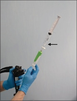
For the wet suction technique, the needle is pre-flushed with saline, and a vacuum syringe is then attached to the needle handle. The saline is aspirated into the syringe (black arrow)
As far as the slow pull technique is concerned, a 2020 meta-analysis showed that fine-needle biopsy (FNB) with a slow pull technique had a similar overall sample compared to wet suction (Fig. 2). However, the slow pull technique had a better CPT [8]. Two more studies were recently published comparing wet suction with slow pull. Specifically, according to the multicenter study by Sharma et al, the wet suction technique compared to the slow pull technique applied to EUS-LB yielded larger histological samples (total fragment length, long fragment length, greater number of fragments, and number of portal tracts) [9]. In addition, the study by Crinò et al showed that, in the subgroup of extra pancreatic lesions, tissue core percentage and tissue integrity score were slightly higher using wet suction. However, diagnostic accuracy was similar in the 2 groups [10].
Figure 2.
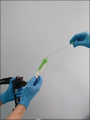
In the slow-pull technique the stylet is slowly withdrawn, while the needle tip is moved inside the target lesion
Needle pass and actuation
Needle pass is defined as the number of times a needle enters the liver parenchyma by puncturing the liver capsule, while actuation refers to the number of back-and-forth movements made in a defined needle pass [11]. Two needle passes are more likely to provide adequate tissue samples, according to the American Association for the Study of Liver Diseases (AASLD) guidelines. There are few studies comparing the number of passes and actuations. However, a recent paper reported that EUS-LB using the 1-pass:3-actuations method produced longer liver cores with more CPTs than a 1:1 technique with an equivalent safety profile [12].
Needle type and size
Apart from the acquisition technique, the type and size of the needle utilized seem to play a decisive role in the quality of the sample. Regarding the needle type, FNB needles commonly used for EUS-LB are the QuickCore needle, ProCore needle, SharkCore needle and Acquire needle (Fig. 3). QuickCore needles are used less frequently, because of difficulty in obtaining a specimen due to both a higher failure rate and a lower yield of adequate specimens [13]. Better performance could be found with the SharkCore and Acquire needles compared to the ProCore needle [14-16]. The studies of Hashimoto et al [15] and Shah et al [14] reported that the Acquire needle could yield longer specimens, more CPTs, and more intact cores compared with the SharkCore needle.
Figure 3.
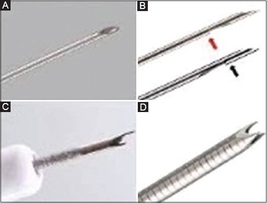
EUS-guided fine-needle tip design: (A) EUS-guided fine-needle aspiration, (B) the 2 versions of “side-fenestrated” needles (ProCore™) (C) the “fork-tip” needle (SharkCore™) (D) the “crown-tip” needle (Acquire™)
EUS, endoscopic ultrasound
A recent meta-analysis was published in January 2022 and aimed primarily to evaluate the value of EUS-LB for parenchymal and focal liver lesions, and secondarily to evaluate factors associated with the performance of EUS-LB. such as the size and type of needle, as well as the presence of adverse effects from the procedure. Thirty-three studies were included in this meta-analysis, including 21 on parenchymal liver diseases, 11 on focal liver lesions and 1 on both diseases. The total number of patients was 2098. The pooled rate of the diagnostic yield of Acquired Franseen-tip needles was significantly higher than that of Sharkcore Fork-tip needles (99% vs. 88%, P=0.047) [13].
The most commonly used needle size in clinical practice is a 19 G. The 22-G needle is an alternative, although 22-G needles seem to give samples that are more fragmented, resulting in poorer sample quality (Fig. 4). The study by Mok et al disclosed that the yield of adequate samples following 22-G needles was 68% while 19-G needles yielded a higher sample adequacy of up to 86% [14]. The superiority of the 19-G needle over the 22-G was also highlighted in studies by Patel et al [17] and Shah et al [18].
Figure 4.
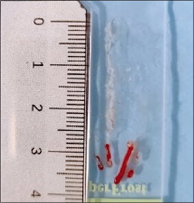
A long white specimen collected during endoscopic ultrasound-guided fine-needle biopsy of the liver
FNA vs. FNB
A meta-analysis by Kahn et al [19] reported no significant difference in the diagnostic yield between FNA and FNB when FNA is accompanied by rapid onsite evaluation (ROSE). However, in the absence of ROSE, FNB is associated with better diagnostic accuracy. Moreover, FNB requires fewer passes to establish the diagnosis. Similar results in terms of sample adequacy, diagnostic accuracy or core sample acquisition were shown in the meta-analysis of Bang et al [20], comparing ProCore needles and standard FNA needles. However, the ProCore needle tends to require fewer passes in order to obtain a diagnosis. A recently published study by Gheorghiu et al reported that EUS-FNB specimens have better histological adequacy with focal liver lesions (greater cellularity and longer tissue aggregates) than EUS-FNA specimens. The diagnostic accuracy of EUS-FNB was 100%, while that of EUS-FNA was 86.7% (P=0.039). This study compared 22-G EUS-FNB needles and 22-G EUS-FNA needles without macroscopic field evaluation [21]. Similarly, a comparison between FNA and FNB needles for LB was also conducted using 19-G-size needles. EUS-LB using a novel 19-G FNB needle yielded longer mean sample lengths, longer total sample length, less post-processing specimen fragmentation, and more CPTs compared to the usual 19-G FNA needle [22].
In spite of the growing body of evidence supporting the use of EUS-LB, a single-center randomized controlled trial and other studies, including a large multicenter retrospective propensity-matched series and 2 meta-analyses, showed a more favorable efficacy profile for percutaneous biopsy as compared to EUS-LB [23-26].
Bile duct
General indications
Biliary strictures can be caused by a variety of etiologies, with malignant strictures being the most common (e.g., bile duct carcinoma, pancreatic cancer, lymphoma and metastasis). Approximately 20-30% of biliary strictures are of benign etiology, such as IgG4 disease, primary sclerosing cholangitis, infection, post-traumatic or postoperative etiologies, and vasculitis. In most cases, the diagnosis of these strictures requires transabdominal imaging, endoscopic retrograde cholangiopancreatography (ERCP) with transpapillary tissue sampling, cholangioscopy-guided biopsy or EUS-guided tissue acquisition.
EUS-guided tissue acquisition vs. cholangioscopy-guided biopsies
According to European Society of Gastrointestinal Endoscopy (ESGE) guidelines, oral cholangioscopy (POC) and/or EUS-guided tissue acquisition are recommended in unspecified biliary strictures [27]. Furthermore, in accord with Asia-Pacific consensus recommendations on endoscopic tissue acquisition for biliary strictures, cholangioscopy-guided biopsy and EUS-guided tissue acquisition can be considered after prior negative conventional tissue sampling. The sensitivity and specificity of POC-guided biopsy are 60% (38-88%) and 98% (83-100%), and of EUS-guided tissue biopsy are 80% (46-100%) and 97% (92-100%), in diagnosing malignant biliary strictures [28]. Similarly, the latest ESGE recommendations cited evidence from studies showing that EUS-guided sampling in indeterminate strictures has a high sensitivity (75-94%) and diagnostic accuracy (79-94%), which are higher than the sensitivity (49-60%) and diagnostic accuracy (60-61%) of ERCP-guided brush cytology [29]. In the case of cholangioscopy-guided biopsies for indeterminate strictures, the sensitivity ranges from 72-94% and the specificity ranges from 87-99% [30-32]. The decision to select a specific method for tissue acquisition in undefined biliary strictures is difficult. It is intertwined with the judgment of the endoscopist, and depends mainly on the location of the lesion, the patient’s clinical picture and the endoscopist’s experience of each technique. POC is preferred for proximal strictures, and EUS-guided sampling for distal strictures [23]. In a large, single-center retrospective trial, the diagnostic sensitivity of EUS-FNA in malignant biliary strictures appeared higher (81%) in distal lesions versus proximal lesions (59%) [33]. A recent meta-analysis reported that, after excluding extrinsic compression by a pancreatic mass, EUS-FNA had 83% diagnostic sensitivity and 100% specificity for distal biliary strictures, and 76% diagnostic sensitivity and 100% specificity for proximal biliary strictures [30].
Seeding
There are data that lead to concern about possible seeding of the needle tract during EUS-guided sampling of potentially resectable proximal or hilar biliary malignancies [34]. Seeding of cancer cells along the needle path has been documented in several cases during percutaneous needle biopsy sampling. However, needle-track seeding appears to be a rare adverse event after EUS-FNA. This could be explained by the small size of EUS-FNA needles and the shorter needle track compared with the percutaneous approach [27]. Chafic et al reported a retrospective, single-center study of 150 patients with cholangiocarcinoma (61 underwent EUS-FNA) and found that preoperative EUS-guided sampling did not affect overall or progression-free patient survival [35]. Needle-track seeding is less concerning in distal biliary malignancy, in which the needle track of transduodenal EUS-FNA is fully resected during pancreaticoduodenectomy [36]. Apparently, the potential risk of tumor seeding has not been adequately studied. Therefore, EUS-FNA should currently be considered as a contraindication for patients with primary biliary malignancies who may be considered for liver transplantation [24,33,37].
Upper gastrointestinal (GI) tract
Upper GI subepithelial tumors (SETs)
GI SETs are sometimes detected casually during routine endoscopic examination. SETs are mostly observed in the stomach, followed in incidence by the esophagus, duodenum and colon. Anatomic position seems to be clinically crucial. Leiomyomas, for example, are commonly detected in the lower esophagus, while GI stromal tumors (GISTs) are typically found in the stomach (Fig. 5) [38]. Even though most SETs are small, grow slowly, and are clinically irrelevant, a subset of these tumors, usually GISTs, are potentially malicious [39,40]. Thus, a proper biopsy specimen with EUS is crucial to establishing an accurate diagnosis. For that purpose, EUS-guided cytology or biopsy methods—namely FNA, Tru-Cut biopsy and FNB—provide favorable diagnostic results. Cytology with immunocytochemical staining may also increase the diagnostic yield for GI SETs, whereas the role of ROSE when using FNB, as in the case of pancreatic masses [40], is uncertain.
Figure 5.
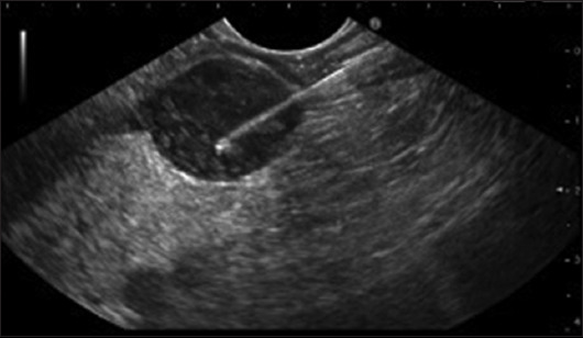
Endoscopic ultrasound-guided fine-needle biopsy of a submucosal gastric lesion originating from the fourth layer, confirmed to be a gastrointestinal stromal tumor
EUS-FNA
Tissue acquisition by EUS-FNA represents an option for the study of SETs with an accuracy of 60-80% [41]. A prospective study applying a 22-G power-shot needle (NA-11J-KB; Olympus Medical Systems, Tokyo, Japan) showed a puncture success rate of 100%, an adequate specimen acquisition rate of 82%, and a diagnostic rate of 82% [42]. Interestingly, a forward-viewing linear EUS endoscope was shown to provide good image quality and shorter examination times in the study of SETs, in comparison to oblique-viewing linear EUS endoscopes (histologic assessment rate 93.4%, sensitivity 92.8%, specificity 100%) [43,44].
In contrast, a study from Europe using 19-G EUS-FNA needles for gastric SETs revealed a feasibility of 46% and a diagnostic accuracy of 52% [45]. In another study, Mekky et al showed that an average of 2.5 EUS-FNA passes were needed to obtain an adequate sample in 83% of cases, with a diagnostic accuracy of 43.3% [46]. Sepe et al conducted an EUS-FNA study for diagnosing GISTs, which revealed a sensitivity of 78.4% [47]. SETs diagnosis using 19-G EUS-FNA needles was proved to obtain better results than 22-G or 25-G needles. Similar results for tissue sampling and diagnostic rates for SETs have been achieved using 22-G and 25-G EUS-FNA needles (sampling rate 100% vs. 100%, sensitivity 55% vs. 64%, positive predictive value 100% vs. 100%, negative predictive value 0% vs. 0%) [48]. Additionally, 25-G needles were proven to be superior to 22-G needles for the diagnosis of small mobile lesions. In a study using 19-G EUS-FNA nitinol needles, adequate cytologic assessment was achieved in 100% of cases with 100% technical success. A histologic accuracy of 95% using 19-G EUS-FNA nitinol needles was comparable to the 90% achieved with EUS-FNB using 19-G Pro-Core needles (Cook Endoscopy, Wilson-Salem, NC, USA) [49]. Diagnostic accuracy in GI SETs seems to increases proportionally with needle passes of EUS-FNA, reaching a plateau after 2.5-4 passes [46,50]. Regarding the safety of EUS-FNA for gastric SETs, a multicenter study revealed an extremely low risk of bleeding (0.46%) and perforation (0%) [51].
EUS-FNB
EUS-FNB was initially introduced using reverse bevel cheese slicer technology [52]. An Asian EUS-guided GI SET tissue acquisition study revealed that EUS-FNB required statistically significantly fewer needle passes than EUS-FNA to yield optimal macroscopic (92% vs. 30% with FNA), histological (75% vs. 20% with FNA) samples, and with a higher diagnostic rate (75% vs. 20% with FNA) [53]. In 2016, a controversial meta-analysis showed that EUS-FNB had moderate diagnostic capability (59.9%) for GI SET [54]. However, a consecutive meta-analysis demonstrated a lower sampling ability of FNA (80.6%) in comparison to FNB (94.9%) that increased when ROSE was used [55]. In the studies of the recent meta-analysis the needles used were predominantly 22 G and the evaluated FNB needle designs included reverse-bevel ProCore (Cook Medical), Acquire (Boston Scientific), and SharkCore (Medtronic). The heterogeneity of the selected studies, differences in the needles that were used, and the accumulation of evidence are probably responsible for the discrepancy of data between these 2 meta-analyses. Two large retrospective multicenter studies demonstrated the superiority of EUS-FNB [56]. Needle size (22 G vs. 19 G ProCore) seems not to be related to FNB sensitivity. However, FNB sensitivity using the Acquire 22 G seems to be higher when visible white tissue cores of >4 mm in length were identified during specimen assessment [57].
EUS-FNB represents a valuable alternative to other sampling methods of SETs, such as bite-on-bite biopsy, as shown in a recent Italian multicenter propensity-matched series, where EUS-FNB outperformed bite-on-bite biopsy in terms of both diagnostic yield and safety profile [58]. Therefore, this technique represents one of the most important sampling methods through EUS [59] currently available.
Mediastinal and abdominal lymph nodes (LNs)
The diagnostic approach to mediastinal and abdominal masses and LNs was drastically changed with the introduction of EUS-FNA and FNB, because it permitted effective and safe tissue acquisition [60,61]. European recommendations for EUS-based tissue acquisition suggest histological assessment of mediastinal and abdominal LNs if the pathological diagnosis could influence the patient’s therapeutic management (Fig. 6) [62].
Figure 6.
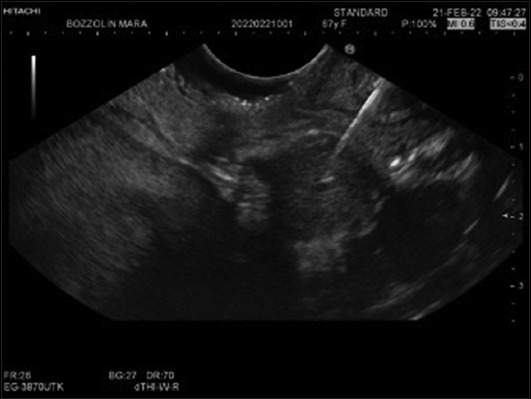
Endoscopic ultrasound-guided fine-needle biopsy of an abdominal lymph node located close to the hepatic hilum. Histology results revealed metastatic neuroendocrine tumor
EUS-guided tissue acquisition
The “fanning technique”, one of the finest sampling techniques, allows the sampling of an extensive part of the lymph node with considerably fewer needle passes [63]. In the “suction technique”, the application of negative pressure, using a simple 5 mL or 10 mL syringe, during the process of suction is able to further facilitate tissue acquisition guided by EUS. In the “slow-pull technique”, controlled pulling of the stylet permits the application of a smooth negative pressure, facilitating the acquisition of cytological specimens [64].
On the other hand, the possibility of ROSE, if it is available, may reduce the number of needle passes needed for the diagnosis. The higher diagnostic sensitivity of EUS-FNA was obtained using ≥7 needle passes. EUS-FNA tissue acquisition is safe, with a very low rate of perforations (0.03-0.15%), related more with the echoendoscope and not the needle itself [65]. Malignant LNs are poorly fibrotic with high cellularity; therefore, the needle type, number of passes, type and technique of suction are very important factors that must be considered prior to performing the procedure.
In a Japanese study, Okasha et al revealed a sensitivity of 92% and a specificity of 100% for EUS-FNA in diagnosing malignant LNs [66]. In a recent meta-analysis that included 26 studies and a total of 2833 LNs, EUS-FNA showed 87% sensitivity and 100% specificity. In this study, a subanalysis demonstrated that sensitivity for abdominal (87%) LNs was mildly superior in comparison with mediastinal LNs (85%), and highlighted the positive impact of ROSE (91% with vs. 85% without ROSE) [67]. Similar results were confirmed in a more recent meta-analysis that studied the results of EUS-FNA for diagnosing abdominal LNs and showed a high sensitivity (94%) and specificity (98%) [68]. A similar meta-analysis regarding mediastinal LNs revealed a pooled sensitivity ranging from 88-91.7%, while specificity was 96.4% [69]. On the other hand, a large clinical trial that included a small number of LNs and compared 20-G FNB needles to 25-G FNA needles revealed a trend towards superior accuracy in the FNB needle group [70].
Another retrospective study including 209 patients undergoing EUS-guided tissue acquisition of LNs with FNB and FNA reported slightly higher sensitivity (75% vs. 67%), accuracy (83% vs. 79%), and specificity (100% vs 94%) [71], respectively. Furthermore, these studies reaffirm the safety of EUS-based LNs tissue acquisition with a very low rate of adverse events (1.6%) [67]. Overall, there is a large amount of data that demonstrates the high specificity, sensitivity and safety of EUS-FNA and EUS-FNB tissue acquisition techniques for the characterization of mediastinal and abdominal LNs.
A recent meta-analysis of 9 studies (1276 patients) found no difference in terms of diagnostic accuracy between EUS-FNB and EUS-FNA (P=0.270) [72]. However, the accuracy of EUS-FNB was significantly higher when it was performed with newer end-cutting needles (P=0.009) and in abdominal LNs (P<0.001) [72]. Moreover, the number of needle passes was significantly lower in the EUS-FNB than in the EUS-FNA group (P=0.010) [72].
Lower GI tract
Only a few studies related to EUS-guided tissue acquisition by FNA or FNB have been published, because of the difficulties related to placement of the echoendoscope above the rectum. Most of the published studies have been confined to rectal or peri-rectal lesions [73]. In a case series of 9 patients, EUS-guided tissue acquisition using a ProCore needle demonstrated an overall diagnostic accuracy of 67%. In another study that included patients with lower GI SETs and non-SET, EUS-FNA/B samples had a diagnostic accuracy of 50% for SETs and 75% for non-SETs. The size of the lesions was the only factor related to diagnostic yield. Hara et al in their study revealed that the diagnostic accuracy of EUS-FNA was 90% in 10 patients with rectal and sigmoid lesions (rectal cancer, endometriosis, GIST) [74]. Finally, Sasaki et al found that the accuracy of EUS-FNA for the diagnosis of submucosal and extrinsic masses of the colon and rectum was 95.5% [75].
Other organs
In addition to the uses of EUS FNA discussed previously, there are reports in the literature of its use in other organs, such as the spleen and adrenal gland, as well as peritoneal, renal and mediastinal masses. Focal lesions in the spleen are not as frequent as those in other solid organs and are found incidentally in various radiologic examinations [76]. Because of their difficult definition, the diagnosis of such masses based only on their clinical and radiologic features is usually challenging [77]. Therefore, the use of EUS in tissue acquisition for the diagnosis of splenic lesions has increased during recent years. In a recent systematic review and meta-analysis, the authors reported an EUS-guided tissue acquisition diagnostic specificity of 77% and sensitivity of 85%, with no major complications [78]. In another meta-analysis, Lisotti et al demonstrated a diagnostic accuracy of 93% and overall accuracy of 88% for tissue acquisition using EUS-FNA in all the articles reviewed. The majority of the studies (80.6%) used 220G needles, while 19-G (11.3%) and 25-G (8.1%) were also used in a minority of the cases, with an overall mean of 2.62 passes of the needle (1.95-3.28) [79]. Other authors confirmed that EUS-FNA technique is more secure in comparison with core needle biopsy [75]. It is difficult to sample tissue from the mediastinum, because of its anatomical proximity to key structures, making safe access to the area a challenge. The standard test to evaluate mediastinal foci, such as lymph nodes, is mediastinoscopy. The technique has sensitivity and specificity of 87% and 100%, respectively [80]. Cost, intraoperative risk and postoperative risk are the main disadvantages of this method. The alternative diagnostic approach is EUS. Patients undergoing EUS-FNA for several mediastinal and lung lesions were included in Lee’s study. The accuracy, sensitivity, specificity, positive predictive value and negative predictive value of EUS-FNA were 75%, 100%, 100%, 67%,and 83%, respectively [81]. The results of an analysis by Srinivasan et al were equivalent. The sensitivity, specificity and accuracy for EUS-FNA of mediastinal LNs in patients with known or suspected lung cancer were 82.35%, 100% and 90%, respectively [82]. The negative predictive value was 80% and the positive predictive value was 100%. Similarly, a study by Shaqib et al showed a sensitivity, specificity, positive predictive value, negative predictive value and diagnostic accuracy of 97.5%, 100%, 100%, 70% and 97.6%, respectively, in the diagnosis of mediastinal lesions [80]. Complications reported in the above studies were minimal.
Sampling of adrenal masses can be performed percutaneously, guided by computed tomography (CT), or transluminally using EUS. With EUS, the left adrenal gland can be accessed transgastrically without affecting vital organs. As a result, side-effects are minimal. The percutaneous approach, on the other hand, has a 12% probability of causing adverse effects [80]. Furthermore, the percutaneous method has drawbacks, including radiation exposure and the requirement to administer contrast. Obtaining material from the right adrenal gland is more difficult, as it is done through the duodenum and several vascular structures are interposed [83]. According to Patel’s meta-analysis, EUS-FNA of the adrenal gland has a pooled sensitivity of 95% and a pooled specificity of 99% in the diagnosis of malignant lesions and is superior to other sampling methods. Furthermore, when compared to other methods, EUS is more efficient, with a pooled technical success rate for obtaining a diagnostic specimen from an adrenal lesion of approximately 94% [84]. In terms of the size of the sampling needle, the 25-G needles appear to be more flexible and are preferred for performing transduodenal FNA [85].
The rapid advances in cancer therapy along with more personalized therapies, even in advanced patients, require a correct histological definition of tumoral peritoneal spread. Diagnostic laparoscopy is considered the gold standard in the diagnosis of peritoneal lesions; nevertheless, it is an invasive surgical procedure. A simple and low-cost bedside procedure, such as abdominal paracentesis, may also be helpful in the diagnosis of malignancy-related ascites. Unfortunately, the sensitivity of ascitic fluid cytology for detecting malignancy is lower than 60% and paracentesis does not provide core tissue [86]. While CT-guided percutaneous biopsy of peritoneal lesions has also been reported, with a sensitivity of 89.5%, this procedure necessitates radiation exposure and is problematic for deeply located target lesions in the abdomen [87]. More recently, EUS-FNA of peritoneal lesions has been shown to yield valuable results [88]; however, it may not provide core tissues for immunohistochemical (IHC) staining, hence the need for EUS-FNB. A recent prospective series showed 63.6% sensitivity, 100% specificity and 66.7% accuracy of EUS-FNB of peritoneal lesions; adequate tissue for IHC stain was found in 25/30 passes (80%). Therefore, EUS-FNB represents a valuable tool in patients with peritoneal disease [89].
Concluding remarks
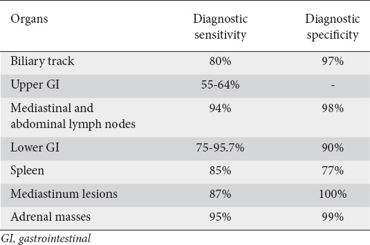
Over the years, the indications for the use of EUS beyond the pancreas have expanded. EUS can provide diagnostic assistance in organs and structures near the GI tract [90,91] (Table 1) and it can also be useful in interventional procedures [92-95]. Using high-resolution imaging of adjacent organs, even small lesions can be sampled in a safe and controlled manner, thus eliminating the need for more invasive methods such as surgery. Further research and investigation of tissue acquisition techniques will continue to improve the diagnostic reach, accuracy and safety of EUS-based methods. The future for EUS-guided tissue acquisition shines bright.
Table 1.
Main applications and clinical results of endoscopic ultrasound-guided sampling of extrapancreatic lesions
Biography
St. Savvas Oncology Hospital of Athens; Greece; University of Utah Health, Salt Lake City, UT, USA; Alexandra General Hospital, Athens, Greece; CUB Erasme Hospital, Université Libre de Bruxelles (ULB), Brussels, Belgium; Medical School, National and Kapodistrian University of Athens, ‘‘Attikon” University General Hospital, Athens, Greece; Humanitas Mater Domini, Castellanza (VA), Italy; University of Foggia, Foggia, Italy
Footnotes
Conflict of Interest: None
References
- 1.Tamanini G, Cominardi A, Brighi N, Fusaroli P, Lisotti A. Endoscopic ultrasound assessment and tissue acquisition of mediastinal and abdominal lymph nodes. World J Gastrointest Oncol. 2021;13:1475–1491. doi: 10.4251/wjgo.v13.i10.1475. [DOI] [PMC free article] [PubMed] [Google Scholar]
- 2.Facciorusso A, Del Prete V, Buccino RV, et al. Comparative efficacy of colonoscope distal attachment devices in increasing rates of adenoma detection:a network meta-analysis. Clin Gastroenterol Hepatol. 2018;16:1209–1219. doi: 10.1016/j.cgh.2017.11.007. [DOI] [PubMed] [Google Scholar]
- 3.Facciorusso A, Di Maso M, Serviddio G, et al. Factors associated with recurrence of advanced colorectal adenoma after endoscopic resection. Clin Gastroenterol Hepatol. 2016;14:1148–1154. doi: 10.1016/j.cgh.2016.03.017. [DOI] [PubMed] [Google Scholar]
- 4.Facciorusso A, Straus Takahashi M, Eyileten Postula C, Buccino VR, Muscatiello N. Efficacy of hemostatic powders in upper gastrointestinal bleeding:A systematic review and meta-analysis. Dig Liver Dis. 2019;51:1633–1640. doi: 10.1016/j.dld.2019.07.001. [DOI] [PubMed] [Google Scholar]
- 5.Neuberger J, Patel J, Caldwell H, et al. Guidelines on the use of liver biopsy in clinical practice from the British Society of Gastroenterology, the Royal College of Radiologists and the Royal College of Pathology. Gut. 2020;69:1382–1403. doi: 10.1136/gutjnl-2020-321299. [DOI] [PMC free article] [PubMed] [Google Scholar]
- 6.Mok SRS, Diehl DL, Johal AS, et al. A prospective pilot comparison of wet and dry heparinized suction for EUS-guided liver biopsy (with videos) Gastrointest Endosc. 2018;88:919–925. doi: 10.1016/j.gie.2018.07.036. [DOI] [PubMed] [Google Scholar]
- 7.Attam R, Arain MA, Bloechl SJ, et al. “Wet suction technique (WEST)”:a novel way to enhance the quality of EUS-FNA aspirate. Results of a prospective, single-blind, randomized, controlled trial using a 22-gauge needle for EUS-FNA of solid lesions. Gastrointest Endosc. 2015;81:1401–1407. doi: 10.1016/j.gie.2014.11.023. [DOI] [PubMed] [Google Scholar]
- 8.Baran B, Kale S, Patil P, et al. Endoscopic ultrasound-guided parenchymal liver biopsy:a systematic review and meta-analysis. Surg Endosc. 2021;35:5546–5557. doi: 10.1007/s00464-020-08053-x. [DOI] [PubMed] [Google Scholar]
- 9.Sharma R, Perisetti A, Gupta S, et al. Blocs-benign liver optimal core study- safety and efficacy of EUS-guided benign liver FNB-core biopsy using wet suction vs. slow pull technique:a randomized multicentric study. Gastrointest Endosc. 2022;95:AB544. [Google Scholar]
- 10.CrinòSF , Conti Bellocchi MC, Di Mitri R, et al. Wet-suction versus slow-pull technique for endoscopic ultrasound-guided fine-needle biopsy:a multicenter, randomized, crossover trial. Endoscopy. 2023;55:225–234. doi: 10.1055/a-1915-1812. [DOI] [PubMed] [Google Scholar]
- 11.Rangwani S, Ardeshna DR, Mumtaz K, Kelly SG, Han SY, Krishna SG. Update on endoscopic ultrasound-guided liver biopsy. World J Gastroenterol. 2022;28:3586–3594. doi: 10.3748/wjg.v28.i28.3586. [DOI] [PMC free article] [PubMed] [Google Scholar]
- 12.Ching-Companioni RA, Johal AS, Confer BD, Forster E, Khara HS, Diehl DL. Single-pass 1-needle actuation versus single-pass 3-needle actuation technique for EUS-guided liver biopsy sampling:a randomized prospective trial (with video) Gastrointest Endosc. 2021;94:551–558. doi: 10.1016/j.gie.2021.03.023. [DOI] [PubMed] [Google Scholar]
- 13.Zeng K, Jiang Z, Yang J, Chen K, Lu Q. Role of endoscopic ultrasound-guided liver biopsy:a meta-analysis. Scand J Gastroenterol. 2022;57:545–557. doi: 10.1080/00365521.2021.2025420. [DOI] [PubMed] [Google Scholar]
- 14.Shah ND, Sasatomi E, Baron TH. Endoscopic ultrasound-guided parenchymal liver biopsy:single center experience of a new dedicated core needle. Clin Gastroenterol Hepatol. 2017;15:784–786. doi: 10.1016/j.cgh.2017.01.011. [DOI] [PubMed] [Google Scholar]
- 15.Hashimoto R, Lee DP, Samarasena JB, et al. Comparison of two specialized histology needles for endoscopic ultrasound (EUS)-guided liver biopsy:a pilot study. Dig Dis Sci. 2021;66:1700–1706. doi: 10.1007/s10620-020-06391-3. [DOI] [PubMed] [Google Scholar]
- 16.Mok SRS, Diehl DL, Johal AS, et al. Endoscopic ultrasound-guided biopsy in chronic liver disease:a randomized comparison of 19-G FNA and 22-G FNB needles. Endosc Int Open. 2019;7:E62–E71. doi: 10.1055/a-0655-7462. [DOI] [PMC free article] [PubMed] [Google Scholar]
- 17.Patel HK, Saxena R, Rush N, et al. A comparative study of 22G versus 19G needles for EUS-guided biopsies for parenchymal liver disease:are thinner needles better? Dig Dis Sci. 2021;66:238–246. doi: 10.1007/s10620-020-06165-x. [DOI] [PubMed] [Google Scholar]
- 18.Shah RM, Schmidt J, John E, Rastegari S, Acharya P, Kedia P. Superior specimen and diagnostic accuracy with endoscopic ultrasound-guided liver biopsies using 19-gauge versus 22-gauge core needles. Clin Endosc. 2021;54:739–744. doi: 10.5946/ce.2020.212. [DOI] [PMC free article] [PubMed] [Google Scholar]
- 19.Khan MA, Grimm IS, Ali B, et al. A meta-analysis of endoscopic ultrasound-fine-needle aspiration compared to endoscopic ultrasound-fine-needle biopsy:diagnostic yield and the value of onsite cytopathological assessment. Endosc Int Open. 2017;5:E363–E375. doi: 10.1055/s-0043-101693. [DOI] [PMC free article] [PubMed] [Google Scholar]
- 20.Bang JY, Hawes R, Varadarajulu S. A meta-analysis comparing ProCore and standard fine-needle aspiration needles for endoscopic ultrasound-guided tissue acquisition. Endoscopy. 2016;48:339–349. doi: 10.1055/s-0034-1393354. [DOI] [PubMed] [Google Scholar]
- 21.Gheorghiu M, Seicean A, Bolboacă SD, et al. Endoscopic ultrasound-guided fine-needle biopsy versus fine-needle aspiration in the diagnosis of focal liver lesions:prospective head-to-head comparison. Diagnostics (Basel) 2022;12:2214. doi: 10.3390/diagnostics12092214. [DOI] [PMC free article] [PubMed] [Google Scholar]
- 22.Ching-Companioni RA, Diehl DL, Johal AS, Confer BD, Khara HS. 19?G aspiration needle versus 19?G core biopsy needle for endoscopic ultrasound-guided liver biopsy:a prospective randomized trial. Endoscopy. 2019;51:1059–1065. doi: 10.1055/a-0956-6922. [DOI] [PubMed] [Google Scholar]
- 23.Bang JY, Ward TJ, Guirguis S, et al. Radiology-guided percutaneous approach is superior to EUS for performing liver biopsies. Gut. 2021;70:2224–2226. doi: 10.1136/gutjnl-2021-324495. [DOI] [PubMed] [Google Scholar]
- 24.Facciorusso A, Ramai D, Conti Bellocchi MC, et al. Diagnostic yield of endoscopic ultrasound-guided liver biopsy in comparison to percutaneous liver biopsy:a two-center experience. Cancers (Basel) 2021;13:306. doi: 10.3390/cancers13123062. [DOI] [PMC free article] [PubMed] [Google Scholar]
- 25.Facciorusso A, CrinòSF , Ramai D, et al. Diagnostic yield of endoscopic ultrasound-guided liver biopsy in comparison to percutaneous liver biopsy:a systematic review and meta-analysis. Expert Rev Gastroenterol Hepatol. 2022;16:51–57. doi: 10.1080/17474124.2022.2020645. [DOI] [PubMed] [Google Scholar]
- 26.Chandan S, Deliwala S, Khan SR, et al. EUS-guided versus percutaneous liver biopsy:a comprehensive review and meta-analysis of outcomes. Endosc Ultrasound. 2022 Oct 5; doi: 10.4103/EUS-D-21-00268. [Online ahead of print] doi:10.4103/EUS-D-21-00268. [DOI] [PMC free article] [PubMed] [Google Scholar]
- 27.Pouw RE, Barret M, Biermann K, et al. Endoscopic tissue sampling - Part 1:Upper gastrointestinal and hepatopancreatobiliary tracts. European Society of Gastrointestinal Endoscopy (ESGE) Guideline. Endoscopy. 2021;53:1174–1188. doi: 10.1055/a-1611-5091. [DOI] [PubMed] [Google Scholar]
- 28.Sun B, Moon JH, Cai Q, et al. Asia-Pacific ERCP Club. Review article:Asia-Pacific consensus recommendations on endoscopic tissue acquisition for biliary strictures. Aliment Pharmacol Ther. 2018;48:138–151. doi: 10.1111/apt.14811. [DOI] [PubMed] [Google Scholar]
- 29.De Moura DTH, Moura EGH, Bernardo WM, et al. Endoscopic retrograde cholangiopancreatography versus endoscopic ultrasound for tissue diagnosis of malignant biliary stricture:Systematic review and meta-analysis. Endosc Ultrasound. 2018;7:10–19. doi: 10.4103/2303-9027.193597. [DOI] [PMC free article] [PubMed] [Google Scholar]
- 30.Sun X, Zhou Z, Tian J, et al. Is single-operator peroral cholangioscopy a useful tool for the diagnosis of indeterminate biliary lesion?A systematic review and meta-analysis. Gastrointest Endosc. 2015;82:79–87. doi: 10.1016/j.gie.2014.12.021. [DOI] [PubMed] [Google Scholar]
- 31.Kulpatcharapong S, Pittayanon R, J Kerr S, Rerknimitr R. Diagnostic performance of different cholangioscopes in patients with biliary strictures:a systematic review. Endoscopy. 2020;52:174–185. doi: 10.1055/a-1083-6105. [DOI] [PubMed] [Google Scholar]
- 32.de Oliveira PVAG, de Moura DTH, Ribeiro IB, et al. Efficacy of digital single-operator cholangioscopy in the visual interpretation of indeterminate biliary strictures:a systematic review and meta-analysis. Surg Endosc. 2020;34:3321–3329. doi: 10.1007/s00464-020-07583-8. [DOI] [PubMed] [Google Scholar]
- 33.Mohamadnejad M, DeWitt JM, Sherman S, et al. Role of EUS for preoperative evaluation of cholangiocarcinoma:a large single-center experience. Gastrointest Endosc. 2011;73:71–78. doi: 10.1016/j.gie.2010.08.050. [DOI] [PubMed] [Google Scholar]
- 34.Heimbach JK, Sanchez W, Rosen CB, Gores GJ. Trans-peritoneal fine needle aspiration biopsy of hilar cholangiocarcinoma is associated with disease dissemination. HPB (Oxford) 2011;13:356–360. doi: 10.1111/j.1477-2574.2011.00298.x. [DOI] [PMC free article] [PubMed] [Google Scholar]
- 35.El Chafic AH, Dewitt J, Leblanc JK, et al. Impact of preoperative endoscopic ultrasound-guided fine needle aspiration on postoperative recurrence and survival in cholangiocarcinoma patients. Endoscopy. 2013;45:883–889. doi: 10.1055/s-0033-1344760. [DOI] [PubMed] [Google Scholar]
- 36.Facciorusso A, CrinòSF , Gkolfakis P, et al. Needle tract seeding after endoscopic ultrasound tissue acquisition of pancreatic lesions:a systematic review and meta-analysis. Diagnostics (Basel) 2022;12:2113. doi: 10.3390/diagnostics12092113. [DOI] [PMC free article] [PubMed] [Google Scholar]
- 37.Baillie J. Distinguishing malignant from benign biliary strictures:can confocal laser endomicroscopy close the gap? Gastrointestinal Endosc. 2015;81:291–293. doi: 10.1016/j.gie.2014.11.037. [DOI] [PubMed] [Google Scholar]
- 38.Polkowski M, Jenssen C, Kaye P, et al. Technical aspects of endoscopic ultrasound (EUS)-guided sampling in gastroenterology:European Society of Gastrointestinal Endoscopy (ESGE) Technical Guideline - March 2017. Endoscopy. 2017;49:989–1006. doi: 10.1055/s-0043-119219. [DOI] [PubMed] [Google Scholar]
- 39.Goto O, Kaise M, Iwakiri K. Advancements in the diagnosis of gastric subepithelial tumors. Gut Liver. 2022;16:321–330. doi: 10.5009/gnl210242. [DOI] [PMC free article] [PubMed] [Google Scholar]
- 40.Facciorusso A, Gkolfakis P, Tziatzios G, et al. Comparison between EUS-guided fine-needle biopsy with or without rapid on-site evaluation for tissue sampling of solid pancreatic lesions:A systematic review and meta-analysis. Endosc Ultrasound. 2022;11:458–465. doi: 10.4103/EUS-D-22-00026. [DOI] [PMC free article] [PubMed] [Google Scholar]
- 41.Moon JS. Endoscopic ultrasound-guided fine needle aspiration in submucosal lesion. Clin Endosc. 2012;45:117–123. doi: 10.5946/ce.2012.45.2.117. [DOI] [PMC free article] [PubMed] [Google Scholar]
- 42.Akahoshi K, Sumida Y, Matsui N, et al. Preoperative diagnosis of gastrointestinal stromal tumor by endoscopic ultrasound-guided fine needle aspiration. World J Gastroenterol. 2007;13:2077–2082. doi: 10.3748/wjg.v13.i14.2077. [DOI] [PMC free article] [PubMed] [Google Scholar]
- 43.Lee S, Seo DW, Choi JH, et al. Evaluation of the feasibility and efficacy of forward-viewing endoscopic ultrasound. Gut Liver. 2015;9:679–684. doi: 10.5009/gnl14394. [DOI] [PMC free article] [PubMed] [Google Scholar]
- 44.Larghi A, Fuccio L, Chiarello G, et al. Fine-needle tissue acquisition from subepithelial lesions using a forward-viewing linear echoendoscope. Endoscopy. 2014;46:39–45. doi: 10.1055/s-0033-1344895. [DOI] [PubMed] [Google Scholar]
- 45.Eckardt AJ, Adler A, Gomes EM, et al. Endosonographic large-bore biopsy of gastric subepithelial tumors:a prospective multicenter study. Eur J Gastroenterol Hepatol. 2012;24:1135–1144. doi: 10.1097/MEG.0b013e328356eae2. [DOI] [PubMed] [Google Scholar]
- 46.Mekky MA, Yamao K, Sawaki A, et al. Diagnostic utility of EUS-guided FNA in patients with gastric submucosal tumors. Gastrointest Endosc. 2010;71:913–919. doi: 10.1016/j.gie.2009.11.044. [DOI] [PubMed] [Google Scholar]
- 47.Sepe PS, Moparty B, Pitman MB, Saltzman JR, Brugge WR. EUS-guided FNA for the diagnosis of GI stromal cell tumors:sensitivity and cytologic yield. Gastrointest Endosc. 2009;70:254–261. doi: 10.1016/j.gie.2008.11.038. [DOI] [PubMed] [Google Scholar]
- 48.Kida M, Araki M, Miyazawa S, et al. Comparison of diagnostic accuracy of endoscopic ultrasound-guided fine-needle aspiration with 22- and 25-gauge needles in the same patients. J Interv Gastroenterol. 2011;1:102–107. doi: 10.4161/jig.1.3.18508. [DOI] [PMC free article] [PubMed] [Google Scholar]
- 49.Varadarajulu S, Bang JY, Hebert-Magee S. Assessment of the technical performance of the flexible 19-gauge EUS-FNA needle. Gastrointest Endosc. 2012;76:336–343. doi: 10.1016/j.gie.2012.04.455. [DOI] [PubMed] [Google Scholar]
- 50.Polkowski M, Larghi A, Weynand B, et al. European Society of Gastrointestinal Endoscopy (ESGE) Learning, techniques, and complications of endoscopic ultrasound (EUS)-guided sampling in gastroenterology:European Society of Gastrointestinal Endoscopy (ESGE) Technical Guideline. Endoscopy. 2012;44:190–206. doi: 10.1055/s-0031-1291543. [DOI] [PubMed] [Google Scholar]
- 51.Hamada T, Yasunaga H, Nakai Y, et al. Rarity of severe bleeding and perforation in endoscopic ultrasound-guided fine needle aspiration for submucosal tumors. Dig Dis Sci. 2013;58:2634–2638. doi: 10.1007/s10620-013-2717-7. [DOI] [PubMed] [Google Scholar]
- 52.Voss M, Hammel P, Molas G, et al. Value of endoscopic ultrasound guided fine needle aspiration biopsy in the diagnosis of solid pancreatic masses. Gut. 2000;46:244–249. doi: 10.1136/gut.46.2.244. [DOI] [PMC free article] [PubMed] [Google Scholar]
- 53.Kim GH, Cho YK, Kim EY, et al. Korean EUS Study Group. Comparison of 22-gauge aspiration needle with 22-gauge biopsy needle in endoscopic ultrasonography-guided subepithelial tumor sampling. Scand J Gastroenterol. 2014;49:347–354. doi: 10.3109/00365521.2013.867361. [DOI] [PubMed] [Google Scholar]
- 54.Zhang XC, Li QL, Yu YF, et al. Diagnostic efficacy of endoscopic ultrasound-guided needle sampling for upper gastrointestinal subepithelial lesions:a meta-analysis. Surg Endosc. 2016;30:2431–2441. doi: 10.1007/s00464-015-4494-1. [DOI] [PubMed] [Google Scholar]
- 55.Facciorusso A, Sunny SP, Del Prete V, Antonino M, Muscatiello N. Comparison between fine-needle biopsy and fine-needle aspiration for EUS-guided sampling of subepithelial lesions:a meta-analysis. Gastrointest Endosc. 2020;91:14–22. doi: 10.1016/j.gie.2019.07.018. [DOI] [PubMed] [Google Scholar]
- 56.Hébert-Magee S, Bae S, Varadarajulu S, et al. The presence of a cytopathologist increases the diagnostic accuracy of endoscopic ultrasound-guided fine needle aspiration cytology for pancreatic adenocarcinoma:a meta-analysis. Cytopathology. 2013;24:159–171. doi: 10.1111/cyt.12071. [DOI] [PMC free article] [PubMed] [Google Scholar]
- 57.Hewitt MJ, McPhail MJ, Possamai L, Dhar A, Vlavianos P, Monahan KJ. EUS-guided FNA for diagnosis of solid pancreatic neoplasms:a meta-analysis. Gastrointest Endosc. 2012;75:319–331. doi: 10.1016/j.gie.2011.08.049. [DOI] [PubMed] [Google Scholar]
- 58.Facciorusso A, Crinò SF, Ramai D, et al. Comparison between endoscopic ultrasound-guided fine-needle biopsy and bite-on-bite jumbo biopsy for sampling of subepithelial lesions. Dig Liver Dis. 2022;54:676–683. doi: 10.1016/j.dld.2022.01.134. [DOI] [PubMed] [Google Scholar]
- 59.Facciorusso A, Kovacevic B, Yang D, et al. Predictors of adverse events after endoscopic ultrasound-guided through-the-needle biopsy of pancreatic cysts:a recursive partitioning analysis. Endoscopy. 2022;54:1158–1168. doi: 10.1055/a-1831-5385. [DOI] [PubMed] [Google Scholar]
- 60.Facciorusso A, CrinòSF , Muscatiello N, et al. Endoscopic ultrasound fine-needle biopsy versus fine-needle aspiration for tissue sampling of abdominal lymph nodes:a propensity score matched multicenter comparative study. Cancers (Basel) 2021;13:4298. doi: 10.3390/cancers13174298. [DOI] [PMC free article] [PubMed] [Google Scholar]
- 61.Gkolfakis P, Crinò SF, Tziatzios G, et al. Comparative diagnostic performance of end-cutting fine-needle biopsy needles for EUS tissue sampling of solid pancreatic masses:a network meta-analysis. Gastrointest Endosc. 2022;95:1067–1077. doi: 10.1016/j.gie.2022.01.019. [DOI] [PubMed] [Google Scholar]
- 62.Fusaroli P, Jenssen C, Hocke M, et al. EFSUMB Guidelines on Interventional Ultrasound (INVUS), Part V - EUS-Guided Therapeutic Interventions (short version) Ultraschall Med. 2016;37:412–420. doi: 10.1055/s-0035-1553742. [DOI] [PubMed] [Google Scholar]
- 63.Bang JY, Magee SH, Ramesh J, Trevino JM, Varadarajulu S. Randomized trial comparing fanning with standard technique for endoscopic ultrasound-guided fine-needle aspiration of solid pancreatic mass lesions. Endoscopy. 2013;45:445–450. doi: 10.1055/s-0032-1326268. [DOI] [PMC free article] [PubMed] [Google Scholar]
- 64.Tarantino I, Di Mitri R, Fabbri C, et al. Is diagnostic accuracy of fine needle aspiration on solid pancreatic lesions aspiration-related? Dig Liver Dis. 2014;46:523–526. doi: 10.1016/j.dld.2014.02.023. [DOI] [PubMed] [Google Scholar]
- 65.Dumonceau JM, Koessler T, van Hooft JE, Fockens P. Endoscopic ultrasonography-guided fine needle aspiration:Relatively low sensitivity in the endosonographer population. World J Gastroenterol. 2012;18:2357–2363. doi: 10.3748/wjg.v18.i19.2357. [DOI] [PMC free article] [PubMed] [Google Scholar]
- 66.Okasha H, Elkholy S, Sayed M, Salman A, Elsherif Y, El-Gemeie E. Endoscopic ultrasound-guided fine-needle aspiration and cytology for differentiating benign from malignant lymph nodes. Arab J Gastroenterol. 2017;18:74–79. doi: 10.1016/j.ajg.2017.05.015. [DOI] [PubMed] [Google Scholar]
- 67.Chen L, Li Y, Gao X, et al. High diagnostic accuracy and safety of endoscopic ultrasound-guided fine-needle aspiration in malignant lymph nodes:a systematic review and meta-analysis. Dig Dis Sci. 2021;66:2763–2775. doi: 10.1007/s10620-020-06554-2. [DOI] [PubMed] [Google Scholar]
- 68.Li C, Shuai Y, Zhou X. Endoscopic ultrasound guided fine needle aspiration for the diagnosis of intra-abdominal lymphadenopathy:a systematic review and meta-analysis. Scand J Gastroenterol. 2020;55:114–122. doi: 10.1080/00365521.2019.1704052. [DOI] [PubMed] [Google Scholar]
- 69.Puli SR, Batapati Krishna Reddy J, Bechtold ML, et al. Endoscopic ultrasound:'it's accuracy in evaluating mediastinal lymphadenopathy? World J Gastroenterol. 2008;14:3028–3037. doi: 10.3748/wjg.14.3028. [DOI] [PMC free article] [PubMed] [Google Scholar]
- 70.van Riet PA, Larghi A, Attili F, et al. A multicenter randomized trial comparing a 25-gauge EUS fine-needle aspiration device with a 20-gauge EUS fine-needle biopsy device. Gastrointest Endosc. 2019;89:329–339. doi: 10.1016/j.gie.2018.10.026. [DOI] [PubMed] [Google Scholar]
- 71.de Moura DTH, McCarty TR, Jirapinyo P, et al. Endoscopic ultrasound fine-needle aspiration versus fine-needle biopsy for lymph node diagnosis:a large multicenter comparative analysis. Clin Endosc. 2020;53:600–610. doi: 10.5946/ce.2019.170. [DOI] [PMC free article] [PubMed] [Google Scholar]
- 72.Facciorusso A, CrinòSF , Gkolfakis P, et al. Endoscopic ultrasound fine-needle biopsy vs fine-needle aspiration for lymph nodes tissue acquisition:a systematic review and meta-analysis. Gastroenterol Rep (Oxf) 2022;10:goac062. doi: 10.1093/gastro/goac062. [DOI] [PMC free article] [PubMed] [Google Scholar]
- 73.Soh JS, Lee HS, Lee S, et al. The clinical usefulness of endoscopic ultrasound-guided fine needle aspiration and biopsy for rectal and perirectal lesions. Intest Res. 2015;13:135–144. doi: 10.5217/ir.2015.13.2.135. [DOI] [PMC free article] [PubMed] [Google Scholar]
- 74.Hara K, Yamao K, Ohashi K, et al. Endoscopic ultrasonography and endoscopic ultrasound-guided fine-needle aspiration biopsy for the diagnosis of lower digestive tract disease. Endoscopy. 2003;35:966–969. doi: 10.1055/s-2003-43473. [DOI] [PubMed] [Google Scholar]
- 75.Sasaki Y, Niwa Y, Hirooka Y, et al. The use of endoscopic ultrasound-guided fine-needle aspiration for investigation of submucosal and extrinsic masses of the colon and rectum. Endoscopy. 2005;37:154–160. doi: 10.1055/s-2004-826152. [DOI] [PubMed] [Google Scholar]
- 76.Sammon J, Twomey M, Crush L, Maher MM, O'Connor OJ. Image-guided percutaneous splenic biopsy and drainage. Semin Intervent Radiol. 2012;29:301–310. doi: 10.1055/s-0032-1330064. [DOI] [PMC free article] [PubMed] [Google Scholar]
- 77.Trenker C, Görg C, Freeman S, et al. WFUMB position paper-incidental findings, how to manage:spleen. Ultrasound Med Biol. 2021;47:2017–2032. doi: 10.1016/j.ultrasmedbio.2021.03.032. [DOI] [PubMed] [Google Scholar]
- 78.Pan X, Huang S, Gan P, et al. Endoscopic ultrasound-guided tissue acquisition for splenic lesions:A systematic review and meta-analysis of diagnostic test accuracy. PLoS One. 2022;17:e0276529. doi: 10.1371/journal.pone.0276529. [DOI] [PMC free article] [PubMed] [Google Scholar]
- 79.Lisotti A, CrinòSF , Mangiavillano B, et al. Diagnostic performance of endoscopic ultrasound-guided tissue acquisition of splenic lesions:systematic review with pooled analysis. Gastroenterol Rep (Oxf) 2022;10:goac022. doi: 10.1093/gastro/goac022. [DOI] [PMC free article] [PubMed] [Google Scholar]
- 80.Saqib M, Maruf M, Bashir S, et al. EUS-FNA, ancillary studies and their clinical utility in patients with mediastinal, pancreatic, and other abdominal lesions. Diagn Cytopathol. 2020;48:1058–1066. doi: 10.1002/dc.24523. [DOI] [PubMed] [Google Scholar]
- 81.Lee YT, Lai LH, Sung JJ, Ko FW, Hui DS. Endoscopic ultrasonography-guided fine-needle aspiration in the management of mediastinal diseases:local experience of a novel investigation. Hong Kong Med J. 2010;16:121–125. [PubMed] [Google Scholar]
- 82.Srinivasan R, Bhutani MS, Thosani N, et al. Clinical impact of EUS-FNA of mediastinal lymph nodes in patients with known or suspected lung cancer or mediastinal lymph nodes of unknown etiology. J Gastrointestin Liver Dis. 2012;21:145–152. [PubMed] [Google Scholar]
- 83.Novotny AG, Reynolds JP, Shah AA, et al. Fine-needle aspiration of adrenal lesions:A 20-year single institution experience with comparison of percutaneous and endoscopic ultrasound guided approaches. Diagn Cytopathol. 2019;47:986–992. doi: 10.1002/dc.24261. [DOI] [PubMed] [Google Scholar]
- 84.Patel S, Jinjuvadia R, Devara A, et al. Performance characteristics of EUS-FNA biopsy for adrenal lesions:A meta-analysis. Endosc Ultrasound. 2019;8:180–187. doi: 10.4103/eus.eus_42_18. [DOI] [PMC free article] [PubMed] [Google Scholar]
- 85.Patil R, Ona MA, Papafragkakis C, Duddempudi S, Anand S, Jamil LH. Endoscopic ultrasound-guided fine-needle aspiration in the diagnosis of adrenal lesions. Ann Gastroenterol. 2016;29:307–311. doi: 10.20524/aog.2016.0047. [DOI] [PMC free article] [PubMed] [Google Scholar]
- 86.Allen VA, Takashima Y, Nayak S, Manahan KJ, Geisler JP. Assessment of false-negative ascites cytology in epithelial ovarian carcinoma:a study of 313 patients. Am J Clin Oncol. 2017;40:175–177. doi: 10.1097/COC.0000000000000119. [DOI] [PubMed] [Google Scholar]
- 87.Pombo F, Rodriguez E, Martin R, Lago M. CT-guided core-needle biopsy in omental pathology. Acta Radiol. 1997;38:978–981. doi: 10.1080/02841859709172113. [DOI] [PubMed] [Google Scholar]
- 88.DeWitt J, LeBlanc J, McHenry L, McGreevy K, Sherman S. Endoscopic ultrasound-guided fine-needle aspiration of ascites. Clin Gastroenterol Hepatol. 2007;5:609–615. doi: 10.1016/j.cgh.2006.11.021. [DOI] [PubMed] [Google Scholar]
- 89.Kongkam P, Orprayoon T, Yooprasert S, et al. Endoscopic ultrasound guided fine needle biopsy (EUS-FNB) from peritoneal lesions:a prospective cohort pilot study. BMC Gastroenterol. 2021;21:400. doi: 10.1186/s12876-021-01953-9. [DOI] [PMC free article] [PubMed] [Google Scholar]
- 90.CrinòSF , Bernardoni L, Manfrin E, Parisi A, Gabbrielli A. Endoscopic ultrasound features of pancreatic schwannoma. Endosc Ultrasound. 2016;5:396–398. doi: 10.4103/2303-9027.195873. [DOI] [PMC free article] [PubMed] [Google Scholar]
- 91.CrinóSF , Brandolese A, Vieceli F, et al. Endoscopic ultrasound features associated with malignancy and aggressiveness of nonhypovascular solid pancreatic lesions:results from a prospective observational study. Ultraschall Med. 2021;42:167–177. doi: 10.1055/a-1014-2766. [DOI] [PubMed] [Google Scholar]
- 92.Facciorusso A, Di Maso M, Serviddio G, Larghi A, Costamagna G, Muscatiello N. Echoendoscopic ethanol ablation of tumor combined with celiac plexus neurolysis in patients with pancreatic adenocarcinoma. J Gastroenterol Hepatol. 2017;32:439–445. doi: 10.1111/jgh.13478. [DOI] [PubMed] [Google Scholar]
- 93.Facciorusso A, Amato A, Crinò SF, et al. i-EUS Group. Definition of a hospital volume threshold to optimize outcomes after drainage of pancreatic fluid collections with lumen-apposing metal stents:a nationwide cohort study. Gastrointest Endosc. 2022;95:1158–1172. doi: 10.1016/j.gie.2021.12.006. [DOI] [PubMed] [Google Scholar]
- 94.Amato A, Tarantino I, Facciorusso A, et al. i-EUS Group. Real-life multicentre study of lumen-apposing metal stent for EUS-guided drainage of pancreatic fluid collections. Gut. 2022;71:1050–1052. doi: 10.1136/gutjnl-2022-326880. [DOI] [PubMed] [Google Scholar]
- 95.Facciorusso A, Crinò SF, Ramai D, et al. Comparative diagnostic performance of different techniques for endoscopic ultrasound-guided fine-needle biopsy of solid pancreatic masses:a network meta-analysis. Gastrointest Endosc. 2023;97:839–848.e5. doi: 10.1016/j.gie.2023.01.024. [DOI] [PubMed] [Google Scholar]


