End-organ dysfunction results in substantial morbidity and mortality in sickle cell disease (SCD).1-3 Chronic kidney disease (CKD), defined as kidney damage or decreased kidney function for ≥3 months, is common in SCD.4 While individuals with SCD have shortened life expectancy, female patients appear to live longer than male patients, although some more recent cohort studies show no differences in survival according to sex.1,5,6 This difference in mortality may be driven by less end-organ damage in females. In mouse models of SCD, males present early development of elevated glomerular filtration rate (GFR), with a subsequent progressive decline in renal function over 20 weeks, findings which are not observed in females.7 Estimated GFR (eGFR) decline is also reportedly faster in male than in female SCD patients.8,9 In this study, we evaluated sex differences in kidney complications and the association of CKD with mortality in SCD. We hypothesized that kidney disease is more prevalent in male patients and is associated with a higher risk of mortality.
We analyzed a previously described pooled cohort from four centers.10 Adult patients with severe SCD genotypes (HbSS, HbSβ0) were evaluated during routine visits to the clinic at 'steady state'. Baseline was defined by first available serum creatinine during the observation period. Only patients with ≥2 creatinine values were evaluated for eGFR decline or CKD progression. Patients with kidney transplant or dialysis requirement were not evaluated for proteinuria or eGFR decline but were included in analyses of baseline CKD and association of CKD with mortality. Each center obtained approval for the study from their Institutional Review Board.
We calculated eGFR using the creatinine-based Chronic Kidney Disease Epidemiology Collaboration (CKD-EPI-2009) equation without adjustment for black race11 and the recent creatinine-based CKD-EPI-2021 equation, which does not include race.12 As CKD-EPI-2021 has not been adequately assessed in SCD, CKD-EPI-2009 was used for primary analyses. We defined CKD as eGFR <90 mL/min/1.73 m2 or proteinuria (≥1+ on urinalysis or urine albumin-creatinine ratio of >300 mg/g) modified from KDIGO (Kidney Disease: Improving Global Outcomes) CKD guidelines.13 Patients with eGFR ≥90 mL/min/1.73 m2 and missing proteinuria data were classified as not having CKD. Hyperfiltration was defined as eGFR >130 mL/min/1.73 m2 for women and >140 mL/min/1.73 m2 for men.14 Progression of CKD was defined as eGFR decline to <90 mL/min/1.73 m2 and ≥25% decline from baseline,15 and rapid kidney function decline was defined as eGFR loss >3.0 mL/min/1.73 m2 annually.16
Continuous variables were summarized by medians and interquartile ranges (IQR), and categorical variables by counts and percentages. A linear mixed effects model with random intercept and random slope for time was used to assess eGFR change over time, adjusted for baseline eGFR, baseline age, main cohort effect, and sex (in the non-stratified analyses). Individual eGFR decline was evaluated from the estimated slope in linear models. Logistic regression modeling, adjusted for baseline eGFR, baseline age and main cohort effect, was used to evaluate the association of sex with rapid eGFR decline. Kaplan-Meier estimates of survival function for age at death were obtained for patients with rapid versus non-rapid eGFR decline and for female versus male patients. Median age at death was obtained from Kaplan-Meier estimates. A Cox regression model evaluated the association of sex, baseline CKD and rapid eGFR decline with age at death. For analyses of age at death, we used age as the time scale and accounted for left truncation using age at baseline (first eGFR measurement) as the left truncation time. In comparisons of sexes, we employed two-sample t test for continuous variables and two-sample proportion test for categorical variables. The interaction between sex and the variable of interest in the mortality analysis was tested to assess if the associations differed according to sex. The interaction of sex and time in the linear mixed effects model was tested to evaluate if eGFR change over time differed according to sex. Analyses were conducted using SAS OnDemand for Academics© 2014 (SAS Institute Inc., Cary, NC, USA).
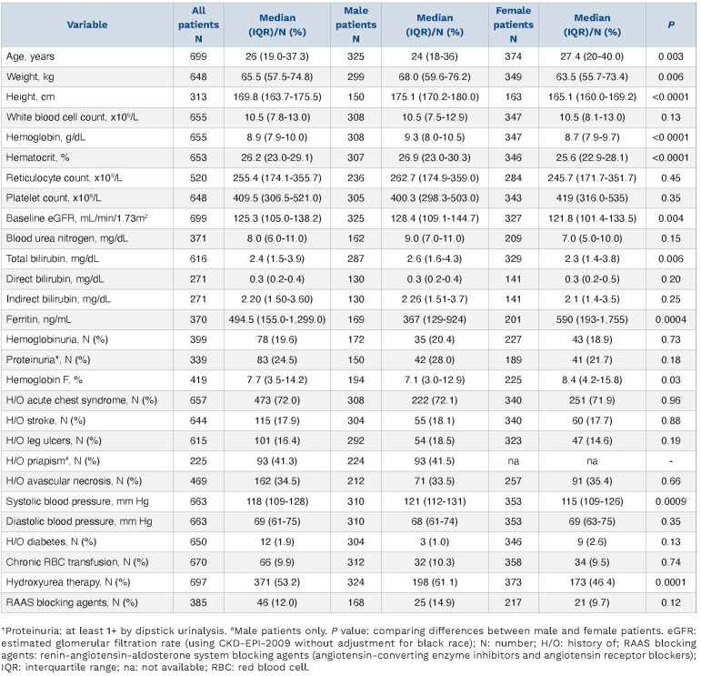
The pooled analysis included 699 individuals (females: 374 [53.5%]) with HbSS and HbSb0 and at least one eGFR value. Baseline laboratory and clinical data, stratified according to sex, are shown in Table 1. Urine microalbumincreatinine ratios were not available in the majority of patients. Proteinuria was present in 83 of 339 (24.5%) patients with available data (1+ proteinuria in 46 patients; 2+ proteinuria in 19 patients; 3+ proteinuria in 12 patients; 4+ proteinuria in 1 patient; and 5 patients with available albumin-creatinine ratio, and levels >300 mg/g were classified as having proteinuria): 21.7% of female versus 28% of male patients. Using CKD-EPI-2009, baseline hyperfiltration was present in 232 of 699 (33.2%) patients, and 36.4% of female versus 29.5% of male patients. At baseline, 173 of 699 (24.7%) patients had CKD (see Online Supplementary Appendix for KDIGO staging), including 3 on dialysis and 2 with kidney transplants. Ninety-eight of 374 (26.2%) female and 75 of 325 (23.1%) male patients had baseline CKD. Twenty-one of 83 patients with proteinuria (25.3%) had baseline eGFR <60 mL/min/1.73 m2, while only 5 of 256 patients without proteinuria (1.95%) had baseline eGFR <60 mL/min/1.73 m2.
Two or more eGFR values were available in 606 patients (excluding kidney transplant or dialysis patients). The median observation period in these patients was 5.20 years (IQR: 1.56, 7.53), with 3128.6 patient-years of observation and a median of 4 (IQR: 2, 10) eGFR values. Progression of CKD occurred in 144 of 606 (23.8%) patients: 83 of 327 (25.4%) female versus 61 of 279 (21.9%) male patients.
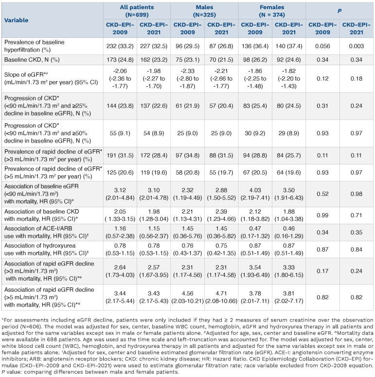
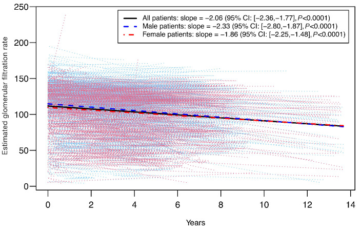
Change in eGFR over time for all patients, adjusted for baseline eGFR, baseline age, sex and cohort, was -2.06 mL/min/1.73 m2 per year (95% confidence interval [CI]: -2.36, -1.77; P<0.0001), with a decline of -1.86 mL/min/1.73 m2 per year (95% CI: -2.25, -1.48; P<0.0001) in females and -2.33 mL/min/1.73 m2 per year (95% CI: -2.80, -1.87; P<0.0001) in males (Table 2, Figure 1). After adjustment for baseline eGFR, age and cohort, no significant associations were observed between eGFR change over time and use of angiotensin converting enzyme inhibitors/angiotensin receptor blockers (ACE-I/ARB) either in all patients (P=0.15) or in male patients (P=0.29). However, change in eGFR over time was significantly associated with use of ACE-I/ARB in female patients (P=0.006; slopes = -1.49 and -2.70 for the group not using and the group using ACE-I/ARB, respectively), suggesting faster eGFR decline in females on ACE-I/ARB than females not taking these agents. No significant associations were observed between change of eGFR over time and hydroxyurea use in all patients (P=0.14), male patients (P=0.16), or female patients (P=0.61). Rapid eGFR decline was observed in 191 of 606 (31.5%) patients, 28.8% female patients and 34.8% male patients. Adjusted for baseline eGFR, baseline age and cohort, there was a trend for association of male sex with rapid eGFR decline (OR: 1.37, 95% CI: 0.96, 1.95; P=0.08). Results obtained using CKD-EPI-2021 and alternative definitions are shown in Table 2.
Table 1.
Baseline demographic, laboratory and clinical variables in pooled patient cohorts with sickle cell disease.
During the observation period, 114 of 698 patients (16.3%), 62 of 373 (16.6%) females and 52 of 325 (16%) males with available data died. The median age at death was 44.8- years overall: 44.8-years for females and 44.5-years for males. Sixty of 173 (34.7%) patients with baseline CKD died versus 54 of 525 (10.3%) patients without CKD. After adjustment for white blood cell (WBC) count, hemoglobin (Hb) and fetal hemoglobin (HbF), CKD was associated with age at death in all patients (hazard ratio [HR]: 2.05, 95% CI: 1.33, 3.15; P=0.0012), and when stratified according to sex, in female patients (HR: 2.12, 95% CI: 1.18, 3.82; P=0.012) and in male patients (HR: 2.21, 95% CI: 1.13, 4.31; P=0.02). Baseline eGFR <90 mL/min/1.73 m2 was significantly associated with age at death in all patients (HR: 3.12, 95% CI: 2.01, 4.84; P<0.0001), and when stratified according to sex, in female patients (HR: 4.03, 95% CI: 2.19, 7.41; P<0.0001) and in male patients (HR: 2.32, 95% CI: 1.19, 4.49; P=0.013). No significant association was observed between sex and age at death, following adjustment for cohort (HR: 1.22, 95% CI: 0.84, 1.78; P=0.29). Neither baseline CKD nor baseline eGFR <90 mL/min/1.73 m2 showed any interaction with sex in the association with mortality. Adjusted for baseline eGFR, neither hydroxyurea use nor use of ACE-I/ARB were significantly associated with risk of death in all patients, male patients or female patients (Table 2).
Table 2.
Biomarkers of kidney function and association of kidney disease with mortality in male and female patients.
Fifty-two of 190 patients (27.4%) with rapid eGFR decline died compared with 46 of 415 patients (11.1%) without rapid eGFR decline. Rapid eGFR decline was associated with age at death in all patients (HR: 2.75, 95% CI: 1.83, 4.14; P<0.0001) following adjustment for sex and cohort, and when stratified according to sex, in female patients (HR: 4.69, 95% CI: 2.63, 8.37; P<0.0001) following adjustment for cohort, but not in male patients (Online Supplementary Figure S1). After adjustment for baseline WBC, Hb, eGFR and use of hydroxyurea, rapid eGFR decline was associated with increased risk of death in all patients (HR: 2.64, 95% CI: 1.73, 4.03; P<0.0001), and when stratified by sex, in female patients (HR: 3.54, 95% CI: 1.93, 6.49; P<0.0001) and male patients (HR: 2.31, 95% CI: 1.17, 4.56; P=0.02). No significant association was observed between sex and risk of death in patients with rapid eGFR decline (HR: 0.77, 95% CI: 0.42, 1.42; P=0.40), but among those with non-rapid eGFR decline, male patients had a significantly higher risk of death than females (HR: 2.20, 95% CI: 1.21, 4.00; P=0.01).
As in our previous report,17 in this pooled analysis hyperfiltration was more prevalent in adult female patients, possibly reflecting earlier declines in eGFR from hyperfiltration to normal range among males. Furthermore, eGFR decline was faster and rapid kidney function decline more common in male patients. However, baseline CKD and progression of CKD were similar in male and female patients, possibly related to the absence of albuminuria assessments in the majority of patients in the pooled analysis, which did not allow a complete assessment of CKD. Although our analyses of interaction of ACE-I/ARB use and time demonstrated eGFR decline was faster in female patients on ACE-I/ARB than in female patients not on such treatment, the number of patients on these agents was only small.
Sex differences in SCD may occur due to lower hemolysis rates,18 higher HbF levels,19 and greater bioavailability of or responsiveness to nitric oxide in females.20 However, no meaningful differences in baseline Hb, HbF or bilirubin were observed between sexes in this pooled analysis, which may relate to the higher proportion of male patients on hydroxyurea compared to females.
Figure 1.
Slope of estimated glomerular filtration rate decline in pooled population with sickle cell disease and stratified according to sex. The change in estimated glomerular filtration rate (eGFR) over time for all patients was -2.06 mL/min/1.73 m2 per year (95% CI: -2.36, -1.77; P<0.0001), for female patients -1.86 mL/min/1.73 m2 per year (95% CI: -2.25, -1.48; P<0.0001), and for male patients -2.33 mL/min/1.73 m2 per year (95% CI: -2.80, -1.87; P<0.0001).
Baseline CKD was associated with age of death in both female and male patients, but no significant association was observed between sex and age at death. Similarly, rapid eGFR decline was significantly associated with increased risk of death in both female and male patients even in adjusted analyses. Although there was no significant association between sex and age at death in patients with rapid eGFR decline, among those with non-rapid eGFR decline, male patients had higher risks of death compared to females. End-organ damage may occur in multiple organ systems simultaneously, with higher mortality seen when multiple organ systems are involved.21 Our study is limited by missing proteinuria data, the lack of albuminuria assessments in the majority of patients, absence of prior longitudinal data from childhood, and exact data on hydroxyurea dosing and adherence. However, it is strengthened by the use of a real-world multicenter cohort with a relatively large sample size given the rarity of the disease under study.
Despite a more rapid eGFR decline and a higher prevalence of rapid kidney function decline in males, mortality associated with kidney disease was not higher in male than female patients with SCD. Further examination of sex-related effects of both kidney disease and multi-organ dysfunction on mortality in SCD is warranted.
Supplementary Material
Funding Statement
Funding: Funding for this study is provided by FDA grant FD006030 (to KIA, JC and VKD) and NIH grant HL159376 (to KIA, SLS and VKD). MJT and AEA-K received support from grants 2015131 and 2012126 from the Doris Duke Charitable Foundation, NIH grants HL68959 and HL079915, and DK110104 (to AEA-K).
References
- 1.Platt OS, Brambilla DJ, Rosse WF, et al. Mortality in sickle cell disease. Life expectancy and risk factors for early death. N Engl J Med. 1994;330(23):1639-1644. [DOI] [PubMed] [Google Scholar]
- 2.Ataga KI, Moore CG, Jones S, et al. Pulmonary hypertension in patients with sickle cell disease: a longitudinal study. Br J Haematol. 2006;134(1):109-115. [DOI] [PubMed] [Google Scholar]
- 3.Elmariah H, Garrett ME, De Castro LM, et al. Factors associated with survival in a contemporary adult sickle cell disease cohort. Am J Hematol. 2014;89(5):530-535. [DOI] [PMC free article] [PubMed] [Google Scholar]
- 4.Group KDIGOKCW. KDIGO 2012 Clinical Practice Guideline for the Evaluation and Management of Chronic Kidney Disease. Kidney Int Suppl. 2013;3(1):1-150. [DOI] [PubMed] [Google Scholar]
- 5.Maitra P, Caughey M, Robinson L, et al. Risk factors for mortality in adult patients with sickle cell disease: a meta-analysis of studies in North America and Europe. Haematologica. 2017;102(4):626-636. [DOI] [PMC free article] [PubMed] [Google Scholar]
- 6.Brewin JN, Nardo-Marino A, Stuart-Smith S, et al. The pleiotropic effects of alpha-thalassemia on HbSS and HbSC sickle cell disease: reduced erythrocyte cation co-transport activity, serum erythropoietin, and transfusion burden, do not translate into increased survival. Am J Hematol. 2022;97(10):1275-1285. [DOI] [PMC free article] [PubMed] [Google Scholar]
- 7.Kasztan M, Fox BM, Lebensburger JD, et al. Hyperfiltration predicts long-term renal outcomes in humanized sickle cell mice. Blood Adv. 2019;3(9):1460-1475. [DOI] [PMC free article] [PubMed] [Google Scholar]
- 8.Asnani M, Serjeant G, Royal-Thomas T, Reid M. Predictors of renal function progression in adults with homozygous sickle cell disease. Br J Haematol. 2016;173(3):461-468. [DOI] [PubMed] [Google Scholar]
- 9.Xu JZ, Garrett ME, Soldano KL, et al. Clinical and metabolomic risk factors associated with rapid renal function decline in sickle cell disease. Am J Hematol. 2018;93(12):1451-1460. [DOI] [PMC free article] [PubMed] [Google Scholar]
- 10.Ataga KI, Zhou Q, Derebail VK, et al. Rapid decline in estimated glomerular filtration rate in sickle cell anemia: results of a multicenter pooled analysis. Haematologica. 2021;106(6):1749-1753. [DOI] [PMC free article] [PubMed] [Google Scholar]
- 11.Levey AS, Stevens LA, Schmid CH, et al. A new equation to estimate glomerular filtration rate. Ann Int Med. 2009;150(9):604-612. [DOI] [PMC free article] [PubMed] [Google Scholar]
- 12.Inker LA, Eneanya ND, Coresh J, et al. New creatinine- and cystatin C-based equations to estimate GFR without race. N Engl J Med. 2021;385(19):1737-1749. [DOI] [PMC free article] [PubMed] [Google Scholar]
- 13.Derebail VK, Ciccone EJ, Zhou Q, Kilgore RR, Cai J, Ataga KI. Progressive decline in estimated GFR in patients with sickle cell disease: an observational cohort study. Am J Kidney Dis. 2019;74(1):47-55. [DOI] [PMC free article] [PubMed] [Google Scholar]
- 14.Haymann JP, Stankovic K, Levy P, et al. Glomerular hyperfiltration in adult sickle cell anemia: a frequent hemolysis associated feature. Clin J Am Soc Nephrol. 2010;5(5):756-761. [DOI] [PMC free article] [PubMed] [Google Scholar]
- 15.Levin A, Stevens PE. Summary of KDIGO 2012 CKD Guideline: behind the scenes, need for guidance, and a framework for moving forward. Kidney Int. 2014;85(1):49-61. [DOI] [PubMed] [Google Scholar]
- 16.Shlipak MG, Katz R, Kestenbaum B, et al. Rapid decline of kidney function increases cardiovascular risk in the elderly. J Am Soc Nephrol. 2009;20(12):2625-2630. [DOI] [PMC free article] [PubMed] [Google Scholar]
- 17.Derebail VK, Zhou Q, Ciccone EJ, Cai J, Ataga KI. Longitudinal study of glomerular hyperfiltration and normalization of estimated glomerular filtration in adults with sickle cell disease. Br J Haematol. 2021;195(1):123-132. [DOI] [PMC free article] [PubMed] [Google Scholar]
- 18.Raslan R, Shah BN, Zhang X, et al. Hemolysis and hemolysis-related complications in females vs. males with sickle cell disease. Am J Hematol. 2018;93(11):E376-E380. [DOI] [PMC free article] [PubMed] [Google Scholar]
- 19.Masese RV, Bulgin D, Knisely MR, et al. Sex-based differences in the manifestations and complications of sickle cell disease: report from the Sickle Cell Disease Implementation Consortium. PloS One. 2021;16(10):e0258638. [DOI] [PMC free article] [PubMed] [Google Scholar]
- 20.Gladwin MT, Schechter AN, Ognibene FP, et al. Divergent nitric oxide bioavailability in men and women with sickle cell disease. Circulation. 2003;107(2):271-278. [DOI] [PubMed] [Google Scholar]
- 21.Chaturvedi S, Ghafuri DL, Jordan N, Kassim A, Rodeghier M, DeBaun MR. Clustering of end-organ disease and earlier mortality in adults with sickle cell disease: a retrospective-prospective cohort study. Am J Hematol. 2018;93(9):1153-1160. [DOI] [PubMed] [Google Scholar]
Associated Data
This section collects any data citations, data availability statements, or supplementary materials included in this article.


