Abstract
Objective.
We aim to more accurately characterize the current distribution and rates of squamous cell carcinoma (SCC) cases across various oral cavity subsites in the United States.
Study Design.
Retrospective cohort.
Setting.
Database study evaluating cancer incidence in the United States from 2001 to 2017.
Methods.
We utilized the US Cancer Statistics Public Use Database, which includes deidentified cancer data reported to the Centers for Disease Control and Prevention’s National Program of Cancer Registries and the National Cancer Institute’s SEER (Surveillance, Epidemiology, and End Results), capturing 97% of newly diagnosed cancers. We restricted our analysis to SCC arising from oral cavity subsites from 2001 to 2017. We calculated trends in annual cancer incidence rates using SEER*Stat, as well as annual and average annual percentage change and joinpoints with the National Cancer Institute’s Joinpoint program.
Results.
Most oral cavity SCC cases arise from the oral tongue (41.7%), followed equally by lip and floor of mouth (each 16.5%), gingival (10.6%), buccal (6.7%), retromolar trigone (5.6%), and hard palate (2.3%) involvement. The overall incidence of oral tongue SCC continues to rise with an average annual percentage change of 1.8% (95% CI, 1.6%−2.1%; P <.001), with a 2.3% increase among women. This increase is seen among males and females of all age groups. Cancers involving the gum, buccal mucosa, and hard palate were also found to be increasing in rate, albeit to a lesser degree and with substantially lower incidence.
Conclusions.
The tongue is the most frequently involved subsite of oral cavity SCC and is increasing in incidence among males and females of all ages.
Keywords: oral cavity, tongue, subsites, squamous cell carcinoma
Oral cavity squamous cell carcinoma (OCSCC) accounts for >90% of malignancies in the oral cavity.1 Globally, it is estimated that there are more than 275,000 to 300,000 new diagnoses of OCSCC and >150,000 subsequent deaths from this disease every year.2,3 In the United States, the annual incidence of these cancers is estimated to be between 4 and 4.3 cases per 100,000.4,5 These oral cavity cancers are classified by the involvement of distinct anatomic subsites—notably, the lip, buccal mucosa, gums (or gingiva/alveolar ridge), anterior two-thirds of the tongue, floor of the mouth, hard palate, and retromolar trigone.6 Specific subsite involvement may be associated with important prognostic implications, as well as treatment-planning considerations.7–12
Similar to the epidemiologic variation in the prevalence of OCSCC observed globally, subsite distribution varies by geographic location secondary to regional variations in environmental exposures such as tobacco and alcohol.13 While there are limited data comprehensively characterizing subsite involvement for OCSCC globally and within the United States, recent literature suggests significant regional heterogeneity. Data from Taiwan, India, and Southeast Asia shows that buccal mucosa is the dominant subsite; this may be primarily attributed to the high rates of betel nut chewing in these areas.7,14,15 Data from Nigeria and Germany demonstrate that the gums and floor of mouth, respectively, are the most common subsites affected in these regions.16,17 However, within the United States, the most frequently involved subsite has been variably reported as oral tongue, floor of mouth, and lip in prior studies.3,12,18
Nonetheless, these studies have been limited due to aggregation of some subsites rather than independent analysis or due to incomplete inclusion of all subsites, such as failing to include squamous cell carcinoma of the lip. A more comprehensive characterization of subsite distribution of OCSCC may help to guide the development of preventative, therapeutic, and surveillance strategies that better inform diagnosis and treatment of this type of malignancy. Here, we present an analysis of subsite distribution of OCSCC from the US Cancer Statistics Public Use Database. In contrast to previous studies, this is among the most comprehensive cancer databases, capturing >97% of all cancer diagnoses within the United States. We report evolving trends in incidence and subsite involvement of OCSCC in the United States.
Methods
We constructed a retrospective cohort of patients diagnosed with OCSCC by utilizing the US Cancer Statistics Public Use Database. This database includes deidentified cancer incidence data reported to the Centers for Disease Control and Prevention’s National Program of Cancer Registries and the National Cancer Institute’s Surveillance, Epidemiology, and End Results (SEER), capturing 97% of newly diagnosed cancers.19 All incident cancer cases are coded within the registry via ICD-O-3 sites and histology codes (International Classification of Disease for Oncology, Third Edition). We restricted our analysis to cases diagnosed between 2001 and 2017 of microscopically confirmed squamous cell carcinoma (ICD-O-3 histology: 8050–8084, 8120–8131) and those involving the following oral cavity sites: lip (C000–006, C008–009, C061), tongue (C020–023, C028–029), gum (C030–031, C039), floor of mouth (C040–041, C048–049), hard palate (C050), buccal mucosa (C060), and retromolar trigone (C062). We calculated trends in annual cancer incidence rates overall and by sex, race, and age. We then used the National Cancer Institute’s Joinpoint program to estimate incidence trends by calculating joinpoints, annual percentage change (APC), and average APC (AAPC). Exemption was obtained for this study from the Institutional Review Board of Washington University in St Louis.
Results
For all ages, the US Cancer Statistics Public Use Database reported 204,350 cases from 2001 to 2017. We found the burden of OCSCC cases to steadily increase during our study period, from 10,616 cases diagnosed in the United States in 2001 to 13,856 cases diagnosed in 2017 (Figure 1). During this period, 62.5% of all cases were among men, while 59.0% of tongue primaries were diagnosed in men. The percentage of cases among women rose from 35.7% in 2001 to 38.9% in 2017. This was largely driven by an increase in cases involving the oral tongue.
Figure 1.
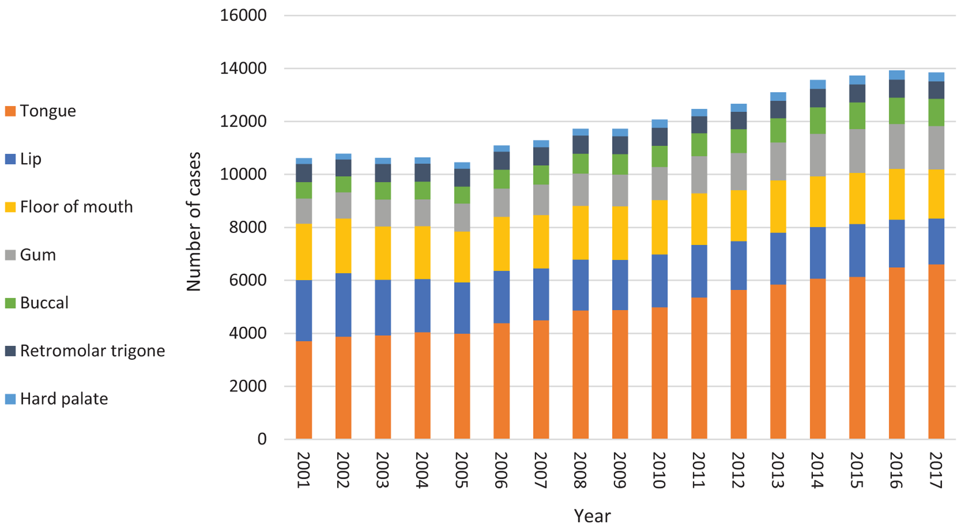
Burden of all oral cavity squamous cell carcinoma cases in the United States by subsite (2001–2017).
The age-adjusted rate of OCSCC decreased over time, dropping from 3.78 per 100,000 in 2001 to 3.54 in 2017. When looking at incidence by sex, we found the age-adjusted rate of OCSCC among men to decrease from 5.48 to 4.42 from 2001 to 2017. In contrast, the rate among women increased over this period (2.40 to 2.57). In terms of incidence by primary subsite, the majority of cases arose from the tongue (41.7%). This was followed equally by primaries of the lip and floor of mouth (each 16.5%), gum (10.6%), buccal mucosa (6.7%), retromolar trigone (5.6%), and hard palate (2.3%).
Across all cases, the rates of lip, floor of mouth, and retromolar trigone primaries decreased significantly over the studied time frame. In contrast, the rates of tongue, gum, hard palate, and buccal cancers increased significantly, though the rise among gum, hard palate, and buccal cancers was less pronounced and associated with substantially lower incidences (Figure 2). Age-adjusted rates among males and females stratified by subsite, including associated AAPCs, are displayed in Figures 3 and 4, respectively. While similar trends were seen among men and women across all subsites, the rising rate of tongue primaries was more profound among women (AAPC, 2.3%) than men (AAPC, 1.5%). Conversely, men had greater increases in rates of gum and buccal primaries.
Figure 2.
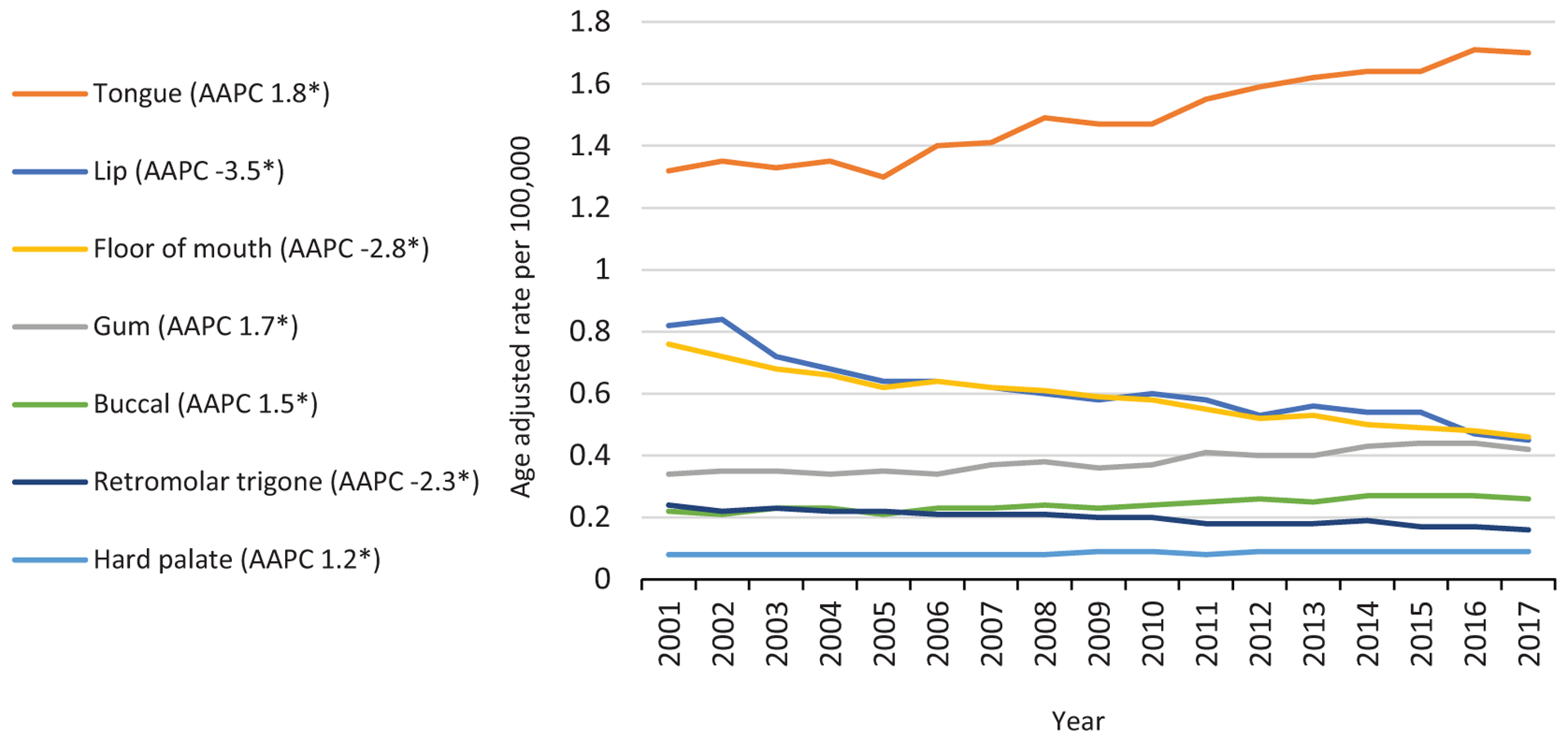
Age-adjusted rate of all oral cavity squamous cell carcinoma cases by subsite (2001–2017). *P<.05. AAPC, average annual percentage change.
Figure 3.
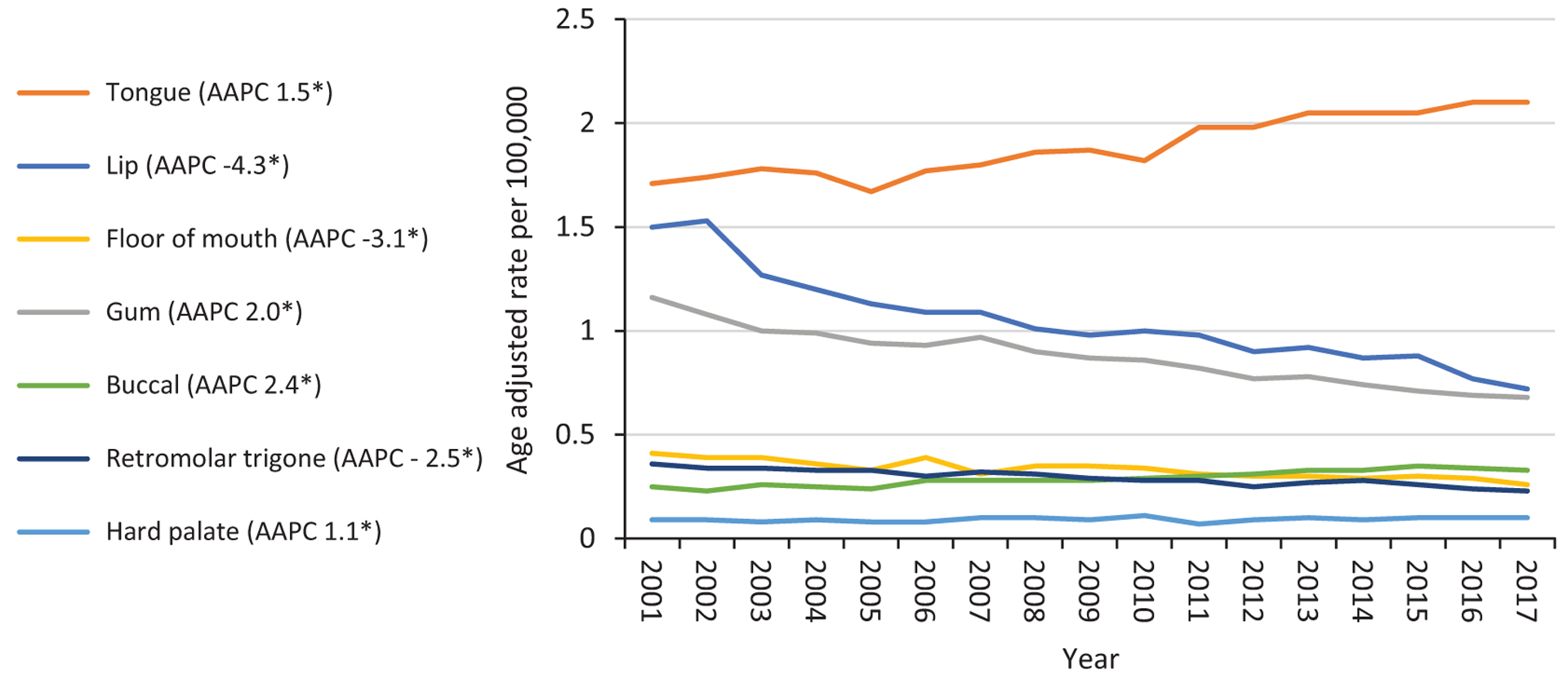
Age-adjusted rate of male oral cavity squamous cell carcinoma cases by subsite (2001–2017). *P<.05. AAPC, average annual percentage change.
Figure 4.
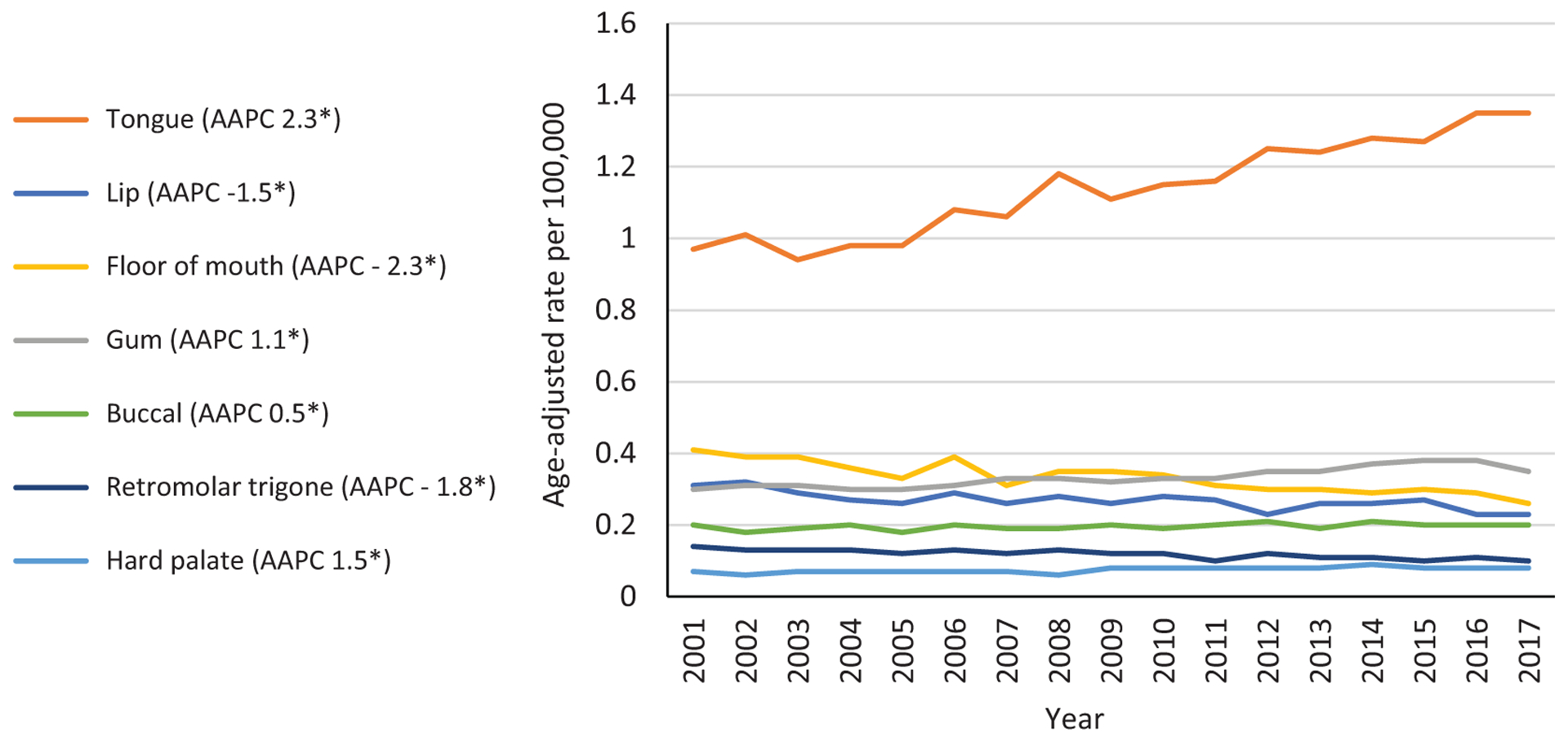
Age-adjusted rate of female oral cavity squamous cell carcinoma cases by subsite (2001–2017). *P<.05. AAPC, average annual percentage change.
Regarding the incidence of oral tongue squamous cell carcinoma (OTSCC) stratified by age, our data suggest a significant increase among men and women; specifically, a significant rise is seen among young patients as well as older patients, though the rise is not uniform across age groups throughout the studied time frame (Figure 5). Among all patients, there was a statistically significant increase in incidence across the entire study interval in those <40 years old (APC, 2.3%; 95% CI, 1.5%−3.0%), 40 to 49 years (APC, 1.7%; 95% CI, 1.0%−2.3%), and ≥80 years (APC, 2.2%; 95% CI, 1.6%−2.7%). Patients who were 50 to 59 years old had significantly higher rates from 2001 to 2009 (APC, 1.3%; 95% CI, 0.6%−2.1%) and 2012 to 2017 (APC, 2.1%; 95% CI, 0.5%−3.6%). In contrast, patients aged 60 to 79 years experienced a significant growth in rates of OTSCC in the later years of the study period, though with the largest percentage change among all age groups. Those aged 60 to 69 years had a significant increase from 2009 to 2017 (APC, 4.6%; 95% CI, 3.2%−4.0%). Those aged 70 to 79 years had a significant increase from 2007 to 2017 (APC, 4.8%; 95% CI, 3.5%−6.1%). When stratified by sex, similar trends are seen among men and women with OTSCC (Table 1).
Figure 5.
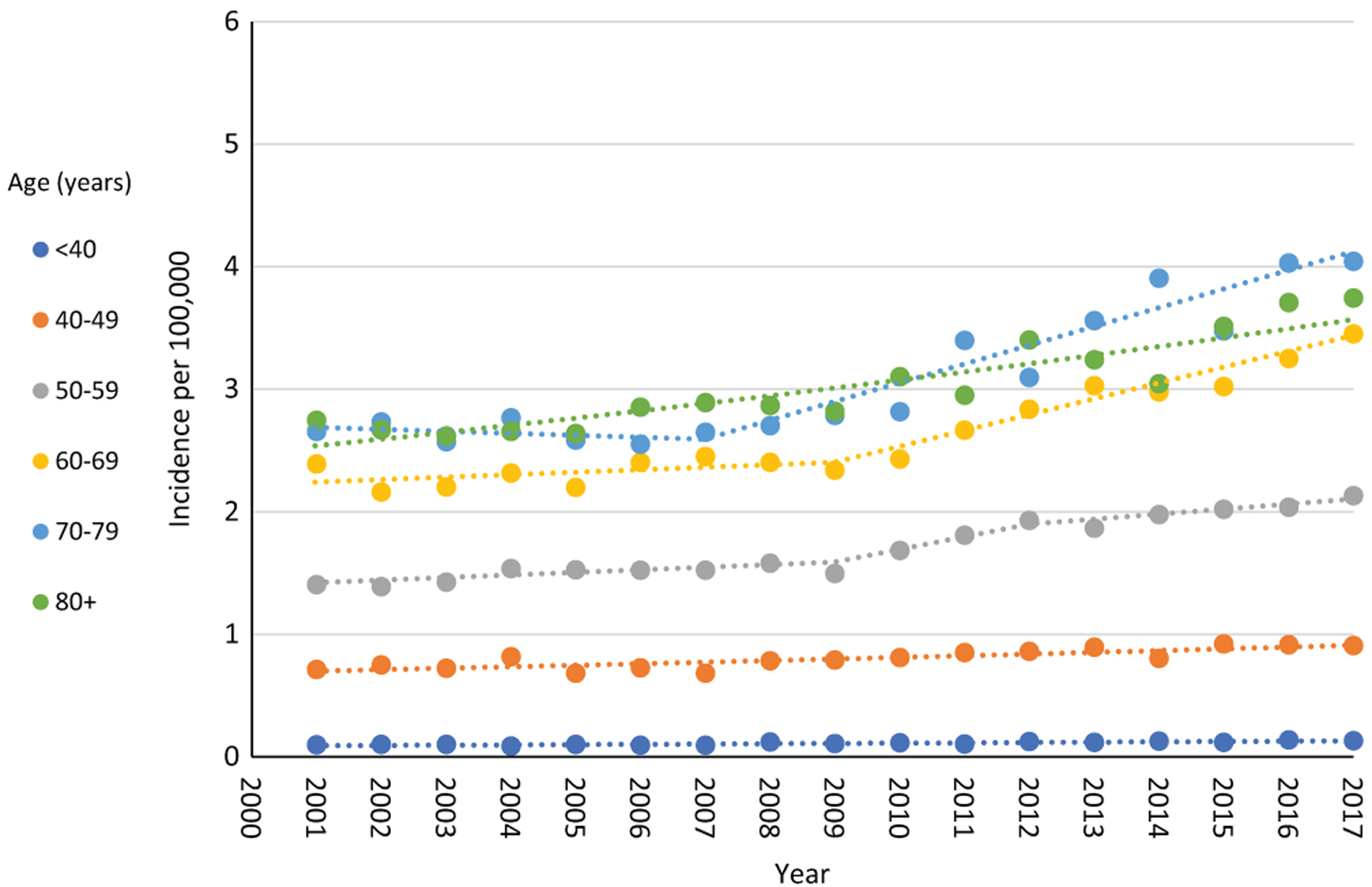
Incidence of all oral tongue squamous cell carcinoma cases by age (2001–2017).
Table 1.
APC for Oral Tongue Squamous Cell Carcinoma Stratified by Age and Sex.
| Male | Female | |||
|---|---|---|---|---|
| APC (95% CI) | Years | Age, y | Years | APC (95% CI) |
| −3.4 (−8.7 to 2.2) | 2001–2006 | <40 | 2001–2017 | 2.7a (1.3 to 4.2) |
| 3.8a (2.1 to 5.6) | 2006–2017 | |||
| 1.3a (0.4 to 2.2) | 2001–2017 | 40–49 | 2001–2017 | 2.3a (1.2 to 3.3) |
| 2.0a (1.6 to 2.4) | 2001–2017 | 50–59 | 2001–2017 | 4.1a (3.4 to 4.9) |
| 60–69 | 2001–2003 | −8.9 (−24.4 to 9.7) | ||
| 0.6 (−1.1 to 2.3) | 2001–2008 | 2003–2006 | 7.8 (−10.5 to 29.9) | |
| 3.5a (2.4 to 4.7) | 2008–2017 | 2006–2009 | −2.5 (−19.1 to 17.5) | |
| 2009–2017 | 6.2a (4.1 to 8.4) | |||
| −0.7 (−3.5 to 2.2) | 2001–2008 | 70–79 | 2001–2005 | −2.9 (−9.2 to 3.9) |
| 5.4a (3.4 to 7.5) | 2008–2017 | 2005–2017 | 4.3a (3.0 to 5.6) | |
| 2.6a (1.7 to 3.4) | 2001–2017 | ≥80 | 2001–2017 | 1.7a (1.1 to 2.4) |
Abbreviation: APC, annual percentage change.
P <.05.
Finally, for OTSCC rates by age and race, a significant increase in incidence was seen among White patients, while no significant rise was appreciated among Hispanic or Black patients (Figure 6, Table 2). The increase among White individuals persisted across all age groups and was consistently noted throughout the study interval in patients ≤59 years (Table 2). Among patients 60 to 69 years, there was a significant but mildly higher incidence from 2001 to 2009 (APC, 1.5%; 95% CI, 0.02%−2.9%) and more substantial growth from 2009 to 2017 (APC, 5.0%; 95% CI, 3.5%−6.6%). The rising rates among those ≥70 years old was significant from 2006 to 2017 (APC, 4.2%; 95% CI, 3.6%−4.9%). These observations emphasize complex interplay between age- and race-related demographics and changes in the incidence of OTSCC.
Figure 6.
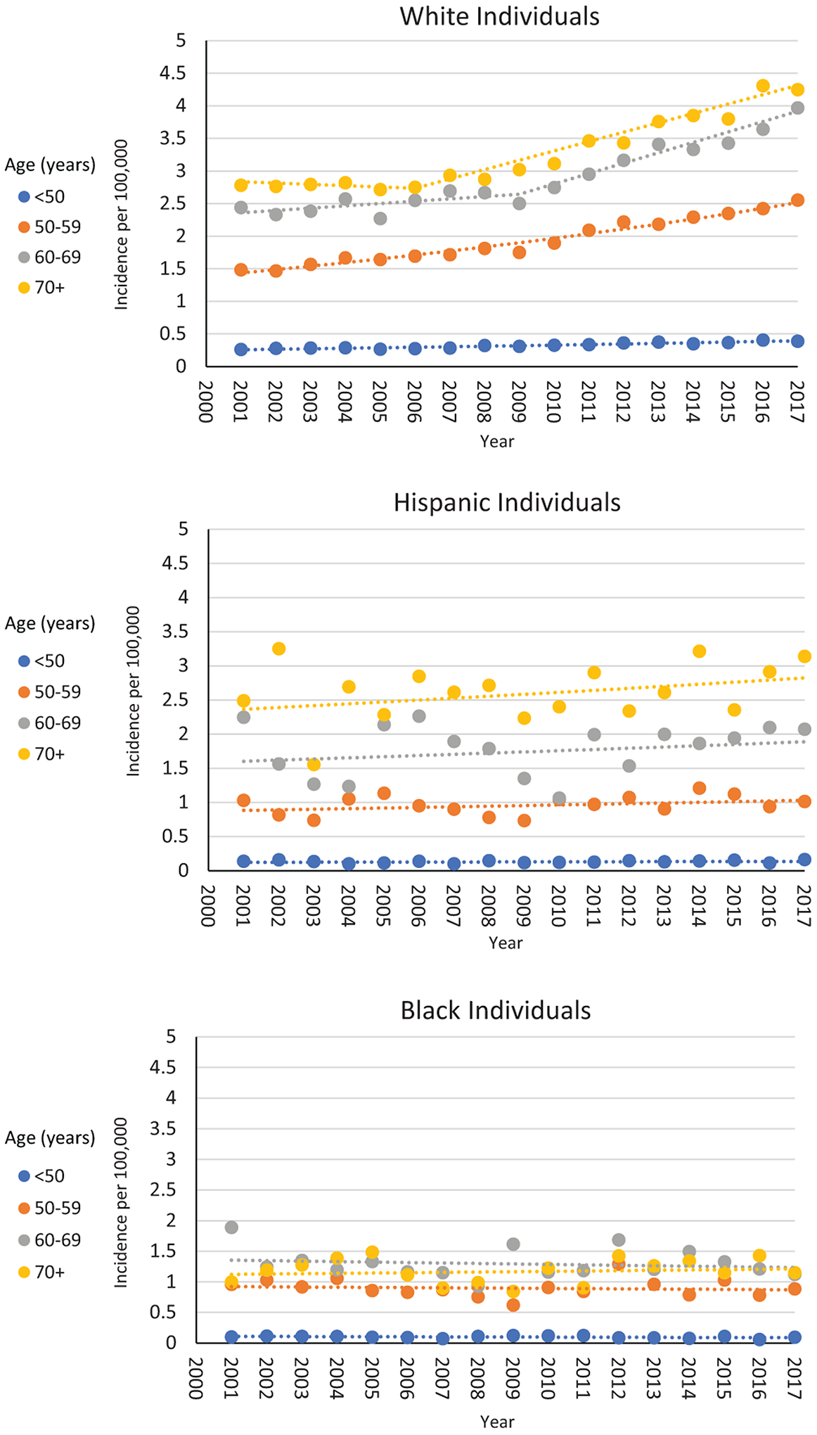
Incidence of all oral tongue squamous cell carcinoma cases by age and race (2001–2017): (A) White, (B) Black, and (C) Hispanic individuals.
Table 2.
APC for Oral Tongue Squamous Cell Carcinoma Stratified by Age and Race.
| APC (95% CI) | ||||
|---|---|---|---|---|
| Age, y | Years | Black | White | Hispanic |
| <50 | 2001–2017 | ‒1.3 (–3.4 to 0.7) | 2.7a (2.2 to 3.2) | 0.7 (–0.9 to 2.4) |
| 50–59 | 2001–2017 | −0.4 (–2.1 to 1.4) | 3.6a (3.2 to 3.9) | 1.0 (–0.6 to 2.5) |
| 60–69 | 2001–2017 | −0.6 (–2.4 to 1.3) | 1.0 (–1.4 to 3.5) | |
| 2001–2009 | 1.5a (0.02 to 2.9) | |||
| 2009–2017 | 5.0a (3.5 to 6.6) | |||
| ≥70 | 2001–2017 | 0.5 (− 1.4 to 2.4) | 1.1 (–0.7 to 2.9) | |
| 2001–2006 | −0.5 (–2.5 to 1.5) | |||
| 2006–2017 | 4.2a (3.6 to 4.9) | |||
Abbreviation: APC, annual percentage change.
P <.05.
Discussion
Most cases of OCSCC arose from the tongue (41.7%), followed by primaries of the lip and floor of mouth (each 16.5%), gum (10.6%), buccal mucosa (6.7%), retromolar trigone (5.6%), and hard palate (2.3%). The rates of OCSCC by subsite have not been well described within the United States, and subsite distribution of OCSCC has been variably reported in the literature. Classical textbook teaching in head and neck oncology emphasizes the lip as the most involved subsite.18,20 However, the tongue and floor of mouth have been cited as the most frequently implicated subsites. In a recent study evaluating the relative incidence of oral cavity cancers within the United States utilizing the National Cancer Database (NCDB), Jacobs et al identified the majority of cases to arise from the oral tongue (41.2%), followed by the floor of mouth (17.2%), alveolar ridge (11.1%), and lip (11.0%).3 Notably, NCDB data are hospital based; therefore, a true incidence is unable to be estimated from these data. Additionally, the relative incidence may not offer a true representation of the subsite distribution given that certain populations, such as nonurban areas, are less well represented by NCDB data. Furthermore, in the study performed by Jacobs et al, analysis was not limited to squamous cell carcinoma but instead included all oral cavity cancers.3
In a recent SEER database study evaluating cases of OCSCC from 1975 to 2013, Farhood et al identified tongue and floor of mouth to be the most common oral cavity subsites involved in OCSCC (each near 34%), followed by gum (14.4%), retromolar trigone (9.1%), buccal mucosa (7.4%), and hard palate (2.6%).12 However, they did not include lip cancers in their analysis. Notably, in contrast to our findings, they reported the relative incidence of tongue and floor of mouth cancers to be similar. The differences in our findings can likely be attributed to the earlier period evaluated by Farhood and colleagues, as we have shown that the proportion of OTSCC continues to rise particularly in more recent decades. Additionally, though SEER provides population-based cancer statistics designed to estimate incidence, the SEER 9 database captures registry data from roughly 9.8% of the US population. In comparison, the US Cancer Statistics Public Use Database is among the most comprehensive cancer databases, capturing >97% of all cancer diagnoses within the United States and therefore provides a more robust and accurate assessment of recent incidence of OCSCC by subsite.
We found the overall rate of OCSCC to decrease over our study period, including that among men. This was largely due to a significant decline in the incidence of lip and floor of mouth squamous cell carcinoma. In contrast, the rate of OCSCC has risen among women, which is primarily secondary to cancers of the oral tongue (AAPC, 2.3%). While the rate of OTSCC grew significantly among men, it was to a slightly lesser degree (AAPC, 1.5%). In addition, when the incidence of OTSCC was stratified by age, we saw a significantly higher incidence across all age groups. Interestingly, this was observed over the entire study period, except for those aged 60 to 79 years, in whom an increase in rates occurred in the latter part of the study period. Furthermore, we found that the rate of OTSCC rose among White patients, with no significant change among Black or Hispanic patients. Again, this was seen among older patients primarily throughout the latter study period. These findings suggest that the increase in OTSCC is largely specific to White individuals, corroborating previous observations that the rising rate of oral tongue cancer appears to disproportionately affect them, which has implications for surveillance and diagnosis within this demographic.13,21
Previous studies found the overall rates of OCSCC to be decreasing in the United States, which is thought to be secondary to declining rates of tobacco use in younger populations.22–24 However, numerous studies have reported an increase in OTSCC, particularly among young White individuals without traditional risk factors, predominantly composed of women.13,21,25–27 Our study corroborates an increase in OTSCC among younger individuals. The cause has been the subject of much investigation. It is hypothesized that a combination of genetic predisposition and environmental exposures may portend a higher risk for these patients, though to date no definitive etiology has been identified.28 Interestingly, this trend does not appear to be specific to the United States, as an increase in OTSCC among younger individuals (<45 years) has been observed globally.29
Our study reveals novel findings regarding subsite incidence trends of OCSCC among various age groups. Patel and colleagues evaluated the incidence of OCSCC and OTSCC utilizing the SEER 9 registries from 1975 to 2007.25 They found the incidence of OTSCC to be decreasing across all ages except the 18- to 44-year-old group, with a greater increase among women vs men. Additionally, they indicated that the incidence of squamous cell carcinoma in all other subsites of the oral cavity was decreasing. These findings differ from the current study in that we found the incidence of OTSCC to be rising across all age groups. However, the increase in older age groups was seen in the later years of the study period (after 2007). Thus, a higher incidence among older patients is appreciated in our analysis due to an inclusion of a more recent studied interval of time. Furthermore, while they reported a decrease in the incidence among other oral cavity subsites, their study was not appropriately powered to analyze trends in each subsite; therefore, the remaining subsites were grouped into 1 category. Although as a much smaller proportion of OCSCC cases, we did see an increase in gum, buccal, and hard palate cancers among men and women. Previous studies have not been appropriately powered to analyze incidence trends within each subsite; thus, these findings are novel and provoke interest into why this may be occurring in conjunction with the trends among oral tongue primaries.
Tota et al utilized SEER data from 1973 to 2012 to evaluate the incidence of OTSCC among White men and women in the United States, and they found the incidence of oral tongue cancers to be significantly increasing among White men and women, particularly among those born after the 1940s.13 Given that we saw an increase among 60- to 79-year-olds in the latter study years, this would roughly align with a similar estimated birth cohort. Interestingly, the rates among ≥80-year-olds did rise across the entire study period. A previous study comparing head and neck cancers diagnosed among younger vs older (>75 years) patients found that cancers involving oral cavity subsites were relatively more common among older patients and that those patients were more likely to be female as females tend to have longer life spans.30 Additionally, overall rates of oral cavity and pharynx cancers are higher among those >75 years old as compared with their younger counterparts.31 Perhaps our findings reflect a higher general risk of oral cavity cancers among the most advanced geriatric populations.
The epidemiology of OCSCC, specifically OTSCC, within the United States is evolving. While previous reports have highlighted the rising rates among young White women, our study, evaluating more recent years, suggests that this higher incidence is also being seen among men as well as older patients. The cause of this shift has been an active area of investigation. To date, no clear genomic or environmental drivers have been established. Unlike the similarly observed increase in oropharyngeal cancer, HPV has not been established to play a causative role in OCSCC.32–36 Moreover, this is the first study to our knowledge to illustrate an associated increase in gum, buccal, and hard palate squamous cell carcinoma, though these subsites represent a much smaller portion of cases overall. Given the drastic decline in lip and floor of mouth cancers and the lack of smoking data, it is difficult to understand what factors may be associated with these trends. Nevertheless, this study characterizes the incidence of OCSCC and describes more recent trends across subsites, highlighting the fact that a rise in OTSCC is occurring not only among young White women but also among men and older patients. Future investigation is certainly warranted to better understand the trends among other oral cavity subsites, as well as underlying causative factors.
Our study has a few limitations. Although the US Cancer Statistics Public Use Database offers robust and comprehensive epidemiologic data, it does not offer information on important patient factors, such as smoking and alcohol history, which are known to be important underlying risk factors for OCSCC. Additionally, given the retrospective nature of our study, there is a risk for site misclassification error. In particular, it is possible that some base of tongue tumors could have been misclassified as tongue cancers. Given the rising incidence of human papillomavirus–related base of tongue cancers, rates of oral tongue cancers could be overestimated due to the miscoding of base of tongue cancers. A study examining a subset of tongue cancers in SEER (2000–2011) found that 5% of tongue cancers were recoded to base of tongue after review, decreasing from 8% to 2% over time.37 Notably, even if tongue cancers were overestimated by 5% in this study, the tongue would still represent the majority of OCSCC cases, and the incidence would be minimally altered. It is also possible that lip cancers may be misclassified as skin cancers, which are not included in this database, representing a caveat in data related to this subsite.
In conclusion, this study offers the most recent and comprehensive analysis of subsite-specific incidence of OCSCC, with the US Cancer Statistics Public Use Database capturing 97% of newly diagnosed cancers in United States. To our knowledge this is the most robust and detailed analysis of national incidence trends evaluating oral cavity cancer by individual subsites across sex, race, and age. Our data revealed the tongue to be the most frequently involved subsite. Furthermore, a significant increase in OTSCC is seen among men and women of all ages, though in more recent years among older patients, specifically among White patients.
Footnotes
This article was presented at the 2021 AAO-HNSF Annual Meeting & OTO Experience; October 4, 2021; Los Angeles, California.
Competing interests: None.
References
- 1.Chi AC, Day TA, Neville BW. Oral cavity and oropharyngeal squamous cell carcinoma—an update. CA Cancer J Clin. 2015; 65(5):401–421. doi: 10.3322/CAAC.21293 [DOI] [PubMed] [Google Scholar]
- 2.D’Cruz AK, Vaish R, Dhar H. Oral cancers: current status. Oral Oncol. 2018;87:64–69. doi: 10.1016/J.ORALONCOLOGY.2018.10.013 [DOI] [PubMed] [Google Scholar]
- 3.Jacobs CD, Barbour AB, Mowery YM. The relative distribution of oral cancer in the United States by subsite. Oral Oncol. 2019; 89:56–58. doi: 10.1016/j.oraloncology.2018.12.017 [DOI] [PubMed] [Google Scholar]
- 4.American Head and Neck Society. Oral cavity cancer: professional version. Accessed November 29, 2021. https://www.ahns.info/resources/oral-cavity-cancer/3/#_Toc400976516
- 5.Nocini R, Lippi G, Mattiuzzi C. Biological and epidemiologic updates on lip and oral cavity cancers. Ann Cancer Epidemiol. 2020;4. doi: 10.21037/ACE.2020.01.01 [DOI] [Google Scholar]
- 6.Chong V Oral cavity cancer. Cancer Imaging. 2005;5(Spec No.A). doi: 10.1102/1470-7330.2005.0029 [DOI] [PMC free article] [PubMed] [Google Scholar]
- 7.Lin NC, Hsien SI, Hsu JT, Chen MYC. Impact on patients with oral squamous cell carcinoma in different anatomical subsites: a single-center study in Taiwan. Sci Rep. 2021;11:15446. doi: 10.1038/s41598-021-95007-5 [DOI] [PMC free article] [PubMed] [Google Scholar]
- 8.Li R, Fakhry C, Koch WM, Gourin CG. The Effect of tumor subsite on short-term outcomes and costs of care after oral cancer surgery. Laryngoscope. 2013;123(7):1652–1659. doi: 10.1002/lary.23952 [DOI] [PubMed] [Google Scholar]
- 9.Zelefsky MJ, Harrison LB, Fass DE, et al. Postoperative radio-therapy for oral cavity cancers: impact of anatomic subsite on treatment outcome. Head Neck. 1990;12(6):470–475. doi: 10.1002/HED.2880120604 [DOI] [PubMed] [Google Scholar]
- 10.Bell RB, Kademani D, Homer L, Dierks EJ, Potter BE. Tongue cancer: is there a difference in survival compared with other subsites in the oral cavity? J Oral Maxillofac Surg. 2007;65(2):229–236. doi: 10.1016/J.JOMS.2005.11.094 [DOI] [PubMed] [Google Scholar]
- 11.Montero PH, Yu C, Palmer FL, et al. Nomograms for preoperative prediction of prognosis in patients with oral cavity squamous cell carcinoma. Cancer. 2014;120(2):214–221. doi: 10.1002/CNCR.28407 [DOI] [PubMed] [Google Scholar]
- 12.Farhood Z, Simpson M, Ward GM, Walker RJ, Osazuwa-Peters N. Does anatomic subsite influence oral cavity cancer mortality? A SEER database analysis. Laryngoscope. 2019;129(6):1400–1406. doi: 10.1002/lary.27490 [DOI] [PubMed] [Google Scholar]
- 13.Tota JE, Anderson WF, Coffey C, et al. Rising incidence of oral tongue cancer among White men and women in the United States, 1973–2012. Oral Oncol. 2017;67(2017):146–152. doi: 10.1016/j.oraloncology.2017.02.019 [DOI] [PubMed] [Google Scholar]
- 14.Tandon P, Dadhich A, Saluja H, Bawane S, Sachdeva S. The prevalence of squamous cell carcinoma in different sites of oral cavity at our Rural Health Care Centre in Loni, Maharashtra—a retrospective 10-year study. Contemp Oncol (Pozn). 2017;21(2): 178–183. doi: 10.5114/WO.2017.68628 [DOI] [PMC free article] [PubMed] [Google Scholar]
- 15.Gupta N, Gupta R, Acharya AK, et al. Changing trends in oral cancer—a global scenario. Nepal J Epidemiol. 2016;6(4):613–619. doi: 10.3126/NJE.V6I4.17255 [DOI] [PMC free article] [PubMed] [Google Scholar]
- 16.Effiom OA, Adeyemo WL, Omitola OG, Ajayi OF, Emmanuel MM, Gbotolorun OM. Oral squamous cell carcinoma: a clinicopathologic review of 233 cases in Lagos, Nigeria. J Oral Maxillofac Surg. 2008;66(8):1595–1599. doi: 10.1016/J.JOMS.2007.12.025 [DOI] [PubMed] [Google Scholar]
- 17.Sundermann BV, Uhlmann L, Hoffmann J, Freier K, Thiele OC. The localization and risk factors of squamous cell carcinoma in the oral cavity: a retrospective study of 1501 cases. J Craniomaxillofac Surg. 2018;46(2):177–182. doi: 10.1016/J.JCMS.2017.10.019 [DOI] [PubMed] [Google Scholar]
- 18.Krolls SO, Hoffman S. Squamous cell carcinoma of the oral soft tissues: a statistical analysis of 14,253 cases by age, sex, and race of patients. J Am Dent Assoc. 1976;92(3):571–574. doi: 10.14219/JADA.ARCHIVE.1976.0556 [DOI] [PubMed] [Google Scholar]
- 19.Centers for Disease Control and Prevention. US Cancer Statistics Public Use Databases. Accessed 2020. http://www.cdc.gov/cancer/uscs/public-use
- 20.Pasha R, Golub JS. Otolaryngology–Head and Neck Surgery: Clinical Reference Guide. 3rd ed. Plural Publishing; 2011:598. [Google Scholar]
- 21.Joseph LJ, Goodman M, Higgins K, et al. Racial disparities in squamous cell carcinoma of the oral tongue among women: a SEER data analysis. Oral Oncol. 2015;51(6):586–592. doi: 10.1016/J.ORALONCOLOGY.2015.03.010 [DOI] [PubMed] [Google Scholar]
- 22.Weatherspoon DJ, Chattopadhyay A, Boroumand S, Garcia I. Oral cavity and oropharyngeal cancer incidence trends and disparities in the United States: 2000–2010. Cancer Epidemiol. 2015;39(4):497–504. doi: 10.1016/j.canep.2015.04.007 [DOI] [PMC free article] [PubMed] [Google Scholar]
- 23.Chaturvedi AK, Engels EA, Anderson WF, Gillison ML. Incidence trends for human papillomavirus-related and -unrelated oral squamous cell carcinomas in the United States. J Clin Oncol. 2008;26(4):612–619. doi: 10.1200/JCO.2007.14.1713 [DOI] [PubMed] [Google Scholar]
- 24.Sturgis EM, Cinciripini PM. Trends in head and neck cancer incidence in relation to smoking prevalence: an emerging epidemic of human papillomavirus-associated cancers? Cancer. 2007;110(7):1429–1435. doi: 10.1002/cncr.22963 [DOI] [PubMed] [Google Scholar]
- 25.Patel SC, Carpenter WR, Tyree S, et al. Increasing incidence of oral tongue squamous cell carcinoma in young White women, age 18 to 44 years. J Clin Oncol. 2011;29(11):1488–1494. doi: 10.1200/JCO.2010.31.7883 [DOI] [PubMed] [Google Scholar]
- 26.Shiboski CH, Schmidt BL, Jordan RCK. Tongue and tonsil carcinoma: increasing trends in the US population ages 20–44 years. Cancer. 2005;103(9):1843–1849. doi: 10.1002/CNCR.20998 [DOI] [PubMed] [Google Scholar]
- 27.Campbell BR, Sanders CB, Netterville JL, et al. Early onset oral tongue squamous cell carcinoma: associated factors and patient outcomes. Head Neck. 2019;41(6):1952–1960. doi: 10.1002/hed.25650 [DOI] [PMC free article] [PubMed] [Google Scholar]
- 28.Heaton CM, Durr ML, Tetsu O, van Zante A, Wang SJ. TP53 and CDKN2a mutations in never-smoker oral tongue squamous cell carcinoma. Laryngoscope. 2014;124(7):E267–E273. doi: 10.1002/lary.24595 [DOI] [PubMed] [Google Scholar]
- 29.Ng JH, Iyer NG, Tan MH, Edgren G. Changing epidemiology of oral squamous cell carcinoma of the tongue: a global study. Head Neck. 2017;39(2):297–304. doi: 10.1002/HED.24589 [DOI] [PubMed] [Google Scholar]
- 30.Sarini J, Fournier C, Lefebvre JL, Bonafos G, Ton Van J, Coche-Dequeant B. Head and neck squamous cell carcinoma in elderly patients: a long-term retrospective review of 273 cases. Arch Otolaryngol Neck Surg. 2001;127(9):1089–1092. doi: 10.1001/ARCHOTOL.127.9.1089 [DOI] [PubMed] [Google Scholar]
- 31.National Cancer Institute. SEER*Explorer application: oral cavity and pharynx cancer. Published 2018. Accessed December 19, 2021. https://seer.cancer.gov/explorer/application.html?site=3&data_type=1&graph_type=2&compareBy=sex&chk_sex_3=3&chk_sex_2=2&rate_type=2&race=1&age_range=166&stage=101&advopt_precision=1&advopt_show_ci=on&advopt_display=1
- 32.Martinez RCP, Sathasivam HP, Cosway B, et al. Clinicopathological features of squamous cell carcinoma of the oral cavity and oropharynx in young patients. Br J Oral Maxillofac Surg. 2018; 56(4):332–337. doi: 10.1016/j.bjoms.2018.03.011 [DOI] [PubMed] [Google Scholar]
- 33.El-Mofty SK, Lu DW. Prevalence of human papillomavirus type 16 DNA in squamous cell carcinoma of the palatine tonsil, and not the oral cavity, in young patients: a distinct clinicopathologic and molecular disease entity. Am J Surg Pathol. 2003;27(11): 1463–1470. doi: 10.1097/00000478-200311000-00010 [DOI] [PubMed] [Google Scholar]
- 34.de Abreu PM, Có ACG, Azevedo PL, et al. Frequency of HPV in oral cavity squamous cell carcinoma. BMC Cancer. 2018;18(1): 324. doi: 10.1186/S12885-018-4247-3 [DOI] [PMC free article] [PubMed] [Google Scholar]
- 35.Kreimer AR, Clifford GM, Boyle P, Franceschi S. Human papillomavirus types in head and neck squamous cell carcinomas worldwide: a systemic review. Cancer Epidemiol Biomarkers Prev. 2005;14(2):467–475. doi: 10.1158/1055-9965.EPI-04-0551 [DOI] [PubMed] [Google Scholar]
- 36.Mork J, Lie AK, Glattre E, et al. Human papillomavirus infection as a risk factor for squamous-cell carcinoma of the head and neck. N Engl J Med. 2001;344(15):1125–1131. doi: 10.1056/NEJM200104123441503 [DOI] [PubMed] [Google Scholar]
- 37.Polednak AP, Phillips C. Cancers coded as tongue not otherwise specified: relevance to surveillance of human papillomavirus–related cancers. J Registry Manag. 2014;41(4):190–195. [PubMed] [Google Scholar]


