Abstract
Soil microbiomes in forest ecosystems act as both nutrient sources and sinks through a range of processes including organic matter decomposition, nutrient cycling, and humic compound incorporation into the soil. Most forest soil microbial diversity studies have been performed in the northern hemisphere, and very little has been done in forests within African continent. This study examined the composition, diversity and distribution of prokaryotes in Kenyan forests top soils using amplicon sequencing of V4-V5 hypervariable region of the 16S rRNA gene. Additionally, soil physicochemical characteristics were measured to identify abiotic drivers of prokaryotic distribution. Different forest soils were found to have statistically distinct microbiome compositions, with Proteobacteria and Crenarchaeota taxa being the most differentially abundant across regions within bacterial and archaeal phyla, respectively. Key bacterial community drivers included pH, Ca, K, Fe, and total N while archaeal diversity was shaped by Na, pH, Ca, total P and total N. To contextualize the prokaryote diversity of Kenyan forest soils on a global scale, the sample set was compared to amplicon data obtained from forest biomes across the globe; displaying them to harbor distinct microbiomes with an over-representation of uncultured taxa such as TK-10 and Ellin6067 genera.
Subject terms: Computational biology and bioinformatics, Ecology, Microbiology, Molecular biology
Introduction
Forests are highly productive components of terrestrial ecosystems1, covering more than 40 million km2 and presenting 30% of the total global land area1. They form part of our most precious natural resources essential to the continued balance and survival of the world’s ecosystem2. Forests act as carbon sinks where soil organic matter is formed from residuals after biomass decomposition1. They play a major role in the global cycling of carbon, as well as organic nitrogen mineralization and conversion of organic phosphorus into inorganic3. Moreover, forests are involved in maintenance of soil structure1, organic matter decomposition3, degradation of pollutants4 and shape soil microbial communities through the symbiotic interaction with primary microbial producers such as mycorrhizal fungi5. Some of bacterial taxa previously shown to dominate forest soil ecosystems and play such key roles include members of the genera Pedobacter and Chitinophaga (Bacteroidetes); Pseudomonas, Variovorax, Ewingella, and Stenotrophomonas (Proteobacteria)1; Burkholderia, Phenylobacterium, and Methylovirgula (Pseudomonadota)6; members of the Rhizobiales and Nitrosopumilus7. Unfortunately, these forest ecosystems have suffered from serious depletion due to anthropogenic activities associated with over-farming, the pulp and paper industry and population encroachment into peri-urban areas, along with other land-use change6. Soil microorganisms are an important component of the forest ecosystem1,5 as they play fundamental roles in most nutrient transformations within forest soils, upon which the stability and sustainable development of forest ecosystems rely8.
The distribution and diversity of soil microbiomes is influenced by numerous aspects such as soil type, physicochemical characteristics, microclimate, vegetation and land-use6. Recent microbial ecology studies have shown that different habitats harbor diverse microbial communities whose succession patterns are shaped by substrate availability, including nutrient pools, physiochemistry and vegetation5.
In addition, factors that modify the microclimate and forest litter chemistry such as forest type, plant species and plant diversity have also been identified as drivers of microbial community composition in forest soils7.
Kenya’s indigenous forests represent some of the most diverse ecosystems in the world, and provide important economic, environmental, recreational, scientific, cultural and spiritual benefits to the nation (Republic of Kenya, 2009)9. Forests play a vital role in the stabilization of soils and ground water, support the conduct of reliable agricultural activity and play a crucial role in protecting water catchments in Kenya besides moderating climate by absorbing greenhouse gases10. In addition, forests such as those of the Taita Hills are hotspots of biodiversity, harboring a wide variety of medicinal plants11. The Forests Act has previously recognized that forests provide the main locus of Kenya’s biological diversity and a major habitat for wildlife, and acknowledges that forests and trees are the main source of domestic fuelwood2. However, these forests have been subjected to land-use changes such as conversion to farmlands, ranches and settlements.
Historically, the majority of forest soil microbial diversity studies have been performed in northern hemisphere countries, with very little focus on the forests of the African continent, even in global studies9,10. To fill this knowledge gap, this study aimed to document the microbial ecology of selected Kenyan forest soil ecosystems, and to study the possible abiotic drivers. The selected forest ecosystems are among Kenyan landscapes endowed with varied climate with different water catchment and soil regime. For instance, the Mt. Kenya, Aberdare and Taita Taveta regions are among Kenya’s water towers. The regions are characterized by a bimodal rainfall patterns which influence the vegetation within each ecoregion. This leads to a variation in moisture content within soil ecosystems further influencing microbial diversity.
Results and discussion
Different forest soils in Kenya have unique physicochemical properties
In this study, 31 soil samples were obtained from forests ecosystems within the Taita Taveta, Nairobi, Western, Aberdare and Mt. Kenya ecoregions (Supplementary Table 1). Samples from the different ecoregions were shown to be significantly different (p-value ≤ 0.01, R2 = 0.45) in terms of soil physicochemical properties, specifically in soil pH, soil texture, macro- and micro-nutrient composition and Enhanced Vegetation Index-2 (EVI2) (Fig. 1a and b, Supplementary Fig. S2a and S2b). Taita Taveta forest soils were highly distinct from those of the Nairobi, Aberdare and Western regions (Fig. 1b). Conversely Nairobi and Western region soils exhibited the least variability (Fig. 1b).
Figure 1.
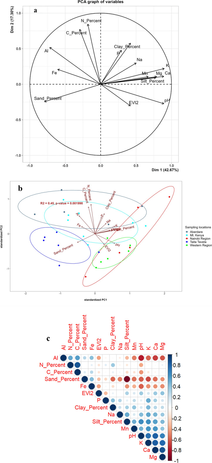
(a) Principal component analysis (PCA) biplot of forest soil samples according to their physicochemical properties. The influence of each variable on sample distribution is represented by the arrows radiating from the center of the PCA plot. (b) The sample clusters corresponding to different ecoregions are highlighted within ellipses of the same color. (c) Pearson correlation plot between measured soil physicochemical properties. Positive and negative correlations are displayed in blue and red shades, respectively, while the size and intensity of matrix circles is proportional to correlation coefficient between variables.
Several soil physicochemical properties were found to be correlated, and thus could be considered as interdependent (Fig. 1c). For instance, the measurement of plant density, vegetation index (EVI2), was positively correlated with all the measured soil nutrients, apart from phosphorus. This is not unexpected, as nutrient-rich forest soils have been repeatedly shown to support high density plant growth12. The soil samples used in this study were collected from 0 to 5 cm depth, which is within the 0–20 cm soil profile characteristically comprising the organic horizon that results from decomposition of litter-derived organic matter and representing a nutrient-rich mixture of processed, plant-derived organic matter12. Low titratable phosphorus concentrations were possibly due to presence of a high content of Al and Fe, which form oxides that fix phosphorus at the low pH’s associated with these soils13. In this study, the pH was positively correlated with EVI2 but negatively correlated with C and N content. This result contradicts a previous study that concluded that at higher soil pH levels, the mineralizable fractions of C and N increased, possibly due to the direct effect of pH on microbial populations and their activities13.
Taxonomic composition of soil microbiomes across Kenyan forest biomes
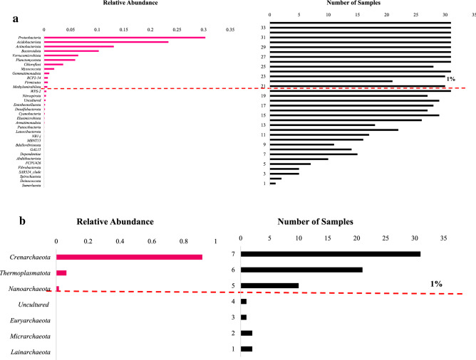
Analysis of Bacterial diversity in forest soil samples indicated the presence of 34 phyla, of which 12 were dominant (i.e. represented by > 1% of ASV reads in at least 87% of the samples). The most abundant of these was Proteobacteria (30.3% mean relative abundance), followed by Acidobacteriota (23.4% mean relative abundance) and Actinobacteria (13.1% mean relative abundance) (Fig. 2a). Actinobacteriota members such as Frankiales and Streptomycetales are known as nitrogen-fixing bacteria and may produce biologically active secondary metabolites14. The dominant bacterial phyla from the current study were consistent with other studies within two forests sites, where bacterial ASVs were assigned to 44 phyla, ten of which; (Proteobacteria, Acidobacteria, Verrucomicrobia, Firmicutes, Actinobacteria, Bacteroidetes, Planctomycetes, Chloroflexi, WD272, and Gemmatimonadetes) comprised more than 90% of the relative abundance in each library15. Our results on bacterial abundance were also consistent with several previous studies where Proteobacteria, Acidobacteria, Verrucomicrobia, Firmicutes, Actinobacteria, Bacteroidetes, Planctomycetes, Chloroflexi were the most abundant phyla15.
Figure 2.
((a) and (b)) Mean relative abundances of prokaryotic ((a) bacteria) ((b) Archaea) phyla across forest soil samples, together with the number of samples in which they were identified. The phyla scoring > 1% mean relative abundance, are dominant and above red dashed line.
In particular, members of Proteobacteria and Acidobacteriota phylum have been reported as ubiquitous and dominant in` soil ecosystems16. Members of these Phyla, such as Anaeromyxobacter, Bradyrhizobium, Azospirillum, Ralstonia, Burkholderia, Brevundimonas Rhodopseudomonas (Proteobacteria), Mycobacterium, Nocardia, Amycolatopsis Thermobispora, Pseudonocardia, Brachybacterium, Frankia, Conexibacter (Actinobacteria), Streptococcus, Lactococcus, and Enterococcus (Firmicutes) have been reported to carry out various key ecological processes such as regulation of biogeochemical cycles, decomposition of biopolymers, exopolysaccharide secretion and plant growth promotion17.
The most dominant taxa at Order level, Rhizobiales (12.8% mean relative abundance), Burkholderiales (6.3% mean relative abundance) and Chitinophagales (6.2% mean relative abundance), were represented across all samples (Supplementary Table 2). The order Chitinophagales contains members that are known to degrade complex organic matter, such as chitin and cellulose18. The orders Rhizobiales, Xanthomonadales and Rhodospirillales found in this study are also well known for nitrogen fixation, mineralization and denitrification activities7. Crenarchaeota was the most abundant Archaeal phylum represented across all samples, with 91.6% mean relative abundance (Fig. 2b). This phylum was further grouped into two classes; Nitrososphaeria (77.1% mean relative abundance) and Bathyarchaeia (0.2% mean relative abundance). Nitrososphaeria includes many ammonia-oxidizing taxa that have been identified previously in forest soil microbiomes19. Other phyla within the Archaeal Domain included Thermoplasmatota (6.4% mean relative abundance), represented within about two thirds of the soil samples, (Supplementary Table 2) while Nanoarchaeota phylum (1.7% mean relative abundance) was represented within about a quarter of the soil samples. These results agree with previous studies where archaeal communities in forest biomes were reported to be dominated by Nitrososphaera20. Members of Nitrososphaera have been described as major contributors to soil biogeochemical processes such as carbon, methane, nitrogen and, sulfur cycle within many ecosystems21.
Alpha- and beta- diversity analysis of soil prokaryotic communities
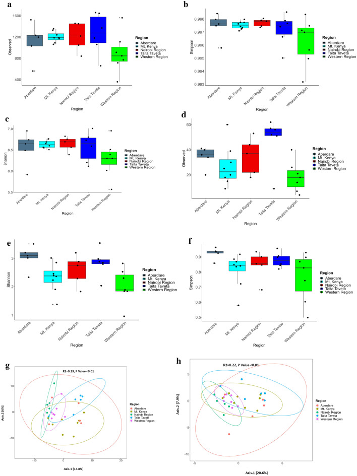
Analysis of sample alpha-diversity showed Western and Taita Taveta regions soils to have significantly different (P ≤ 0.01) levels of Archaeal richness, while Western and Aberdare regions soils displayed significantly different Shannon diversity index (P = 0.02) (Fig. 3d–f). Although there were no significant differences between bacterial communities displayed within various forests ecosystems (Fig. 3a–c) soil samples under bamboo vegetation cover within Mt. Kenya and Aberdare regions displayed lower diversity than the other ecoregions. Soils collected from the Taita Taveta region (Vuria and Ngangao) were shown to have the highest number of observed prokaryotic taxa. These forests are characterized by a montane climate vegetation with thick ground cover22. The high number of ASVs could be attributed to a broad range of bacterial micro-habitats associated with high nutrient availability besides other specific microbial diversity drivers such as plant density and vegetation index that positively influenced bacterial abundance23. There was high prokaryotic variability observed within each region, an indication of distinct microhabitats and microclimates in each forest region covered (Fig. 3 g,h).
Figure 3.
(a–f) Alpha Diversity of soil prokaryotic communities. (a–c) represent diversity indices (Observed, Shannon and Inverse Simpson) of Bacterial communities while (d–f) represent diversity indices (Observed, Shannon and Inverse Simpson) of Archaea communities within soil samples collected from the five regions in Kenya. Figure 3 (g,h) Principal Coordinate Analysis of prokaryotic diversity based on Bray–Curtis index within soil samples. (g) represents beta diversity of Bacterial community structure while (h) represents beta diversity of Archaea community structure within soil samples collected from the five regions in Kenya.
Beta-diversity analysis of soil samples from these regions showed a significant difference (P < 0.01) on bacterial and archaeal community structure (Bacteria R2 = 0.22; Archaea R2 = 0.24) (Fig. 3 g,h). Notably, the microbial composition of samples from the Taita Taveta region showed a lower degree of overlap with other regions, which mimics the soil chemistry differences observed between the regions. Taita Hills comprise the northernmost part of the Precambrian Eastern-Arc Mountain range, known for its rich biodiversity24 and recognized as one of the world’s 25 biodiversity hot-spots25.
The highly significant (P < 0.01) richness and Shannon diversity index values for samples from Western region forests could be attributed to the tropical nature of forests within this region such as sample K21 (Kakamega forest) which is considered an important biodiversity reservoir and the only remaining Guinea-Congolian tropical rain forest in Kenya26. Kakamega forest is the largest moist lowland forest ecosystem in Kenya, and has similar characteristics to Central Africa equatorial forests22.
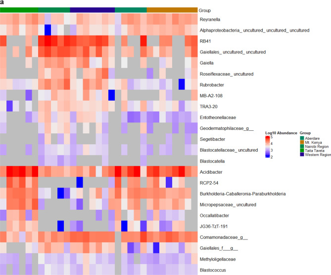
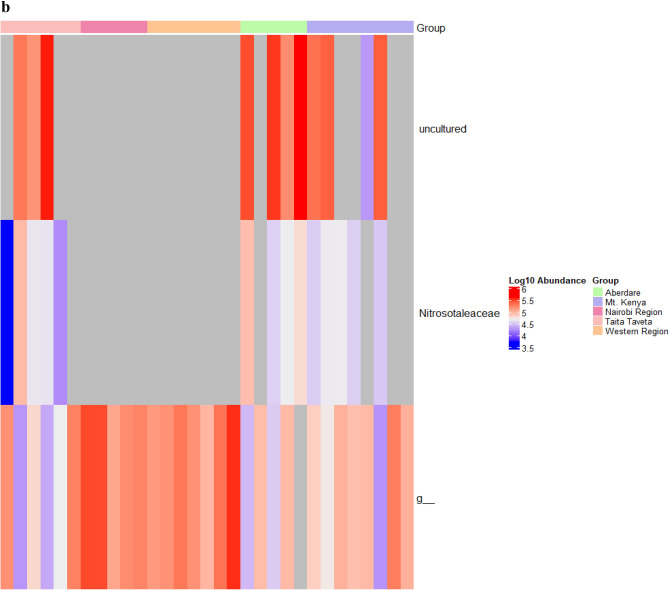
To explore further the differences in the soil microbiome structure between the different forest areas, linear discriminant analysis (LDA) effect size (LEfSe) was used to detect prokaryotic taxa that were differentially abundant within and between soil samples. In a comparison of samples from the five forest regions (Aberdare, Mt. Kenya, Nairobi, Taita Taveta and Western), several taxa were identified as differentially abundant (P adj. < 0.01): 13 genera in Taita Taveta, 21 in Nairobi, 1 in Mt. Kenya, 2 in Western and 5 in Aberdare region (Fig. 4a). The LEfSe algorithm identified several differentially abundant archaeal taxa (P adj. < 0.01) within the three regions (Aberdare, Nairobi and Taita Taveta) each having a taxon (Fig. 4b). The genus Acidibacter was over-represented in Taita forest soils, possibly due to the low soil pH observed in this region. IMCC26256 was over-represented in western region. Burkholderia-Caballeronia-Paraburkholderia taxa, which typically have a very broad ecological diversity and metabolic versatility27 were the most abundant in Aberdare Forest soils, RB41 in Mt. Kenya while Rhodovastum was the most abundant in Mt. Kenya region soil samples (Fig. 4a).
Figure 4.
Taxa, at genus level, that are over-represented in different regions based on the LEfSe analysis. (a) represents LEfSe analysis of Bacterial communities while (b) represents LEfSe analysis of Archaea communities within soil samples collected from the five regions in Kenya. The log10 abundance colour scheme represents LDA score. The colors represent the group in which that taxon was found to be more abundant compared to the other groups. f_ indicates that the ASV was not able to be classified to a family while g_ indicates that the ASV was not able to be classified to a genus. The heat maps were generated using Microbiome Marker R package56.
Environmental drivers of soil microbiomes in Kenyan forest soils
A stepwise model-building approach for constrained ordination models was used to assess the potential environmental drivers of the prokaryotic communities within forest ecosystems. Canonical correspondence analysis (CCA) ordination plots showed that bacterial and archaeal community structures were significantly affected by several soil physicochemical characteristics (P < 0.01). Soil pH, Ca, K, Fe and %N were shown as key drivers of bacterial community structure, while Na, pH, Ca, P and %N were important factors in shaping archaeal community structure within forest soils (Fig. 5a,b). The significant effect of nitrogen to community structure is consistent with the composition of soil microbiomes described in this study, which were dominated by taxa potentially involved in nitrogen fixation such as Cyanobacteria and Nitrospirota (Supplementary Fig. S2a). Fe concentration and soil texture are known to be major factors in shaping bacterial community structures in some soils28. Soil pH possibly affected the thermodynamics and kinetics of microbial respiration, thus shaping the microbial communities’ composition and function.
Figure 5.
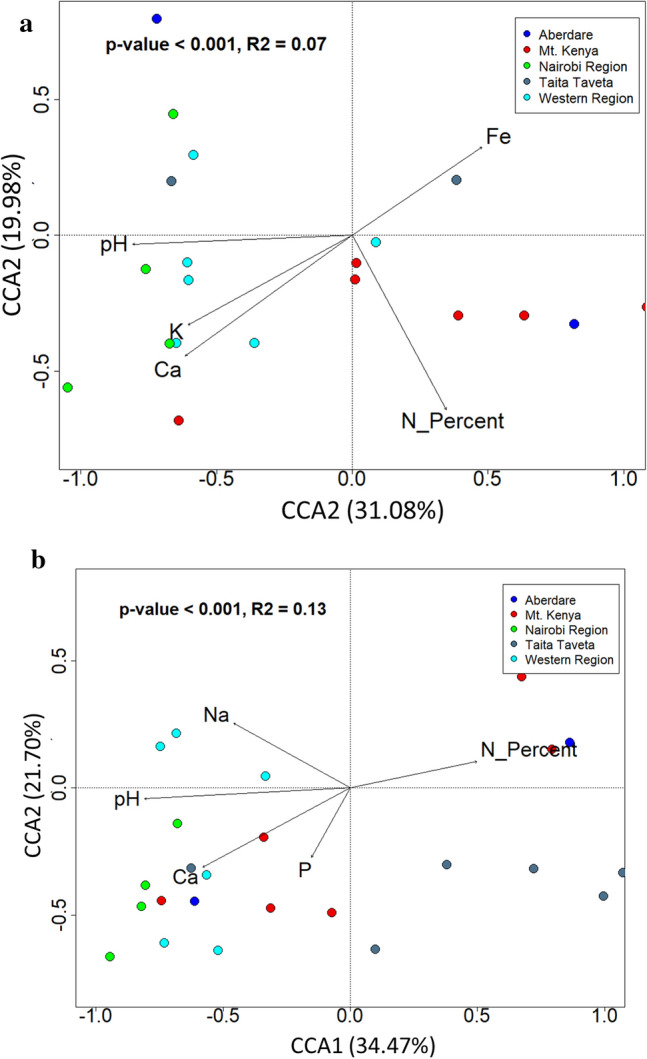
(a,b) Canonical correspondence analysis (CCA) plots showing the effect of soil physicochemical characteristics and plant density index on bacterial and archaeal communities at 99% significance. The percentage explained by various soil characteristics is expressed in the CCA1 and CCA2 axes and samples were color-coded on the plots according to forest regions. (a) represents CCA of Bacterial communities while (b) represents CCA of Archaea communities within soil samples collected from the five regions in Kenya.
The “uniqueness” of Kenyan forest microbiomes
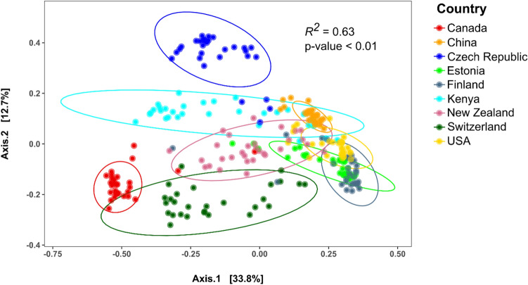
In order to address the question of whether Kenyan forest soils harbor unique microbiome compositions, the phylogenetic datasets used in this study were compared with datasets on forest soil microbiomes from other countries across the globe (Supplementary Table 3). Comparisons of the beta-diversity scores between these datasets, based on Bray–Curtis index (Fig. 6), revealed community structures of forest soil microbiomes which were, to some extent, distinguishable by the country of origin (R2 = 0.63; p-value < 0.01). The Kenyan dataset formed a distinct group with some degree of overlap with soil microbiomes from China, the Czech Republic, New Zealand and the USA. This overlap could be a result of common plant cover between the sampled areas in the different countries. Some forests in Kenya are known to harbor globally distributed plant species such as bamboo (A. alpina), indigenous plant species found within forests with highest floral diversity such as (Coffea fadenii, Juniperus procera—African pencil cedar, Podocarpus falcatus, latifolius, Tabernaemontana stapfiana, Ocotea usambarensis, Macaranga conglomerata, and Psychotria petit. Forests harboring moderate floral diversity included Podocarpus, Dombeya, Croton megalocarpus, while dryland species included Acacia species such as A. tortilis, A. melifera, A. abyssinica, and A. polyacantha. Plantation species included Eucalyptus grandis, E. saligna, E. camaldulensis and E. urophylla29. It is also worth noting that the Kenyan dataset exhibited the highest variability of beta-diversity scores, which reflect the variety of ecoregions sampled in this study.
Figure 6.
Principal Component Analysis (PCoA) ordination of the Bray–Curtis distance between samples from different country datasets. Samples are colored according to country of origin. The PERMANOVA significance results on differences in beta-diversity according to country of origin are displayed in the plot. Ellipses around the clusters of samples represent the predicted distribution of points within each country group at a 95% confidence interval.
The significant compositional differences between national datasets were reflected in the LDA comparison results, which identified 178 taxa differently distributed across the datasets (Supplementary Table 4). Fourteen of these were over-represented in Kenyan forest soils, including the Archeal genus Nitrososphaera. Other over-represented genera of potential ecological relevance to Kenya forest soils included Bradyrhizobium, which is positively associated with soil health30 and Chitinophaga, members of chitinolytic Myxobacteria known to control fungal populations in soils31. It is also worth noting that several of the over-represented taxa in the Kenyan soil dataset belonged to uncultured groups of bacteria, including members of uncultured genera TK-10 and Ellin606, an indication of Kenyan forest soils may harbor a catalogue of novel taxa. During development of bio-conservation strategies in these forest regions, consideration of these distinct microbiomes with unique taxa should be taken into account.
Materials and methods
Study site and sample collection
This study was part of an ongoing consortium project that focused on a primary-scale survey of soil chemistry and microbiology across a range of regional and climatic zones in sub-Saharan Africa9.
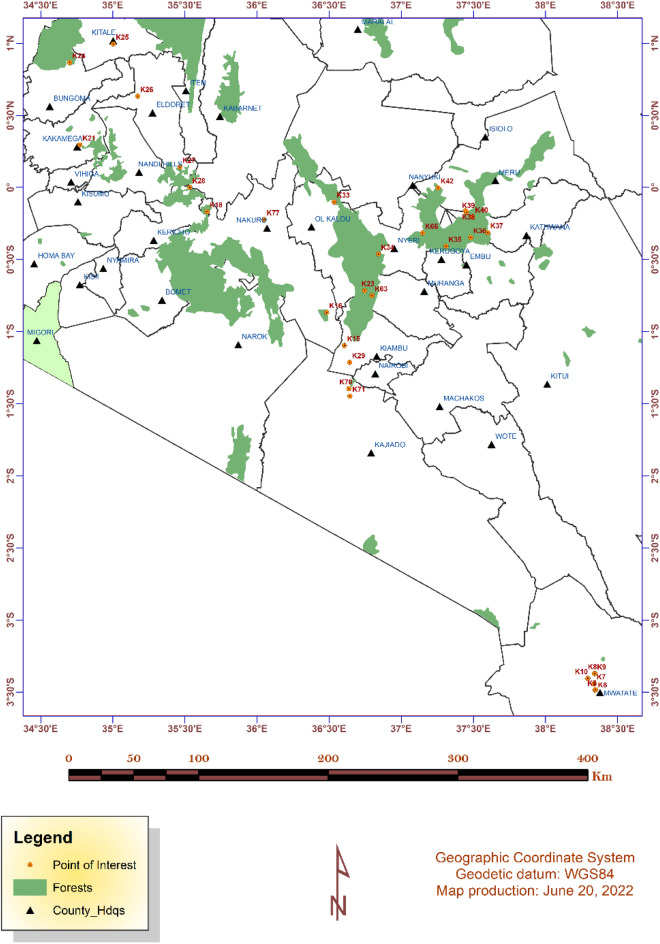
In Kenya, a microbiome survey of the soils across selected forest ecosystems was carried out based on a census for forest regions (http://kws.go.ke/content/overview-0). Data capture at each sampling site included GPS location, elevation, vegetation at the time of sample collection, slope, general soil description and general site description. To accurately show the sampled forest sites to scale, a map was constructed using the GPS coordinates captured from the forests during fieldwork using ArcGIS 10.8.1 (Environmental Systems Research Institute software application, 2020), https://www.esri.com/en-us/arcgis/products/arcgis-platform/overview; which was used to visualize and display the sample sites. The layers for towns, rivers, lakes and roads were added from ArcGIS Online database to enrich the thematic map as shown in (Fig. 7). The distribution and characteristics of the selected forests used in this study are summarized in Supplementary Table 1.
Figure 7.
Kenyan forest sites where soil samples were collected. The samples are indicated sequentially from K5 to K77 within the map. The point of interest represents each sampling site. The map was created using ArcGIS 10.8.1 (Environmental Systems Research Institute software application, 2020); https://www.esri.com/en-us/arcgis/products/arcgis-platform/overview.
Sampling was done by recovering 4 × 200 g topsoil samples (0–5 cm depth) at approximately 50 m spacing at each site. Each working sample was obtained by scooping a composite of 4 × 50 g pseudo-replicate samples, recovered from the corners of a one square meter virtual quadrat. Each sample was collected in a separate labelled Whirl Pak bags and stored at 4 °C prior to shipment to University of Pretoria (South Africa) for nucleic acid extraction and soil physicochemical analysis.
These samples were later grouped into regions depending on geographical location on the Kenyan map as follows: Aberdare (Sample K23, K33, K34, K63 and K77); Mt. Kenya (K35, K36, K37, K38, K39, K40, K42 and K66); Nairobi (K15, K16, K29, K70 and K71); Taita Taveta (K5, K6, K7, K8, K9 and K10) and Western region (K18, K21, K24, K25, K26, K27 and K28).
Soil physicochemical characteristics
Soil physicochemical characteristics (Supplementary Table 1) were determined using protocols outlined by AgriLASA (2004). Soil pH was measured using the slurry method at a 1:2.5 soil/water ratio, and the pH of the supernatant was recorded with a calibrated bench top pH meter (Crison Basic, + 20, Crison, Barcelona, Spain). The concentrations of soluble and exchangeable of sodium (Na), potassium (K), carbon (C), magnesium (Mg), and phosphorus (P) were determined using Mehlich 3 test32. The extractable ion concentration was quantified using ICP-OES (Inductively Coupled Plasma Optical Emission Spectrometry, Spectro Genesis, SPECTRO Analytical Instruments GmbH & Co. KG, Germany). Soil particle size distribution (sand/silt/clay percent) was measured using the Bouyoucos method33. Total nitrogen (TN) and soil organic carbon (TOC) were measured using the catalyzed high temperature combustion method (Dumas method)34. The Enhanced Vegetation Index-2 (EVI2) was obtained from the NASA Land Processes Distributed Active Archive Center’s (LP DAAC) VIIRS Vegetation Indices dataset35 at a 500-m resolution.
Prokaryotic DNA extraction and 16SrRNA gene sequencing
Total DNA was extracted from soil samples using the DNeasy PowerSoil Kit (QIAGEN, USA) following the manufacturer's instructions with the following modifications; the elution buffer C6 was pre-heated to 55ºC for 10 min before the final elution step, and the DNA was eluted using 70 μl of the elution buffer. After extraction, DNA concentration and purity were checked using the Nanodrop 2000 (ThermoFisher, USA) and agarose gel electrophoresis. The DNA samples were sent to MRDNA laboratories (www.mrdnalab.com, Shallowater, TX, USA) for sequencing of the V4/V5 16S rRNA gene, using the 515F (5'-GTGYCAGCMGCCGCGGTAA-3') and 909 R (5'-CCCCGYCAATTCMTTTRAGT-3') primers, according to9,36. Before library preparation, the regions of interest were amplified using the HotStarTaq Plus Master Mix Kit (Qiagen, USA) and subsequently purified using calibrated Ampure XP beads (Beckman Coulter Life Sciences, USA). Sequencing was performed at MR DNA (www.mrdnalab.com, Shallowater, TX, USA) on MiSeq instrument following the manufacturer’s guidelines.
Sequence analysis and taxonomic classification
The generated raw amplicon sequence reads were filtered, trimmed, and clustered into unique amplicon sequence variants (ASVs) using the QIIME2 pipeline37. Briefly, raw sequences were demultiplexed, quality checked and a feature table constructed using Divisive Amplicon Denoising Algorithm 2 (DADA2) pipeline38 inbuilt within QIIME239.The raw sequences were denoised and chimeras removed. Sequences which were < 200 base pairs after phred20- base quality trimming, with ambiguous base calls, and those with homopolymer runs exceeding 6 bp, were removed. The forward and reverse reads were truncated at 324 base pairs. This was followed by calculation of denoising statistics, picking of representative sequences and creation of ASVs feature table. Sequencing processing resulted in a total of 1,944,316 high quality sequence reads, which were clustered into 41,901 ASVs at 3% genetic distance.
Representative sequences were aligned using MAFFT39 and highly variable regions were masked to reduce the noise in phylogenetic analysis40. Phylogenetic trees were created and rooted at midpoint on QIIME2. Taxonomic classification of ASVs was done using QIIME feature-classifier39 against the untrained SILVA 138.1 (release 2022.2)41. Demultiplexed high-quality sequence reads were deposited in the National Centre for Biotechnology Information (NCBI) Sequence Read Archive (SRA), as Bio Project ID: PRJNA851255 and study accession numbers available for download at http://www.ncbi.nlm.nih.gov/bioproject/851255. In addition, the metadata, soil chemistry data, input files Qiime and R analysis scripts were deposited at https://zenodo.org/ and a DOI-10.5281/zenodo.7827433 available using the link; https://doi.org/10.5281/zenodo.7827432.
Data processing of amplicon datasets from other countries.
Sequence datasets from selected forests around the globe were downloaded from publicly available databases (accession numbers at Supplementary Table 3) and processed using the QIIME2 pipeline as described above. Raw reads from the downloaded datasets spanned the 16S rRNA gene hypervariable regions v3-v4, v4, and v4-v5, depending on the study. To keep the sample sizes between countries comparable, a subset of between 28 to 30 samples was chosen for each dataset. To accommodate the variable quality scores of the different datasets, quality threshold was set to 20 and all reads were truncated at 220 bps. After DADA2 processing, the resulting representative sequence file and ASV table were merged with the Kenyan dataset. Read counts for the combined dataset ranged from 10877 to 346157 reads (Supplementary Fig. S1). The merged representative sequence file was taxonomically annotated using the un-trained SILVA database 138.1 (release 2022.2)41.
Statistical analysis
ASVs from QIIME2 were modified for use with the package phyloseq (version 1.36.0) in RStudio42. The taxonomy table was merged with the feature table, and the relative abundance and bar plots were plotted using the ggplot2 package (version 3.3.5)42. The normality of the dataset was first tested with the Shapiro–Wilk test43. The Kruskal–Wallis Rank Sum test44 was subsequently used to calculate the significance of mean differences in soil variables between forest soil samples (adj. p. value < 0.01). Tukey post hoc analysis test41 were used to compare significant differences between regions where soil environmental variables were normally distributed (adj. p. value < 0.01).
Significant differences in soil physicochemical characteristics were calculated using the stats package version 3.6.2 in RStudio version 4.0.341. The distribution of soil physicochemical variables across different forest sites was calculated on log-standardized data using the “decostand” function from vegan package (version 2.5.7)45, which performs principal component analysis of the data (PCA)45. The resulting distance matrix between samples was plotted in a PCA graph, with the projected direction and magnitude of the distribution for each variable plotted in a separate loading plot. The hmisc (version 4.5) package41 was subsequently used to calculate strong significant Pearson correlations44 between variables (adj. p-value < 0.01), which were plotted in a bubble graph using the corrplot (version 0.9) package41. Biodiversity metrics (alpha diversity) and community structure dissimilarity (beta diversity) were calculated using the vegan (version 2.5.7)46 and phyloseq (version 1.16.2)47 packages in RStudio. Observed richness, Inverse Simson48 and the Shannon indexes49 were used as metrics for alpha-diversity48. The prokaryotic ASV table was split into Archaea and Bacteria using the “subset_taxa” function in phyloseq before calculating the diversity indexes. Differences in alpha-diversity between designated regions were assessed as described for the soil physicochemical variables. Beta-diversity index of each soil sample was calculated from the Centered log-ratio transformation (CLR) ASV tables using the “vegdist” function in vegan, based on Bray-Cutis distance estimation method50. Ordination of the beta-diversity scores was plotted on a principal component analysis plot (PCoA)51, and the significance of beta-diversity dissimilarity between forest regions was calculated using Permutational Multivariate Analyses of Variance (PERMANOVA)52 with 999 permutations. Comparison of beta-diversity distribution between the samples of different countries datasets was also performed using the methodology described above.
The environmental drivers of prokaryotic community structure were estimated using Redundancy analysis (RDA)53. The soil physicochemical dataset was z-score standardized and tested for multicollinearity using the “vif” function from the car (version 3.0.11) package54. The best models for explanatory variables were calculated using forward step-wise regression model selection method with the ordistep() function in the vegan package, with 1000 permutations, and significant variables with vif values above 10 were removed. The significance of the best fitted models and each predictor variables in the model were calculated using the ANOVA permutation test with 1000 permutations55. The relative taxonomic abundances of prokaryotic taxa were compared between regions using Linear Discriminant Analysis (LDA) effect size (LEfSe) algorithm56 for high-dimensional biomarker discovery and explanation of differentially abundant organisms. This analysis was implemented using the package Microbiome Marker in RStudio56. Differences were analyzed using Kruskal–Wallis sum-rank test57 to detect significant differentially abundant taxa at genus level (adj. p. value < 0.01). The biological consistency was investigated using a set of pairwise tests among genera using the Wilcoxon rank-sum test58,59, with an LDA threshold of 2. The same LDA method was used to detect differently abundant taxa across the country datasets.
Supplementary Information
Acknowledgements
The authors gratefully acknowledge the receipt of financial support from USAID, The Oppenheimer Foundation and the University of Pretoria. We thank the following institutions for their assistance in this research. The National Commission for Science, Technology and Innovation (NACOSTI), National Environmental Management Authority (NEMA) and Kenya Wildlife Service (KWS) for permission to conduct research on microbial communities in Kenyan soils and access permit to the sampling areas. More appreciation goes to Taita Taveta University for hosting the consortium research activities.
Author contributions
Conceptualization, design of the experiments and methodology: H.I.B., D.A.C., L.A.O., A.K.K., E.N.K. Performed the experiments: L.A.O., A.K.K., E.N.K., M.W.M., H.I.B., D.A.C., F.A.N., J.O’.K. Data analysis; curation, formal analysis, validation, and visualization: L.A.O., A.K.K., E.N.K., K.W.M., P.B.N.M.L. Contributed reagents/materials/sequencing costs/analysis tools: H.I.B., D.A.C. Manuscript writing, review and editing: L.A.O., A.K.K., E.N.K., M.W.M., H.I.B., D.A.C., F.A.N., J.O.’K., K.W.M., P.B.N.M.L. All authors read and approved the final manuscript.
Data availability
The demultiplexed high-quality sequence reads has been deposited in the National Centre for Biotechnology Information (NCBI) Sequence Read Archive (SRA), as Bio Project ID: PRJNA851255 and study accession numbers available for download at http://www.ncbi.nlm.nih.gov/bioproject/851255. This Whole Genome Shotgun project has been deposited at DDBJ/EMBL/GenBank under the Bioproject PRJNA291812. The metadata, soil chemistry data, input files for Qiime and R analysis scripts were deposited at https://zenodo.org/ and a DOI-10.5281/zenodo.7827433 available using the link; 10.5281/zenodo.7827432.
Competing interests
The authors declare no competing interests.
Footnotes
Publisher's note
Springer Nature remains neutral with regard to jurisdictional claims in published maps and institutional affiliations.
Supplementary Information
The online version contains supplementary material available at 10.1038/s41598-023-33993-4.
References
- 1.Lladó S, López-Mondéjar R, Baldrian P. Drivers of microbial community structure in forest soils. Appl. Microbiol. Biotechnol. 2018;102:4331–4338. doi: 10.1007/s00253-018-8950-4. [DOI] [PubMed] [Google Scholar]
- 2.Act, F. Forests Act 2005, Act No. 7 of 2005. (FAO, Nairobi Kenya, 2005).
- 3.Grosso F, Iovieno P, Alfani A, De Nicola F. Structure and activity of soil microbial communities in three Mediterranean forests. Appl. Soil Ecol. 2018;130:280–287. doi: 10.1016/j.apsoil.2018.07.007. [DOI] [Google Scholar]
- 4.Thakur S, et al. Recent progress in sodium alginate based sustainable hydrogels for environmental applications. J. Clean. Prod. 2018;198:143–159. doi: 10.1016/j.jclepro.2018.06.259. [DOI] [Google Scholar]
- 5.Knelman JE, et al. Interspecific plant interactions reflected in soil bacterial community structure and nitrogen cycling in primary succession. Front. Microbiol. 2018;9:128. doi: 10.3389/fmicb.2018.00128. [DOI] [PMC free article] [PubMed] [Google Scholar]
- 6.Liu T, et al. Soil organic matter, nitrogen and pH driven change in bacterial community following forest conversion. For. Ecol. Manag. 2020;477:118473. doi: 10.1016/j.foreco.2020.118473. [DOI] [Google Scholar]
- 7.Hoppe B, et al. A pyrosequencing insight into sprawling bacterial diversity and community dynamics in decaying deadwood logs of Fagus sylvatica and Picea abies. Sci. Rep. 2015;5:1–9. doi: 10.1038/srep09456. [DOI] [PMC free article] [PubMed] [Google Scholar]
- 8.Fierer N. Embracing the unknown: Disentangling the complexities of the soil microbiome. Nat. Rev. Microbiol. 2017;15:579–590. doi: 10.1038/nrmicro.2017.87. [DOI] [PubMed] [Google Scholar]
- 9.Cowan DA, et al. Biogeographical survey of soil microbiomes across sub-Saharan Africa: Structure, drivers, and predicted climate-driven changes. Microbiome. 2022;10:131. doi: 10.1186/s40168-022-01297-w. [DOI] [PMC free article] [PubMed] [Google Scholar]
- 10.Maquia IS, et al. Mining the microbiome of key species from African savanna woodlands: Potential for soil health improvement and plant growth promotion. Microorganisms. 2020;8:1291. doi: 10.3390/microorganisms8091291. [DOI] [PMC free article] [PubMed] [Google Scholar]
- 11.Watuma BM, et al. An annotated checklist of the vascular plants of Taita Hills, Eastern Arc Mountain. PhytoKeys. 2022;191:1. doi: 10.3897/phytokeys.191.73714. [DOI] [PMC free article] [PubMed] [Google Scholar]
- 12.López-Mondéjar R, Voříšková J, Větrovský T, Baldrian P. The bacterial community inhabiting temperate deciduous forests is vertically stratified and undergoes seasonal dynamics. Soil Biol. Biochem. 2015;87:43–50. doi: 10.1016/j.soilbio.2015.04.008. [DOI] [Google Scholar]
- 13.Neina D. The role of soil pH in plant nutrition and soil remediation. Appl. Environ. Soil Sci. 2019;2019:1–9. doi: 10.1155/2019/5794869. [DOI] [Google Scholar]
- 14.Chen P, et al. Community composition and metabolic potential of endophytic Actinobacteria from coastal salt marsh plants in Jiangsu, China. Front. Microbiol. 2019;10:1063. doi: 10.3389/fmicb.2019.01063. [DOI] [PMC free article] [PubMed] [Google Scholar]
- 15.Wei H, et al. Contrasting soil bacterial community, diversity, and function in two forests in China. Front. Microbiol. 2018;9:1693. doi: 10.3389/fmicb.2018.01693. [DOI] [PMC free article] [PubMed] [Google Scholar]
- 16.Lauber CL, Hamady M, Knight R, Fierer N. Pyrosequencing-based assessment of soil pH as a predictor of soil bacterial community structure at the continental scale. Appl. Environ. Microbiol. 2009;75:5111–5120. doi: 10.1128/AEM.00335-09. [DOI] [PMC free article] [PubMed] [Google Scholar]
- 17.Kalam S, et al. Recent understanding of soil acidobacteria and their ecological significance: A critical review. Front. Microbiol. 2020 doi: 10.3389/fmicb.2020.580024. [DOI] [PMC free article] [PubMed] [Google Scholar]
- 18.Rosenberg EJT. The Family Chitinophagaceae. Springer; 2014. pp. 493–495. [Google Scholar]
- 19.Wu R-N, Meng H, Wang Y-F, Lan W, Gu J-D. A more comprehensive community of ammonia-oxidizing archaea (AOA) revealed by genomic DNA and RNA analyses of amoA gene in subtropical acidic forest soils. Microb. Ecol. 2017;74:910–922. doi: 10.1007/s00248-017-1045-4. [DOI] [PubMed] [Google Scholar]
- 20.Saghaï A, et al. Diversity of archaea and niche preferences among putative ammonia-oxidizing Nitrososphaeria dominating across European arable soils. Environ. Microbiol. 2022;24:341–356. doi: 10.1111/1462-2920.15830. [DOI] [PubMed] [Google Scholar]
- 21.Karanja EN, et al. Diversity and structure of prokaryotic communities within organic and conventional farming systems in central highlands of Kenya. PloS One. 2020;15:e0236574. doi: 10.1371/journal.pone.0236574. [DOI] [PMC free article] [PubMed] [Google Scholar]
- 22.Pellikka P, et al. Impact of land cover change on aboveground carbon stocks in Afromontane landscape in Kenya. Appl. Geogr. 2018;94:178–189. doi: 10.1016/j.apgeog.2018.03.017. [DOI] [Google Scholar]
- 23.Lladó S, López-Mondéjar R, Baldrian P. Forest soil bacteria: Diversity, involvement in ecosystem processes, and response to global change. Microbiol. Mol. Biol. Rev. 2017;81:e00063–16. doi: 10.1128/MMBR.00063-16. [DOI] [PMC free article] [PubMed] [Google Scholar]
- 24.Platts PJ, et al. Delimiting tropical mountain ecoregions for conservation. Environ. Conserv. 2011;38:312–324. doi: 10.1017/S0376892911000191. [DOI] [Google Scholar]
- 25.Myers N, Mittermeier RA, Mittermeier CG, Da Fonseca GA, Kent J. Biodiversity hotspots for conservation priorities. Nature. 2000;403:853–858. doi: 10.1038/35002501. [DOI] [PubMed] [Google Scholar]
- 26.Lung, T. & Schaab, G. Change-detection in Western Kenya: the documentation of fragmentation and disturbance for Kakamega forest and associated forest areas by means of remotelysensed imagery. ISPRS archives35 (2004).
- 27.Hussain S, et al. Significance of Fe (II) and environmental factors on carbon-fixing bacterial community in two paddy soils. Ecotoxicol. Environ. Saf. 2019;182:109456. doi: 10.1016/j.ecoenv.2019.109456. [DOI] [PubMed] [Google Scholar]
- 28.Maundu, P. & Tengnäs, B. Useful trees and shrubs for Kenya. ICRAF Technical handbook series (2005).
- 29.VanInsberghe D, et al. Non-symbiotic Bradyrhizobium ecotypes dominate North American forest soils. ISME J. 2015;9:2435–2441. doi: 10.1038/ismej.2015.54. [DOI] [PMC free article] [PubMed] [Google Scholar]
- 30.Sharma S, et al. Biocontrol potential of chitinases produced by newly isolated Chitinophaga sp. S167. World J. Microbiol. Biotechnol. 2020;36:1–15. doi: 10.1007/s11274-020-02864-9. [DOI] [PubMed] [Google Scholar]
- 31.Mehlich A. Mehlich 3 soil test extractant: A modification of Mehlich 2 extractant. Commun. Soil Sci. Plant Anal. 1984;15:1409–1416. doi: 10.1080/00103628409367568. [DOI] [Google Scholar]
- 32.Bouyoucos GJ. Hydrometer method improved for making particle size analyses of soils 1. Agron. J. 1962;54:464–465. doi: 10.2134/agronj1962.00021962005400050028x. [DOI] [Google Scholar]
- 33.Bremner JM. Nitrogen-total. Methods Soil Anal. 1996;5:1085–1121. [Google Scholar]
- 34.Didan, A. VIIRS/NPP Vegetation Indices 16-Day L3 Global 500m SIN Grid V001. NASA EOSDIS Land Processes DAAC: Oak Ridge, TN, USA (2018).
- 35.Caporaso JG, et al. Ultra-high-throughput microbial community analysis on the Illumina HiSeq and MiSeq platforms. ISME J. 2012;6:1621–1624. doi: 10.1038/ismej.2012.8. [DOI] [PMC free article] [PubMed] [Google Scholar]
- 36.Dacal M, et al. Climate change legacies contrastingly affect the resistance and resilience of soil microbial communities and multifunctionality to extreme drought. Funct. Ecol. 2022;36:908–920. doi: 10.1111/1365-2435.14000. [DOI] [Google Scholar]
- 37.Callahan BJ, et al. DADA2: High-resolution sample inference from Illumina amplicon data. Nat. Methods. 2016;13:581–583. doi: 10.1038/nmeth.3869. [DOI] [PMC free article] [PubMed] [Google Scholar]
- 38.Bolyen E, et al. Reproducible, interactive, scalable and extensible microbiome data science using QIIME 2. Nat. Biotechnol. 2019;37:852–857. doi: 10.1038/s41587-019-0209-9. [DOI] [PMC free article] [PubMed] [Google Scholar]
- 39.Rozewicki J, Li S, Amada KM, Standley DM, Katoh K. MAFFT-DASH: Integrated protein sequence and structural alignment. Nucleic Acids Res. 2019;47:W5–W10. doi: 10.1093/nar/gkz342. [DOI] [PMC free article] [PubMed] [Google Scholar]
- 40.Price MN, Dehal PS, Arkin AP. FastTree 2–approximately maximum-likelihood trees for large alignments. PLoS ONE. 2010;5:e9490. doi: 10.1371/journal.pone.0009490. [DOI] [PMC free article] [PubMed] [Google Scholar]
- 41.RStudio, T. RStudio: integrated development for R. Rstudio Team, PBC, Boston, MA URLhttp://www.rstudio.com (2020).
- 42.McMurdie PJ, Holmes S. phyloseq: An R package for reproducible interactive analysis and graphics of microbiome census data. PLoS ONE. 2013;8:e61217. doi: 10.1371/journal.pone.0061217. [DOI] [PMC free article] [PubMed] [Google Scholar]
- 43.Hollander M, Wolfe DA. Nonparametric Statistical Methods. Inc; 1973. [Google Scholar]
- 44.Armstrong, R. A. & Hilton, A. The use of analysis of variance (ANOVA) in applied microbiology. (2004).
- 45.Jari, O. et al. Peter R. Minchin, RB O’Hara, Gavin L. Simpson, Peter Solymos, M., Henry H. Stevens, Eduard Szoecs and Helene Wagner Oksanen Jari, & Oksanen Jari. vegan: Community Ecology Package. https://CRAN.R-project.org/package=vegan (2020).
- 46.Chao A, et al. Rarefaction and extrapolation with Hill numbers: A framework for sampling and estimation in species diversity studies. Ecol. Monogr. 2014;84:45–67. doi: 10.1890/13-0133.1. [DOI] [Google Scholar]
- 47.Royston JP. An extension of Shapiro and Wilk's W test for normality to large samples. J. Roy. Stat. Soc.: Ser. C (Appl. Stat.) 1982;31:115–124. [Google Scholar]
- 48.Shannon CE. The Mathematical Theory of Communication, by CE Shannon (and Recent Contributions to the Mathematical Theory of Communication), W. Weaver. University of illinois Press Champaign; 1949. [Google Scholar]
- 49.Bray JR, Curtis JT. An ordination of the upland forest communities of southern Wisconsin. Ecol. Monogr. 1957;27:326–349. doi: 10.2307/1942268. [DOI] [Google Scholar]
- 50.Jolliffe IT. Principal Component Analysis for Special Types of Data. Springer; 2002. [Google Scholar]
- 51.Anderson MJ. Permutational multivariate analysis of variance. Dept. Stat. Univ. Auckland. 2005;26:32–46. [Google Scholar]
- 52.McArdle BH, Anderson MJ. Fitting multivariate models to community data: A comment on distance-based redundancy analysis. Ecology. 2001;82:290–297. doi: 10.1890/0012-9658(2001)082[0290:FMMTCD]2.0.CO;2. [DOI] [Google Scholar]
- 53.Dormann CF, et al. Collinearity: A review of methods to deal with it and a simulation study evaluating their performance. Ecography. 2013;36:27–46. doi: 10.1111/j.1600-0587.2012.07348.x. [DOI] [Google Scholar]
- 54.Legendre P, Oksanen J, ter Braak CJ. Testing the significance of canonical axes in redundancy analysis. Methods Ecol. Evol. 2011;2:269–277. doi: 10.1111/j.2041-210X.2010.00078.x. [DOI] [Google Scholar]
- 55.Segata N, et al. Metagenomic biomarker discovery and explanation. Genome Biol. 2011;12:1–18. doi: 10.1186/gb-2011-12-6-r60. [DOI] [PMC free article] [PubMed] [Google Scholar]
- 56.Cao Y, et al. microbiomeMarker: An R/Bioconductor package for microbiome marker identification and visualization. Bioinformatics. 2022;38:4027–4029. doi: 10.1093/bioinformatics/btac438. [DOI] [PubMed] [Google Scholar]
- 57.Kruskal WH, Wallis WA. Use of ranks in one-criterion variance analysis. J. Am. Stat. Assoc. 1952;47:583–621. doi: 10.1080/01621459.1952.10483441. [DOI] [Google Scholar]
- 58.Wilcoxon F. Breakthroughs in Statistics. Springer; 1992. pp. 196–202. [Google Scholar]
- 59.Mann HB, Whitney DR. On a test of whether one of two random variables is stochastically larger than the other. Ann. Math. Stat. 1947;18:50–60. doi: 10.1214/aoms/1177730491. [DOI] [Google Scholar]
Associated Data
This section collects any data citations, data availability statements, or supplementary materials included in this article.
Supplementary Materials
Data Availability Statement
The demultiplexed high-quality sequence reads has been deposited in the National Centre for Biotechnology Information (NCBI) Sequence Read Archive (SRA), as Bio Project ID: PRJNA851255 and study accession numbers available for download at http://www.ncbi.nlm.nih.gov/bioproject/851255. This Whole Genome Shotgun project has been deposited at DDBJ/EMBL/GenBank under the Bioproject PRJNA291812. The metadata, soil chemistry data, input files for Qiime and R analysis scripts were deposited at https://zenodo.org/ and a DOI-10.5281/zenodo.7827433 available using the link; 10.5281/zenodo.7827432.


