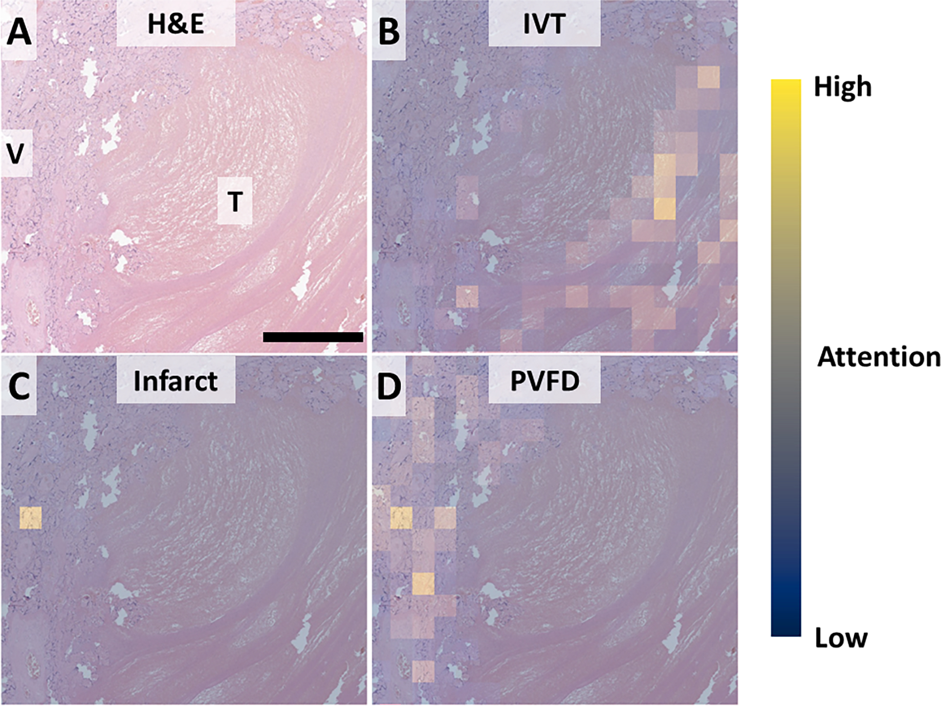
Figure 7, attention maps for understanding inappropriate classification.

A: Low power view showing the interface between thrombus (IVT) and rim of compressed villi (V). B: Attention for IVT is appropriately highest within the thrombus and overlapping lines of Zahn. C: Attention for infarct highlighting expected a single very high-attention focus. D: Expected rim of PVFD between thrombus and normal tissue. Scale bar: 250 um. Attention: Blue – low, Yellow – high.
