

Abstract
Humans are exposed to large numbers of electrophiles from diet, the environment, and endogenous physiological processes. Adducts formed at the N-terminal valine of hemoglobin are often used as biomarkers of human exposure to electrophilic compounds. We have previously reported the formation of hemoglobin N-terminal valine adducts (added mass, 106.042 Da) in the blood of human smokers and non-smokers and identified their structure as 4-hydroxybenzyl-Val. In the present work, mass spectrometry-based proteomics was utilized to identify additional sites for 4-hydroxybenzyl adduct formation at internal nucleophilic amino acid side chains within hemoglobin. Hemoglobin isolated from human blood was treated with para-quinone methide (para-QM), followed by global nanoLC-MS/MS and targeted nanoLC-MS/MS to identify amino acid residues containing the 4-hydroxybenzyl modification. Our experiments revealed the formation of 4-hydroxybenzyl adducts at αHis20, αTyr24, αTyr42, αHis45, βSer72, βThr84, βThr87, βSer89, βHis92, βCys93, βCys112, βThr123, and βHis143 residues (in addition to N-terminal valine) through characteristic MS/MS spectra. These amino acid side chains had variable reactivity towards para-QM, with αHis45, αTyr42, βCys93, βHis92, and βSer72 forming the largest numbers of adducts upon exposure to para-QM. Two additional mechanisms for formation of 4-hydroxybenzyl adducts in humans were investigated: exposure to 4-hydroxybenzaldehyde (4-HBA) followed by reduction and UV-mediated reactions of hemoglobin with tyrosine. Exposure of hemoglobin to 5-fold molar excess of 4-HBA followed by reduction with sodium cyanoborohydride produced 4-hydroxybenzyl adducts at several amino acid side chains, of which αHis20, αTyr24, αTyr42, αHis45, βSer44, βThr84, and βHis92 were verified in targeted mass spectrometry experiments. Similarly, exposure of human blood to ultraviolet radiation produced 4-hydroxybenzyl adducts at αHis20, αTyr24, αTyr42, αHis45, βSer44, βThr84, and βSer89. Overall, our results reveal that 4-hydroxybenzyl adducts form at multiple nucleophilic sites of hemoglobin and that para-QM is the most likely source of these adducts in humans.
Keywords: adducts, hemoglobin adducts, quinone methide, benzaldehyde, adductomics
Graphical Abstract

Introduction
Over their lifetimes, humans are exposed to large numbers of endogenous and exogenous electrophilic compounds, resulting in the formation of covalent adducts at nucleophilic sites of cellular biomolecules.1,2 These electrophiles range from products of normal cellular metabolism3 and dietary components4,5 to the metabolic byproducts of commensal microbiota6 and environmental contaminants.7,8 Characterization of protein and DNA adducts (adductomics) in humans is of considerable importance for disclosing risk factors to human health as adducts are associated with an increased risk of cancer9 and other chronic diseases.10–12
Protein adducts are useful biomarkers of exposure to electrophiles due to their longevity as compared to DNA lesions, since the latter can be efficiently removed via cellular repair mechanisms.2,13 Specifically, hemoglobin adducts are commonly used in human exposome studies due to the high abundance of hemoglobin in blood, ready blood sample availability, and the relatively long lifetime of hemoglobin (120 days).14 Adducts to the N-terminal valine of hemoglobin are commonly used as biomarkers of human exposure due to the high solvent accessibility of this site and its low pKa value as compared to other potential nucleophilic sites within the protein.15–17 The Tӧrnqvist group has developed an untargeted methodology for identifying novel N-terminal valine hemoglobin adducts (FIRE).18–20 In this approach, the N-terminal valine residue of human hemoglobin is derivatized and cleaved via modified Edman degradation to yield a fluorescein isothiohydantoin-valine derivative, which can subsequently be analyzed by HPLC-ESI-MS/MS.18–20 Studies using hemoglobin isolated from human subjects revealed a number of adducts at the N-terminal valine of hemoglobin resulting from methylation, carboxymethylation, and ethylation, as well as modification by ethyl vinyl ketone, acrylic acid, glyoxal, methylglyoxal, and 1-octen-3-one.18,21 Our recent work using the FIRE approach determined that the third-most abundant N-terminal valine adduct (106.042 Da, corresponding to the elemental formula C7H6O) corresponds to the addition of a 4-hydroxybenzyl group.22 Although the exact source of this adduct in humans remains unknown, we proposed that they could be a result of para-quinone methide (para-QM)23–25 or 4-hydroxybenzaldehyde (HBA)26 reactions with the N-terminal valine of hemoglobin.
While the solvent exposed N-terminal valine residue of Hb is frequently modified upon exposure to electrophiles, side chains of many amino acids including cysteine, lysine, and histidine are inherently nucleophilic, rendering them as potential sites for adduct formation.27,28 For example, Cys34 of human serum albumin is a likely adduction site upon exposure to electrophiles29,30 and has been previously used to monitor human exposures.31 We have shown that Cys145 of human O6-alkylguanine DNA alkyltransferase (AGT) is the preferred modification site upon exposure to 1,2,3,4-diepoxybutane32 and antitumor nitrogen mustards,33,34 while cis-diamminedichloroplatinum (II) (cisplatin) targets several nucleophilic residues within the AGT protein including Glu110, Lys125, Cys145, His146, Arg147, and Cys150.35 Sulfinamide adducts at βCys93 of hemoglobin are associated with exposure to tobacco smoke components and grilled meat byproducts such as 2-amino-9H-pyrido[2,3-b]indole 4-aminobiphenyl (HONH-ABP).36,37 Further, α,β-unsaturated aldehyde byproducts of lipid peroxidation have been shown to form adducts at histidine side chains in hemoglobin.38
The primary aims of the present study were to characterize additional sites of 4-hydroxybenzyl adduct formation in hemoglobin and to help elucidate their potential source(s) in humans. To help control the reaction of para-QM with hemoglobin, the molecule itself was eschewed in favor of a precursor that could be converted into the highly reactive para-QM when desired. A stable synthetic para-QM precursor,39,40 [4-(bromomethyl)phenoxy]-tert-butyl-dimethylsilane, was activated by potassium fluoride to form para-QM in situ (Scheme 1A). Bottom-up global and targeted proteomics nanoLC-MS/MS workflows were used to identify the sites containing 4-hydroxybenzyl adducts. Alternate sources of 4-hydroxybenzyl adducts in humans were also investigated through the reactions of hemoglobin with 4-hydroxybenzaldehyde (4-HBA) and through exposure of whole blood to UV light with and without addition of tyrosine, a possible precursor of para-QM (Scheme 1B, 1C).41
Scheme 1:

Proposed reaction mechanisms for the formation of adducts with histidine side chains using three potential sources of a 4-hydroxybenzyl adduct. A) Formation of para-QM from para-QM precursor with potassium fluoride and subsequent reaction. B) Reductive amination of 4-hydroxybenzaldehyde with sodium cyanoborohydride. C) Generation of para-QM upon reaction of tyrosine in a peptide with ultraviolet light and subsequent adduct formation.
Experimental Procedures
Materials and instrumentation
Human blood with added potassium EDTA was purchased from Biochemed Services (Winchester, VA). [4-(Bromomethyl)phenoxy]-tert-butyl-dimethylsilane (para-QM precursor) was synthesized in our laboratory according to established protocols.39,40 4-Hydroxybenzaldehyde, sodium cyanoborohydride, potassium chloride, potassium phosphate, trifluoroacetic acid, and oxidized L-glutathione were purchased from Millipore Sigma (Burlington, MA). Potassium fluoride was obtained from Mallinkrodt (Staines-upon-Thames, UK). Sodium chloride and calcium chloride were procured from Thermo Fisher Scientific (Waltham, MA). Trypsin was purchased from Promega Corporation (Madison, WI). L-Tyrosine was provided by C. Eddington. Formic acid was purchased from Honeywell Fluka (Mexico City, MX). LC-MS grade acetonitrile and water were obtained from Thermo Fisher Scientific (Waltham, MA).
All nanoLC-MS/MS analyses were performed on a QExactive Orbitrap Mass Spectrometer interfaced to a Dionex Ultimate 3000 nanoLC System set to nanoflow mode. Liquid chromatography was carried out using a reverse-phase C18 nanospray column (250 μm × 34 cm) manually packed with Luna 5μm C18 solid-phase media (Phenomenex). Photoreactions were performed in an RMR-600 Photochemical Reactor from Rayonet (Branford, CT).
Isolation of hemoglobin from blood
Hemoglobin was isolated from fresh human blood according to published protocols.37 Briefly, 10 mL of human blood was centrifuged at 800 g for 15 min at 4 °C to separate the erythrocytes from the plasma. The plasma was decanted from the pelleted erythrocytes, which were then subjected to three washes with an equal volume of cold Ringer’s solution (250 mM NaCl, 10 mM KCl, 3 mM CaCl2, pH = 7.4) followed by further centrifugation. To isolate hemoglobin, erythrocytes were suspended in an equal volume of distilled water and subjected to 5 min of sonication. Samples were then centrifuged at 15,000 rpm for 15 min to pellet cellular debris, reserving the hemoglobin-containing supernatant for further experiments. The concentration of HbO2 in the supernatant was ascertained via absorption at λ = 542 nm (ε = 57.1 M−1cm−1).
Hemoglobin Reactions with para-QM
Aliquots of human hemoglobin (64 mg) were diluted to 100 μL with 100 mM potassium phosphate buffer (pH= 7.4) containing 50 mM KF. The final hemoglobin concentration was 10 mM. Para-QM precursor was dissolved in DMSO, and aliquots were added to the hemoglobin samples (n = 3) to achieve the final concentrations of 1 mM, 5 mM, 10 mM, or 50 mM QM, followed by incubation overnight at 37 °C. Following treatment with para-QM, hemoglobin samples were added to Amicon Ultra centrifugal filters (Millipore-Sigma) with a 10 kDa cutoff and centrifugated at 14,000 g for 10 min. A solution of triethyammonium bicarbonate (TEAB, 25 mM pH = 8.0) was then added to the spin columns, followed by two additional rounds of buffer exchange.
Hemoglobin Reactions with 4-HBA
4-Hydroxybenzaldehyde (4-HBA) was dissolved in ethanol to obtain a 1 M stock solution. In two sets of three replicates, hemoglobin aliquots and 4-HBA stock were added to distilled water to give final concentrations of 10 mM hemoglobin and 50 mM 4-HBA, respectively. Samples were incubated at 37 °C overnight. Following treatment, one set of samples was supplemented with sodium cyanoborohydride to a concentration of 10 mM and incubated at room temperature for 30 min. Samples were buffer exchanged into TEAB buffer three times using 10K filters as described above.
Exposure of human blood to ultraviolet radiation
Four quartz cuvettes were each filled with 300 μL of whole blood and subdivided into two groups of 2. One set of cuvettes was also supplemented with 250 μL of a saturated solution of L-tyrosine in Ringer’s solution, while the other was supplemented with 250 μL of Ringer’s solution. The solutions were exposed to ultraviolet light (254 nm) at room temperature for one hour. As another negative control, a set of cuvettes containing 250 μL blood and 250 μL Ringer’s solution were incubated in the dark for one hour at room temperature. Following incubation, the samples were transferred to Eppendorf vials and centrifuged at 800 g for 10 min to pellet the erythrocytes in solution. The erythrocytes were then washed three times in Ringer’s solution before being lysed in an equivalent volume of distilled water accompanied by 5 min of sonication. The samples were then centrifuged at 15,000 rpm for 15 min to pellet cellular debris, reserving the supernatant for mass spectrometry analysis.
Sample processing for proteomics analyses
Buffer-exchanged hemoglobin samples were treated with a 10-fold molar excess iodoacetamide in TEAB buffer in the dark at room temperature for 30 min. Following incubation, aliquots of protein (50 μg) were taken and buffer exchanged three times into TEAB buffer. Each sample was then supplemented with trypsin at a ratio of 1:20 w/w and incubated overnight at 37 °C. Proteolytic digestion was terminated via the addition of formic acid to 10%, after which the samples were desalted via C18 spin columns and evaporated to dryness under vacuum.
Global nanoLC-MS/MS analyses
Tryptic digests were reconstituted in buffer A (0.1% FA in water) and analyzed on an Orbitrap QExactive Mass Spectrometer interfaced with a reverse-phase HPLC system operating in nanoflow mode. The nanoLC column was eluted at a flow rate of 300 nL/min. Samples were analyzed using a gradient of 5 – 22 % buffer B (0.1% FA in acetonitrile) over 71 min, followed by 22 – 33 % over 5 min, 33 – 90% over 5 minutes, a 90% buffer B wash for 4 min, and finally a 90 – 4% decrease in buffer B over 2 minutes followed by a 4 min equilibration at 4% B.
Peptides were analyzed in the positive ion mode using Full MS/dd-MS2. The instrument was operated at 70,000 resolution with an AGC target of 1 e6, a maximum IT of 30 ms, and a scan range of 300 to 2000 m/z. Tandem mass (MS2) spectra were captured at 17,500 resolution, AGC target of 5e4, maximum IT of 50 ms, an isolation window of 2.0 m/z and a normalized collision energy of 30. Data were collected in the centroid mode. Control hemoglobin samples were used to create an exclusion list of unmodified hemoglobin peptides.42
Raw mass spectrometry data were analyzed using Proteome Discoverer 2.2. The data was searched against the SwissProt human proteome version 2017–10-25 containing the conventional hemoglobin FASTA sequence supplemented with hemoglobin alpha and beta subunit FASTA files with the N-terminal methionine residues removed to account for the N-terminal cleavage common in eukaryotic post-translational modification.43 Variable modifications included oxidation at methionine, carbamidomethyl modification of cysteine, and a 106.042 Da modification at cysteine, histidine, lysine, arginine, serine, threonine, tyrosine, and N-termini of peptides corresponding to the putative 4-hydroxybenzyl group (C7H6O) with 10 ppm mass tolerance. In addition, a Percolator step44 was added with a strict target FDR of 0.01, a relaxed target FDR of 0.05, and a validation based on the q-value to ensure confident identification of peptide modifications.
Targeted nanoLC-MS/MS analysis
Peptides identified in the global proteomics study were used to create an inclusion list containing high-confidence peptides modified with para-QM (+106.042 Da) alongside the corresponding unmodified peptides. This inclusion list was used for targeted analysis of these peptides in tryptic digests using joint PRM and full MS-SIM experiments. The instrument was operated in the positive ion mode, and samples were analyzed using the same nanoLC column and gradient as above. PRM experiments were conducted with a resolution of 70,000, an AGC target of 2e5, a maximum IT of 250 ms, an isolation window of 1.0 m/z and a normalized collision energy of 25. The accompanying Full MS-SIM experiment was run at a resolution of 70,000, an AGC target of 3e6, a maximum IT of 200ms, and a scan range of 300 to 2000 m/z.
The resulting targeted mass spectrometry data was analyzed using Skyline.45 The FASTA files of human hemoglobin alpha and beta subunits were imported into Skyline and used to build a curated target lists of peptides of interest with variable 4-hydroxybenzyl modification at cysteine, histidine, lysine, serine, threonine, and tyrosine residues (added mass of 106.042 Da). Magellan storage files (MSFs) generated by Proteome Discoverer containing the identified MS2 spectra of the hemoglobin-para-QM global data were used in Skyline to build a spectral library that the targeted data could be compared against. Peak areas of the three most abundant product ions of each precursor peptide were measured at each concentration of added para-QM and exported for analysis. Fraction of modified side chains following the addition of reactive species (para-QM precursor + KF, 4-HO-BA, or UV ± Tyr) served as a proxy measurement for the reactivity of that side chain towards the added electrophiles. The extent of adduct formation at each site was expressed as a percentage of total peptide detected as follows:
Predicted physiochemical properties of hemoglobin side chains
A consensus sequence for sites identified as having 4-hydroxybenzyl adducts was generated using ggseqlogo in R.46 Briefly, a list of polypeptides containing the modified side chain with 7 residues before and after the adducted site were uploaded into this tool, after which the consensus sequence was generated.
To estimate the pKa values of the amino acid side chains in human hemoglobin, the H++ automated server (version 3.2)47–49 was used with a normal human hemoglobin NMR structure 2h3550 obtained from the RCSB Protein Data Bank.51 In the determination of the side chain pKa values, the external dielectric constant was set to the default value of 80, and the external salinity to 0.15 M, and the internal dielectric constant to 4.
The relative accessible surface areas of hemoglobin side chains were obtained from the hemoglobin NMR structure 2h35 using the PISA webserver.52 These values were subsequently normalized using the theoretical maximal allowed solvent accessibilities of amino acid side chains calculated in Tien et al.53
Results
Global proteomic analysis of hemoglobin exposed to para-QM shows adduct formation at nucleophilic side chains
To identify amino acid residues within human hemoglobin that form adducts with para-QM, hemoglobin freshly isolated from human blood was incubated with a five-fold molar excess of para-QM precursor in the presence of potassium fluoride to release para-QM (Scheme 1A). Para-QM treated hemoglobin was digested to peptides with trypsin, and the resulting peptides were analyzed by global nanoLC-MS/MS as described in the Methods section. The resulting mass spectrometry data were processed using Proteome Discover. We utilized a variable modification of 106.042 Da (hydroxybenzyl group) and searched for the presence of this modification at various nucleophilic side chains as well as the N-termini of the protein. The hemoglobin molecule is a tetramer of two alpha and beta subunits; each of the two subunits has its own N-terminal valine residue which could accommodate a 4-hydroxybenzyl adduct. Analysis of the MS2 spectra of tryptic peptides obtained from para-QM treated hemoglobin showed spectral matches consistent with 4-hydroxybenzyl adduct (106.042 Da) at the N-termini of both alpha and beta subunits of hemoglobin (Figure 1A, 1B). This is in agreement with our previous work which employed the FIRE procedure to detect 4-hydroxybenzyl adducts at the N-terminal valine of human hemoglobin, but which could not distinguish between subunits of the protein.22
Figure 1:

Tandem mass spectra of A) Hemoglobin alpha subunit N-terminal peptide with N-terminal 4-hydroxybenzyl adduct, B) Hemoglobin beta subunit N-terminal peptide with N-terminal 4-hydroxybenzyl adduct, C) 4-hydroxybenzaldehyde adduct of histidine 45 in alpha subunit, D) 4-hydroxybenzaldehyde adduct of cysteine 93 in beta subunit. Spectra in A) and B) were sourced from Proteome Discoverer 2.2, C) and D) were taken from Skyline v20.2
Proteomics analyses allowed for 91% alpha subunit polypeptide sequence coverage and 95% beta subunit polypeptide sequence coverage of human hemoglobin (Table S1). In addition to 4-hydroxybenzyl adduct formation at N-terminal valines, global proteomics analyses led to preliminary identification of seventy-eight potentially adducted amino acid side chains. All MS/MS spectra were manually interrogated, and only those containing the requisite b- and y-ions for conclusive identification of the peptide and confident placement of the adduct were retained for further examination. This initial filtering resulted in twenty-nine potentially modified peptides (Table S2).
Targeted proteomic analysis verifies the presence of para-QM adducts at cysteine, histidine, lysine, serine, threonine, and tyrosine side chains
To verify the presence of 4-hydroxybenzyl adducts at sites of the protein preliminary identified in global proteomics experiments, targeted mass spectrometry analyses were conducted. Samples were re-analyzed in the parallel reaction mode (PRM) using an inclusion list of para-QM modified peptides obtained from global proteomics experiments (Table S2). Raw MS/MS data were searched in Skyline45 against a target list constructed from the peptides of the alpha and beta subunits of human hemoglobin with and without 4-hydroxybenzyl modification at the amino acid residues of interest. Assignments of peptides to the target list were confirmed using a spectral library constructed using MSF files generated in Proteome Discoverer during the global mass spectrometry analysis of hemoglobin exposed to excess para-QM. By using strict selection criteria wherein the exact position of the putative adduct can be localized using the b and y series in the MS/MS spectra, we were able to confirm 4-hydroxybenzyl modification of 14 amino acid side chains in the interior of the alpha and beta subunits of the protein. In samples treated with para-QM, mass spectrometry evidence was obtained for 4-hydroxybenzyl modification of cysteines (βCys93, βCys112), histidines (αHis20, αHis45, βHis92, βHis143), serines (βSer44, βSer72, βSer89), threonines (βThr84, βThr87, βThr123), and tyrosines (αTyr24, αTyr42) within the protein (Table 1B, Figures S1A–S1Q). Each of these modified peptides contains the 4-hydroxybenzyl adduct assigned to a specific amino acid residue using characteristic b- and y-ion series (Figure 1C, 1D). Hypothetical chemical structures of the adducts are shown in Scheme 2. Interestingly, preliminary analyses suggested that para-QM modified residues were localized in several discrete clusters within the alpha and beta subunits of the protein (Figure 2).
Table 1:
Hemoglobin amino acid residues showing 4-hydroxybenzyl adduct formation before and after treatment with para-QM, 4-hydroxybenzaldehyde, and UV irradiation. A) Levels of endogenous adducts in the control hemoglobin samples. B) Levels of adduct formation relative to control samples following incubation with potential 4-hydroxybenzyl adduct sources. Red highlighted values indicate increased adduct formation following treatment, green highlighted values indicate a loss of adduct following treatment. Residues annotated with an asterisk showed significant statistical differences to controls. The N-terminal peptides were not included in targeted experiments.
|
A)
| |||||
| Subunit | Residue |
Endogenous Adducted Side chains (%)
|
|||
| Mean | Std. Dev. | ||||
|
| |||||
| α | His20 | 8.7E-02 | 8.8E-02 | ||
| Tyr24 | 2.2E-01 | 3.0E-01 | |||
| Tyr42 | 1.8E-01 | 2.1E-01 | |||
| His45 | 1.2E-01 | 1.2E-01 | |||
|
| |||||
| β | Ser44 | 1.2E-01 | 2.7E-01 | ||
| Ser72 | 8.6E-02 | 8.0E-02 | |||
| Thr84 | 7.3E-03 | 8.8E-03 | |||
| Thr87 | 6.1E-03 | 8.5E-03 | |||
| Ser89 | 1.2E-02 | 1.5E-02 | |||
| His92 | 1.5E-02 | 1.7E-02 | |||
| Cys93 | 9.4E-03 | 1.1E-02 | |||
| Cys112 | 6.4E-01 | 9.2E-01 | |||
| Thr123 | 1.1E-03 | 1.4E-03 | |||
| His143 | 3.2E-04 | 7.2E-04 | |||
|
B)
| |||||
| Subunit | Residue |
% Adducted Side chain
|
|||
| para-QM (5-fold excess) | 4-HBA (5-fold excess) | UV | UV + Y | ||
|
| |||||
| α | His20 | 12.9 * | 1.0 | 8.8E-01 | 1.3 * |
| Tyr24 | 10.6 * | 5.8E-01 | 1.2E-02 | 9.8E-01 | |
| Tyr42 | 22.6 | 2.7 | 2.4E-02 | 5.8E-02 | |
| His45 | 24.0 | 2.5 | 4.1E-02 | 3.6E-02 | |
|
| |||||
| β | Ser44 | < 1E-04 | 2.9 | 2.1 | 2.2 |
| Ser72 | 24.6 * | < 1E-04 | < 1E-04 | < 1E-04 | |
| Thr84 | 16.6 * | 2.6 | 6.4E-02 | 5.2E-02 | |
| Thr87 | 16.4 * | < 1E-04 | < 1E-04 | < 1E-04 | |
| Ser89 | 5.6 | < 1E-04 | 1.5E-01 | 5.1E-02 * | |
| His92 | 27.1 * | 1.3 | < 1E-04 | < 1E-04 | |
| Cys93 | 32.8 * | < 1E-04 | < 1E-04 | < 1E-04 | |
| Cys112 | 13.3 * | < 1E-04 | < 1E-04 | < 1E-04 | |
| Thr123 | 1.8 * | < 1E-04 | < 1E-04 | < 1E-04 | |
| His143 | 9.80 * | < 1E-04 | < 1E-04 | < 1E-04 | |
p < 0.05
Scheme 2:

Putative structures of 4-hydroxybenzyl amino acid adducts.
Figure 2:
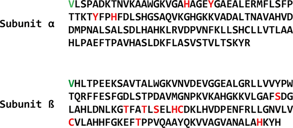
4-Hydroxylbenzyl adducted side chains identified in global proteomics analysis and confirmed via targeted mass spectrometry, highlighted in red. N-terminal valines are highlighted in green.
As expected, control samples of human blood not pre-treated with para-QM contained background levels of 4-hydroxybenzyl adducts (Table 1A). Adduct amounts in untreated samples ranged from 3.2×10⁻⁴ % modification of βHis143 to 0.64 % modification of βCys112. In our previous study that examined blood samples from smokers and nonsmokers (N = 6 per group), N-terminal valine 4-hydroxybenzyl adducts were present at levels of 380 ± 160 pmol/g of hemoglobin, which corresponds to approximately 3.55 × 10⁻⁴ % to 8.71 × 10⁻⁴ % modification of the N-terminal valine.20 These results suggest that at least some of the internal amino acid side chains of hemoglobin are more reactive towards para-QM than the N-terminal valines. Although the number of 4-hydroxybenzyl adducts at βSer44 was estimated to be as high as 0.12%, adduct numbers did not increase following incubation with para-QM and were highly variable between the replicates (results not shown). These results suggest that 4-hydroxybenzyl adducts at βSer44 may not be stable during tryptic digestion and other sample processing steps.
Differential reactivity of the hemoglobin side chains towards para-QM
To compare the relative reactivity of various hemoglobin amino acid side chains toward para-QM, hemoglobin was treated with increasing amounts of para-QM, and the adducted peptides were analyzed using the targeted PRM assay. Percent adduct formation at each site was quantified directly from HPLC-ESI-MS/MS peak areas corresponding to 4-hydroxybenzyl-modified and intact peptides as described in Materials and Methods.
Hemoglobin amino acid side chains showed concentration dependent formation of 4-hydroxybenzyl adduct and exhibited variable degrees of reactivity toward para-QM, with some residues showing a linear relationship between adduct formation and amount of para-QM added (Figure 3A) and other residues showing saturation of adduct levels at lower amounts of added para-QM (Figure 3B). On the alpha subunit of hemoglobin, the αHis45 and αTyr42 residues were less reactive than αHis20 and αTyr24 at lower exposure levels (0.1–0.5-fold excess para-QM), but ultimately had higher levels of adduct formation after treatment with a 5-fold excess of para-QM (23– 24%) (Figure 3A). These results suggest the contribution of both kinetic and thermodynamic factors to 4-hydroxybenzyl adduct yield at individual sites within the protein.
Figure 3:

Side chain adduct occupancy of residues in hemoglobin with titration of increasing amounts of para-QM. Adduct formation on side chain residues follows either a A) linear or B) saturation relationship with increasing addition of para-QM.
Among amino acid side chains that exhibited 4-hydroxybenzyl adduct formation with para-QM within the beta subunit of hemoglobin, most exhibited saturation of adduct formation at lower concentrations of added para-QM (approximately 5μM), with the exceptions of βSer72, βThr123, and βHis143 which showed a linear relationship between adduct formation and added para-QM (Figure 3A). The highest reactivity was observed at βCys93, βHis92, and βSer72. Following treatment with 5-fold molar excess of para-QM, 33%, 27%, and 26% of these residues were modified, respectively (Figure 3B). Under the same reaction conditions, other residues showed varying levels of reactivity, with 17% of βThr84, 16% of βThr87, 12% of βCys112, 10% of βHis143, 6% of βSer89, and 2% of βThr123. The residues also show different modification affinities, with βCys93 and βHis92 showing saturation at lower concentrations of para-QM than βCys112, βThr84, βThr87, and βSer89, (Figure 3B, Table 1). Overall, these results indicate that amino acid side chains within the protein show differential reactivity towards p-QM depending on exposure levels.
4-Hydroxybenzyl adducts form at side chains characterized by low pKa values and/or high degrees of solvent accessibility
Visualizing para-QM modified side chains within the three-dimensional structure of hemoglobin reveals that the 4-hydroxybenzylated sites preferentially occupy solvent exposed regions within the alpha helices of the protein (Figure 4). Relative reactivity of these sites correlates with the calculated relative solvent accessibilities of the side chain residues (Figures 5A and 5B), where 62.5% of highlighted residues show 50% solvent accessibility or greater (Table 1). Interestingly, less than 50% of the dissociable side chains have favorable pKa values at physiological pH (Figures 4C and 4D), suggesting that steric accessibility of these residues plays a more important role in their reactivity towards para-QM. In considering the protein structure further, it is apparent that these reactive side chains occur in discrete clusters around the protein molecule (Figure 4). Each cluster contains at least one residue with high affinity towards para-QM. For example, several side chains showing increased reactivity towards para-QM are found in and around βCys93, the most reactive residue in the protein beta subunit (Table 1B, Figure 4D). This provides preliminary evidence for 4-hydroxybemzyl adduct migration along the protein as previously reported for ortho-QM adducts on DNA.54
Figure 4:

A) NMR structure of human hemoglobin50 (PDB ID: 2h35) with 4-hydroxybenzyl adducted side chains shown in red. Alpha and beta subunits are grey, with the N-terminal valine residues shown in green and heme molecules in blue. Clusters of adduct sites in alpha subunits are shown in subfigures B) and C), clusters of adduct sites in beta subunits are shown in subfigures D) and E).
Figure 5:

Biophysical properties of N-terminal valine and amino acid side chains within the A) alpha subunit and B) beta subunit. Labelled residues correspond to nucleophilic sidechains. Relative solvent accessibilities are scaled relative to the theoretical values52. pKa values were not calculated for non-ionizable values. Physiological pH is designated by the horizontal red line.
4-Hydroxybenzyl adduct formation in hemoglobin treated with 4-hydroxybenzaldehyde
4-Hydroxybenzaldehyde (4-HBA) is found in the common flavoring agent vanillin and is present in many other foods.55 To determine whether 4-HBA can potentially serve as a source of 4-hydroxybenzyl adducts, human hemoglobin was reacted with five-fold molar excess of 4-HBA. Global proteomic analysis of tryptic digests revealed no adducts (results not shown). However, 4-hydroxybenzyl adduct formation was observed in both hemoglobin subunits when the 4-hydroxybenzaldehyde treated hemoglobin was subjected to reductive amination with sodium cyanoborohydride (Scheme 1B). The sixty-four sites observed in 4-HBA/NaCNBH4 treated hemoglobin using global proteomics analysis showed a high degree of overlap with those observed upon incubation with excess para-QM (Figure S2A). Most of the 4-hydroxybenzylated sites (77%) were shared between the two treatments (Figure S2B). However, adducts at αHis122, αSer124, αLys127 were observed exclusively upon 4-hydroxybenzaldehyde treatment and were found near the C-terminus of the hemoglobin alpha subunit.
Peptides corresponding to 4-hydroxybenzaldehyde modification of αHis122, αSer124, and αLys127 were added to the previously chosen sites in an inclusion list for targeted mass spectrometry analysis of hemoglobin treated with 4-hydroxybenzaldehyde and sodium cyanoborohydride. Targeted analysis of the hemoglobin treated with 4-hydroxybenzaldehyde/sodium cyanoborohydride provided evidence for 4-hydroxybenzyl modifications at αHis20, αTyr24, αTyr42, αHis45, βSer44, βThr84, and βHis92 residues (Table 1B). While βSer44 was not validated in the targeted analysis of para-QM-treated samples, the other six adducts associated with exposure to 4-hydroxybenzaldehyde/sodium cyanoborohydride were seen in both targeted experiments. Overall, 4-hydroxybenzyl adduct levels were higher in 4-hydroxybenzaldehyde treated samples as compared to controls, but the results did not reach statistical significance due to high variability between replicates (Table 1B). The efficiency of 4-hydroxybenzyl adduct formation in 4-hydroxybenzaldehyde treated hemoglobin was far lower than that seen in para-QM-treated samples (Table 1B), suggesting that 4-hydroxybenzaldehyde is less reactive towards nucleophilic side chains of hemoglobin as compared to para-QM.
4-Hydroxybenzyl adduct formation in hemoglobin exposed to UV light
A recent report documented the formation of para-QM upon UV irradiation of tyrosine.41 In this mechanism, para-QM is released from the side chain of Tyr via a free radical mechanism to leave behind a glycine residue (Scheme 1C). To determine whether UV light can lead to the formation of 4-hydroxybenzyl adducts on hemoglobin, human blood was exposed to ultraviolet radiation C (254 nm) with and without the addition of external L-tyrosine. Global proteomics experiments revealed 32 amino acid side chains potentially carrying a 4-hydroxybenzyl modification following exposure to ultraviolet radiation, these adducts formed with or without the addition of tyrosine (Figure S3A). Of these potential 4-hydroxybenzylated sites, 84% were also detected in the global analysis of hemoglobin exposed to para-QM. Three of the five new adducts (αHis122, αSer124, αLys127) detected in this experiment were also seen in the global analysis of the hemoglobin treated with 4-hydroxybenzaldehyde (Table 1B, Figure S2B). To validate our initial results, these 32 peptides were combined with the original list of peptides in Table 2 to generate an inclusion list for validation via targeted mass spectrometry. Targeted MS experiments confirmed that UV irradiation induced 4-hydroxybenzyl adducts at multiple sites across alpha and beta subunits of hemoglobin including αHis20, αTyr24, αTyr42, αHis45, βSer44, βThr84, and βSer89 (Table 1B). As was the case for 4-hydroxybenzaldehyde, the numbers of 4-hydroxybenzyl adducts in human blood exposed to ultraviolet radiation were much lower than in experiments with para-QM (Table 1B). αHis20 and βSer89 showed significant increases in abundance relative to their control samples. Interestingly, the addition of free L-tyrosine did not significantly impact the formation of UV-induced 4-hydroxybenzyl adducts in hemoglobin, as the adduct levels with and without the addition of extra tyrosine were statistically equivalent (Figure S3B). These results suggest that tyrosine residues present within the protein, rather than free tyrosine, are the source of the observed 4-hydroxybenzyl adducts. To test this hypothesis, global proteomics data were searched against hemoglobin FASTA files in which tyrosines were replaced with glycines. As mentioned above, the release of para-QM from a side chain results in the formation of glycine at the same site (Scheme 1C). In support of this hypothesis, MS-based proteomics analyses revealed peptides in hemoglobin containing potential glycine “scars” in place of tyrosine (Figure S4). Two of the tyrosine residues that show glycine substitution are αTyr24 and αTyr42. Both Tyr residues had decreased amounts of 4-hydroxybenzyl adduct formation in UV-irradiated samples relative to their control samples and exhibited a slight increase upon the addition of external tyrosine to the reaction (Table 1B). Overall, this is consistent with a loss of tyrosine at these sites in hemoglobin alpha subunits and a local release of para-QM that is available for binding to neighboring amino acid residues. This effect is also seen at αHis45, where UV exposure results in a decrease in adduct formation relative to the control samples. Adduct formation at αHis45 stemming from para-QM release from αTyr42 could be undetected in our analyses due to incomplete b- and y- ion series in the MS2 spectrum. Of the residues identified as having increased 4-hydroxybenzyl adduct formation following UV exposure, βSer44 and βThr89 are in proximity to a tyrosine (11.4 and 7.2 angstroms away, respectively) (Figure S5). The remaining residues were found to be on the exterior of the protein. Further detailed investigations are needed to examine UV-mediated release of para-QM as a possible source of para-hydroxybenzyl adducts in hemoglobin.
Discussion
Characterization of 4-hydroxybenzyl adducts in hemoglobin treated with para-QM
While covalent adducts formed at the N-terminal valine of hemoglobin are widely used for human exposome studies,2,56 internal nucleophilic amino acid side chains are also reactive towards electrophiles.27,36,38 This study examined the formation of 4-hydroxybenzyl adducts at internal residues within human hemoglobin. We proposed that examination of the patterns of hemoglobin side chains susceptible to 4-hydroxybenzyl adduct formation may help identify the possible sources of such adducts (para-QM, para-benzaldehyde, UV radiation) in humans because these electrophiles form protein adducts by different mechanisms and may exhibit distinct site specificity within the protein.57
para-QM preferentially modifies solvent exposed Cys, His, Ser, Thr, and Tyr residues of human hemoglobin
Our MS based proteomics analyses revealed the formation of 4-hydroxybenzyl adducts at several side chains in hemoglobin following treatment with para-QM (Figure 2). Four of the preferentially adducted amino acid side chains are located within the alpha subunit of the protein (αHis20, αTyr24, αTyr42, αHis45), with additional ten adducts found on the beta subunit (βSer44, βSer72, βThr84, βThr87, βSer89, βHis92, βCys93, βCys112, βThr123, βHis143). Previous studies reported the preferential formation of hemoglobin adducts on cysteine residues of hemoglobin (Cys93 and Cys112) by other electrophiles.58–61 Although our experiments confirmed the formation of 4-hydroxybenzyl adducts at these two sites (Figures S1J and S1L), they were not the only reactive side chains on the hemoglobin molecule. We found that 4-hydroxybenzyl adducts readily formed at histidine, serine, and tyrosine side chains of hemoglobin following treatment with para-QM (Table 1 and Scheme 2). Hemoglobin adducts at histidine and lysine side chains have been previously reported e.g. upon reactions with 2-octenal and methylene diphenyl diisocyanate.38,62
To help explain the observed site specificity for adduct formation at specific residues within the protein, local environment of the preferentially modified hemoglobin side chains was considered. Interrogating the consensus sequences46 of the adducted side chains within 7 residues of the reaction sites shows a very diverse set surrounding side chains which are unlikely to contribute to the increased nucleophilicity of the reactive side chains (Figure S6). This is reflected in the highly variable pKa of the adducted amino acid side chains (Figures 5C and 5D), with many residues showing high pKa values relative to the physiological pH (7.4). At the same time, many of the preferentially modified residues show increased solvent accessibility, which could increase their reactivity towards electrophiles.63 In addition, the adducted residues are clustered into islands located in solvent exposed regions of the protein, with each cluster containing at least one side chain residue that is particularly reactive towards para-QM (i.e. αHis45, βHis92/βCys93) (Figures 4B–4D). While there is some evidence for migration of QM-nucleobase adducts along DNA chains,64 it is likely that the clusters of adducts are the results of pre-association of the para-QM precursors with hemoglobin at especially accessible regions of the protein.65
Potential sources of 4-hydroxybenzyl adducts in hemoglobin
The presence of 4-hydroxybenzyl adducted side chains in untreated hemoglobin demonstrates potential formation of these adducts at physiological conditions (Table 1A). Out prime motivation for treating hemoglobin with electrophiles was to determine which (if any) of these could be a source of 4-hydroxybenzyl side chain adducts in vivo.
Para-QM is an extremely reactive electrophilic species that is unlikely to survive long under physiological conditions.66,67 Therefore, para-QM itself is unlikely the source of 4-hydroxybenzyl adducts in humans unless released in situ from a suitable precursor. Therefore, we investigated other potential sources of 4-hydroxybenzyl adducts in humans. 4-Hydroxybenzaldehyde (4-HBA) was considered as a potential candidate due to its relative stability in an aqueous environment as compared to QMs and its ability to induce 4-hydroxybenzyl-protein adducts under reducing conditions.23,68 4-HBA compound is found in many natural sources, most notably in vanilla,55 a flavoring nearly ubiquitous in the modern world. 4-Hydroxybenzaldehyde is a major constituent of the aromatic compounds in vanilla extract55,69 and is created during the curing of the vanilla pods. The addition of 4-hydroxybenzaldehyde to hemoglobin appears to result in the formation of 4-hydrozybenzyl adducts only following a subsequent reduction step (Figure S3A), implying that a reducing environment is necessary for adduct formation. This is in agreement with our previous work showing the formation of the 4-hydroxybenzyl adduct to valine after incubation of valine with 4-hydroxybenzaldehyde and a reducing agent. 22
Exposure of hemoglobin to excess 4-hydroxybenzaldehyde and subsequent reduction resulted in a small number of side chain adducts relative to those formed upon exposure to para-QM (Table 1B), including histidine adducts formed through Schiff base formation and reduction (Scheme 2) as well as adduct formation on the hydroxyl groups of serine, threonine, and tyrosine. We speculate that this may occur through an acid-catalyzed loss of water with subsequent reduction (Scheme S1), though further work is needed to explore the nature of this adduct. While the levels of 4-hydroxybenzaldehyde-hemoglobin adducts increased in treated samples relative to controls, these differences were not statistically significant.
Another possible route for 4-hydroxybenzyl adduct formation via para-QM involves in situ formation of para-QM in blood via reactions of free tyrosine or tyrosine side chains of proteins with ultraviolet radiation.41 However, global proteomics analysis of hemoglobin peptides obtained upon UV irradiation of human blood in the presence or the absence of free Tyr showed relatively few reproducible sites corresponding with those seen in the endogenous samples (Table 1). Future studies should explore adduct formation via oxidation of excess p-cresol in the liver to para-QM via cytochrome P450 or peroxidase enzymes.70 Recent studies revealed a pathway by which gut microbiota high in Clostridia can metabolize excess L-tyrosine in the gut into p-cresol,71 which could give rise to p-QM and 4-hydroxybenzyl adducts upon disruption of gut microbiota.
In summary, this investigation provides the first evidence that exposure to para-QM leads to 4-hydroxybenzyl adduct formation on the thiols, hydroxyl groups, amino, and imidazole amino acid side chains of human hemoglobin (Table 1, Figure 4). In addition, 4-hydroxybenzyl adducts at these side chains are also observed with exposure to 4-hydroxybenzaldehyde under reducing conditions. Our new results reveal several novel reactive sites for electrophilic attack within hemoglobin and expands the utility of hemoglobin as a record-keeping molecule of the human exposome, allowing for the identification of adducts throughout the protein. We also demonstrate that mapping the adduction sites within hemoglobin via mass spectrometry-based proteomics can help identify possible electrophilic precursors of known N-terminal valine adducts because each electrophile produces a characteristic pattern of amino acid side chain adducts within the protein.
Supplementary Material
Acknowledgements
The authors would like to acknowledge Robert Carlson (University of Minnesota Cancer Center) and Jenna Fernandez (University of Minnesota) for their assistance in the construction of figures.
Funding Information
This study was partially sponsored by NIH grants to N.T. (R01-CA100670, R01-CA095039) and special funding for collaborative studies from Faculty of Stockholm University (Sweden). Andrew T. Rajczewski was supported by Biotechnology Training Grant: NIH T32GM008347. Lorena Ndreu and Isabella Karlsson were supported by the Swedish Research Council for Sustainable Development (FORMAS 2017-01511) and Birgit and Hellmuth Hertz’ foundation (Royal Physiographic Society of Lund).
Footnotes
Supporting Information
Peptides detected in hemoglobin treated with excess para-QM; peptides selected for targeted proteomic analysis; MS/MS spectra of peptides with 4-hydroxybenzyl adducts; numbers of adducts following 4-HBA treatment with and without reduction; adducted sites following 4-HBA treatment; adducted sites following UV exposure; levels of adduct formation with and without added tyrosine; MS/MS spectra of peptides with αTyr24→αGly24 and αTyr42→αGly42; protein structures depicting closest tyrosine residues to adducted residues; consensus sequence of adducted side chains; potential mechanism for 4-hydroxybenzaldehyde adduct formation on serine/threonine/tyrosine
References
- 1.Farmer PB (2004) DNA and protein adducts as markers of genotoxicity. Toxicol. Lett. 149, 3–9. [DOI] [PubMed] [Google Scholar]
- 2.Törnqvist M, Fred C, Haglund J, Helleberg H, Paulsson B, and Rydberg P (2002) Protein adducts: quantitative and qualitative aspects of their formation, analysis and applications. J. Chromatogr. B 778, 279–308. [DOI] [PubMed] [Google Scholar]
- 3.Niemela O, Parkkila S, Ylaherttuala S, Halsted C, Witztum JL, Lanca A, and Israel Y (1994) Covalent protein adducts in the liver as a result of ethanol-metabolism and lipid-peroxidation. Lab. Invest. 70, 537–546. [PubMed] [Google Scholar]
- 4.Zhu Y, Duan X, Qin N, Lv J, Wu G, and Wei F (2019) Health risk from dietary exposure to polycyclic aromatic hydrocarbons (PAHs) in a typical high cancer incidence area in southwest China. Sci. Total. Environ. 649, 731–738. [DOI] [PubMed] [Google Scholar]
- 5.Polachova A, Gramblicka T, Parizek O, Sram RJ, Stupak M, Hajslova J, and Pulkrabova J (2020) Estimation of human exposure to polycyclic aromatic hydrocarbons (PAHs) based on the dietary and outdoor atmospheric monitoring in the Czech Republic. Environ. Res. 182, 108977. [DOI] [PubMed] [Google Scholar]
- 6.Kautiainen A, Midtvedt T, and Törnqvist M (1993) Intestinal bacteria and endogenous production of malonaldehyde and alkylators in mice. Carcinogenesis 14, 2633–2636. [DOI] [PubMed] [Google Scholar]
- 7.Blondin O, and Viau C (1992) Benzo[a]pyrene-blood protein adducts in wild woodchucks used as biological sentinels of environmental polycyclic aromatic-hydrocarbons contamination. Arch. Environ. Contam. Toxicol. 23, 310–315. [DOI] [PubMed] [Google Scholar]
- 8.Hemminki K, Grzybowska E, Chorazy M, Twardowska-Saucha K, Sroczynski J, Putman K, Randerath K, Phillips D, Hewer A, et al. (1990) DNA adducts in humans environmentally exposed to aromatic compounds in an industrial area of Poland. Carcinogenesis 11, 1229–1231. [DOI] [PubMed] [Google Scholar]
- 9.Bartsch H, and Nair J (2006) Chronic inflammation and oxidative stress in the genesis and perpetuation of cancer: role of lipid peroxidation, DNA damage, and repair. Langenbecks Arch. Surg. 391, 499–510. [DOI] [PubMed] [Google Scholar]
- 10.De Flora S, Izzotti A, Randerath K, Randerath E, Bartsch H, Nair J, Balansky R, Van Schooten F, Degan P, et al. (1996) DNA adducts and chronic degenerative diseases. Pathogenetic relevance and implications in preventive medicine. Mutat. Res. 366, 197–238. [PubMed] [Google Scholar]
- 11.Nair J, Gansauge F, Beger H, Dolara P, Winde G, and Bartsch H (2006) Increased etheno-DNA adducts in affected tissues of patients suffering from Crohn’s disease, ulcerative colitis, and chronic pancreatitis. Antioxid. Redox. Signal. 8, 1003–1010. [DOI] [PubMed] [Google Scholar]
- 12.Setshedi M, Wands JR, and Monte SM (2010) Acetaldehyde adducts in alcoholic liver disease. Oxid. Med. Cell Longev. 3, 178–185. [DOI] [PMC free article] [PubMed] [Google Scholar]
- 13.Skipper PL, Peng XC, Soohoo CK, and Tannenbaum SR (1994) Protein adducts as biomarkers of human carcinogen exposure. Drug Metab. Rev. 26, 111–124. [DOI] [PubMed] [Google Scholar]
- 14.Troester MA, Lindstrom AB, Kupper LL, Waidyanatha S, and Rappaport SM (2000) Stability of hemoglobin and albumin adducts of benzene oxide and 1,4-benzoquinone after administration of benzene to F344 rats. Toxicol. Sci. 54, 88–94. [DOI] [PubMed] [Google Scholar]
- 15.Rosen CB, and Francis MB (2017) Targeting the N terminus for site-selective protein modification. Nat. Chem. Biol. 13, 697–705. [DOI] [PubMed] [Google Scholar]
- 16.Sereda TJ, Mant CT, Quinn AM, and Hodges RS (1993) Effect of the α-amino group on peptide retention behaviour in reversed-phase chromatography determination of the pKa values of the α-amino group of 19 different N-terminal amino acid residues. J. Chromatogr. A 646, 17–30. [DOI] [PubMed] [Google Scholar]
- 17.Carlsson H, and Tornqvist M (2017) An adductomic approach to identify electrophiles in vivo. Basic Clin. Pharmacol. Toxicol. 121 Suppl 3, 44–54. [DOI] [PubMed] [Google Scholar]
- 18.Carlsson H, and Törnqvist M (2016) Strategy for identifying unknown hemoglobin adducts using adductome LC-MS/MS data: Identification of adducts corresponding to acrylic acid, glyoxal, methylglyoxal, and 1-octen-3-one. Food. Chem. Toxicol. 92, 94–103. [DOI] [PubMed] [Google Scholar]
- 19.Carlsson H, von Stedingk H, Nilsson U, and Törnqvist M (2014) LC–MS/MS screening strategy for unknown adducts to N-terminal valine in hemoglobin applied to smokers and nonsmokers. Chem. Res. Toxicol. 27, 2062–2070. [DOI] [PubMed] [Google Scholar]
- 20.Von Stedingk H, Rydberg P, and Törnqvist M (2010) A new modified Edman procedure for analysis of N-terminal valine adducts in hemoglobin by LC–MS/MS. J. Chromatogr. B 878, 2483–2490. [DOI] [PubMed] [Google Scholar]
- 21.Carlsson H, Motwani HV, Osterman Golkar S, and Törnqvist M (2015) Characterization of a hemoglobin adduct from ethyl vinyl ketone detected in human blood samples. Chem. Res. Toxicol. 28, 2120–2129. [DOI] [PubMed] [Google Scholar]
- 22.Degner A, Carlsson H, Karlsson I, Eriksson J, Pujari SS, Tretyakova NY, and Törnqvist M (2018) Discovery of novel N-(4-hydroxybenzyl)valine hemoglobin adducts in human blood. Chem. Res. Toxicol. 31, 1305–1314. [DOI] [PMC free article] [PubMed] [Google Scholar]
- 23.Bolton JL, and Thompson JA (2009) Formation and Reactions of Xenobiotic Quinone Methides in Biology, In Reactive Intermediates in Chemistry and Biology: Quinone Methides (Rokita SE, Ed.) pp 329–356, John Wiley & Sons, Inc., Hoboken, New Jersey. [Google Scholar]
- 24.Elgawish MS, Kishikawa N, Helal MA, Ohyama K, and Kuroda N (2015) Molecular modeling and spectroscopic study of quinone–protein adducts: insight into toxicity, selectivity, and reversibility. Toxicol. Res. 4, 843–847. [Google Scholar]
- 25.Gaikwad NW, and Bodell WJ (2001) Formation of DNA adducts by microsomal and peroxidase activation of p-cresol: role of quinone methide in DNA adduct formation. Chem. Biol. Interact. 138, 217–229. [DOI] [PubMed] [Google Scholar]
- 26.Matthews R, Massey V, and Sweeley CC (1975) Identification of p-hydroxybenzaldehyde as the ligand in the green form of old yellow enzyme. J. Biol. Chem. 250, 9294–9298. [PubMed] [Google Scholar]
- 27.Isom AL, Barnes S, Wilson L, Kirk M, Coward L, and Darley-Usmar V (2004) Modification of cytochrome c by 4-hydroxy-2-nonenal: evidence for histidine, lysine, and arginine-aldehyde adducts. J. Am. Soc. Mass. Spectrom. 15, 1136–1147. [DOI] [PubMed] [Google Scholar]
- 28.Segerbäck D (1983) Alkylation of DNA and hemoglobin in the mouse following exposure to ethene and ethene oxide. Chem. Biol. Interact. 45, 139–151. [DOI] [PubMed] [Google Scholar]
- 29.Grigoryan H, Edmands W, Lu SS, Yano Y, Regazzoni L, Iavarone AT, Williams ER, and Rappaport SM (2016) Adductomics pipeline for untargeted analysis of modifications to Cys34 of human serum albumin. Anal. Chem. 88, 10504–10512. [DOI] [PMC free article] [PubMed] [Google Scholar]
- 30.Lu SS, Grigoryan H, Edmands WM, Hu W, Iavarone AT, Hubbard A, Rothman N, Vermeulen R, Lan Q, et al. (2017) Profiling the serum albumin Cys34 adductome of solid fuel users in Xuanwei and Fuyuan, China. Environ. Sci. Technol. 51, 46–57. [DOI] [PMC free article] [PubMed] [Google Scholar]
- 31.Rappaport SM, Li H, Grigoryan H, Funk WE, and Williams ER (2012) Adductomics: characterizing exposures to reactive electrophiles. Toxicol. Lett. 213, 83–90. [DOI] [PMC free article] [PubMed] [Google Scholar]
- 32.Michaelson-Richie ED, Loeber RL, Codreanu SG, Ming X, Liebler DC, Campbell C, and Tretyakova NY (2010) DNA−Protein cross-linking by 1,2,3,4-diepoxybutane. J. Proteome Res. 9, 4356–4367. [DOI] [PMC free article] [PubMed] [Google Scholar]
- 33.Loeber RL, Michaelson-Richie ED, Codreanu SG, Liebler DC, Campbell CR, and Tretyakova NY (2009) Proteomic analysis of DNA-protein cross-linking by antitumor nitrogen mustards. Chem. Res. Toxicol. 22, 1151–1162. [DOI] [PMC free article] [PubMed] [Google Scholar]
- 34.Groehler A 4th, Villalta PW, Campbell C, and Tretyakova N (2016) Covalent DNA-protein cross-linking by phosphoramide mustard and nornitrogen mustard in human cells. Chem. Res. Toxicol. 29, 190–202. [DOI] [PMC free article] [PubMed] [Google Scholar]
- 35.Ming X, Michaelson-Richie ED, Groehler A. S. t., Villalta PW, Campbell C, and Tretyakova NY (2020) Cross-linking of the DNA repair protein O6-alkylguanine DNA alkyltransferase to DNA in the presence of cisplatin. DNA Repair 89, 102840. [DOI] [PMC free article] [PubMed] [Google Scholar]
- 36.Cai T, Bellamri M, Ming X, Koh W-P, Yu MC, and Turesky RJ (2017) Quantification of hemoglobin and white blood cell DNA adducts of the tobacco carcinogens 2-amino-9H-pyrido[2,3-b]indole and 4-aminobiphenyl formed in humans by nanoflow liquid chromatography/ion trap multistage mass spectrometry. Chem. Res. Toxicol. 30, 1333–1343. [DOI] [PMC free article] [PubMed] [Google Scholar]
- 37.Pathak KV, Chiu T-L, Amin EA, and Turesky RJ (2016) Methemoglobin formation and characterization of hemoglobin adducts of carcinogenic aromatic amines and heterocyclic aromatic amines. Chem. Res. Toxicol. 29, 255–269. [DOI] [PMC free article] [PubMed] [Google Scholar]
- 38.Yoshitake J, Shibata T, Shimayama C, and Uchida K (2019) 2-Alkenal modification of hemoglobin: Identification of a novel hemoglobin-specific alkanoic acid-histidine adduct. Redox. Biol. 23, 101115. [DOI] [PMC free article] [PubMed] [Google Scholar]
- 39.Huang C, Liu Y, and Rokita SE (2016) Targeting duplex DNA with the reversible reactivity of quinone methides. Signal Transduct. Target. Ther. 1, 1–5. [DOI] [PMC free article] [PubMed] [Google Scholar]
- 40.Rokita SE, Yang J, Pande P, and Greenberg WA (1997) Quinone methide alkylation of deoxycytidine. J. Org. Chem. 62, 3010–3012. [DOI] [PubMed] [Google Scholar]
- 41.Kang H, Tolbert TJ, and Schöneich C (2019) Photoinduced tyrosine side chain fragmentation in IgG4-Fc: Mechanisms and solvent isotope effects. Mol. Pharm. 16, 258–272. [DOI] [PubMed] [Google Scholar]
- 42.Hodge K, Have ST, Hutton L, and Lamond AI (2013) Cleaning up the masses: Exclusion lists to reduce contamination with HPLC-MS/MS. J. Proteomics 88, 92–103. [DOI] [PMC free article] [PubMed] [Google Scholar]
- 43.Wingfield PT (2017) N-Terminal methionine processing. Curr. Protoc. Protein. Sci. 88, 6.14.11–16.14.13. [DOI] [PMC free article] [PubMed] [Google Scholar]
- 44.The M, MacCoss MJ, Noble WS, and Käll L (2016) Fast and accurate protein false discovery rates on large-scale proteomics data sets with Percolator 3.0. J. Am. Soc. Mass. Spectrom. 27, 1719–1727. [DOI] [PMC free article] [PubMed] [Google Scholar]
- 45.Pino LK, Searle BC, Bollinger JG, Nunn B, MacLean B, and MacCoss MJ (2020) The Skyline ecosystem: Informatics for quantitative mass spectrometry proteomics. Mass Spectrom. Rev. 39, 229–244. [DOI] [PMC free article] [PubMed] [Google Scholar]
- 46.Wagih O (2017) ggseqlogo: a versatile R package for drawing sequence logos. Bioinformatics 33, 3645–3647. [DOI] [PubMed] [Google Scholar]
- 47.Anandakrishnan R, Aguilar B, and Onufriev AV (2012) H++ 3.0: Automating pK prediction and the preparation of biomolecular structures for atomistic molecular modeling and simulations. Nucleic Acids Res. 40, W537–541. [DOI] [PMC free article] [PubMed] [Google Scholar]
- 48.Gordon JC, Myers JB, Folta T, Shoja V, Heath LS, and Onufriev A (2005) H++: A server for estimating p K as and adding missing hydrogens to macromolecules. Nucleic Acids Res. 33, W368–W371. [DOI] [PMC free article] [PubMed] [Google Scholar]
- 49.Myers J, Grothaus G, Narayanan S, and Onufriev A (2006) A simple clustering algorithm can be accurate enough for use in calculations of pKs in macromolecules. Proteins 63, 928–938. [DOI] [PubMed] [Google Scholar]
- 50.Xu Y, Zheng Y, Fan JS, and Yang D (2006) A new strategy for structure determination of large proteins in solution without deuteration. Nat. Methods 3, 931–937. [DOI] [PubMed] [Google Scholar]
- 51.Berman H, Henrick K, and Nakamura H (2003) Announcing the worldwide Protein Data Bank. Nat. Struct. Mol. Biol. 10, 980–980. [DOI] [PubMed] [Google Scholar]
- 52.Krissinel E, and Henrick K (2007) Inference of macromolecular assemblies from crystalline state. J. Mol. Biol. 372, 774–797. [DOI] [PubMed] [Google Scholar]
- 53.Tien MZ, Meyer AG, Sydykova DK, Spielman SJ, and Wilke CO (2013) Maximum allowed solvent accessibilites of residues in proteins. PloS One 8, e80635–e80635. [DOI] [PMC free article] [PubMed] [Google Scholar]
- 54.Deeyaa BD, and Rokita SE (2020) Migratory ability of quinone methide-generating acridine conjugates in DNA. Org. Biomol. Chem. 18, 1671–1678. [DOI] [PubMed] [Google Scholar]
- 55.Gallage NJ, and Møller BL (2018) Vanilla: The Most Popular Flavour, In Biotechnology of Natural Products (Schwab W, Lange BM, and Wüst M, Eds.) pp 3–24, Springer International Publishing, Cham. [Google Scholar]
- 56.Aasa J, Vryonidis E, Abramsson-Zetterberg L, Törnqvist M. Internal Doses of Glycidol in Children and Estimation of Associated Cancer Risk. Toxics. 2019. Feb 1;7(1):7. doi: 10.3390/toxics7010007. [DOI] [PMC free article] [PubMed] [Google Scholar]
- 57.Favinha AG, Barreiro DS, Martins JN, O’Toole P, and Pauleta SR (2020) Acrylamide-hemoglobin adduct: A spectroscopic study. Spectrochim. Acta A Mol. Biomol. Spectrosc. 241, 118644. [DOI] [PubMed] [Google Scholar]
- 58.Kassa T, Strader MB, Nakagawa A, Zapol WM, and Alayash AI (2017) Targeting βCys93 in hemoglobin S with an antisickling agent possessing dual allosteric and antioxidant effects. Metallomics 9, 1260–1270. [DOI] [PMC free article] [PubMed] [Google Scholar]
- 59.Miranda JJ (2000) Highly reactive cysteine residues in rodent hemoglobins. Biochem. Biophys. Res. Commun. 275, 517–523. [DOI] [PubMed] [Google Scholar]
- 60.Nakagawa A, Lui FE, Wassaf D, Yefidoff-Freedman R, Casalena D, Palmer MA, Meadows J, Mozzarelli A, Ronda L, et al. (2014) Identification of a small molecule that increases hemoglobin oxygen affinity and reduces SS erythrocyte sickling. ACS Chem. Biol. 9, 2318–2325. [DOI] [PMC free article] [PubMed] [Google Scholar]
- 61.Wang Q, Sun L, Ji S, Zhao D, Liu J, Su Z, and Hu T (2014) Reversible protection of Cys-93 (β) by PEG alters the structural and functional properties of the PEGylated hemoglobin. Biochim. Biophys. Acta. 1844, 1201–1207. [DOI] [PubMed] [Google Scholar]
- 62.Gries W, and Leng G (2013) Analytical determination of specific 4,4’-methylene diphenyl diisocyanate hemoglobin adducts in human blood. Anal. Bioanal. Chem. 405, 7205–7213. [DOI] [PubMed] [Google Scholar]
- 63.Gould NS, Evans P, Martinez-Acedo P, Marino SM, Gladyshev VN, Carroll KS, and Ischiropoulos H (2015) Site-specific proteomic mapping identifies selectively modified regulatory cysteine residues in functionally distinct protein networks. Chem. Biol. 22, 965–975. [DOI] [PMC free article] [PubMed] [Google Scholar]
- 64.Deeyaa BD, and Rokita SE (2020) Migratory ability of quinone methide-generating acridine conjugates in DNA. Org Biomol Chem 18, 1671–1678. [DOI] [PubMed] [Google Scholar]
- 65.Montero EI, Benedetti BT, Mangrum JB, Oehlsen MJ, Qu Y, and Farrell NP (2007) Pre-association of polynuclear platinum anticancer agents on a protein, human serum albumin. Implications for drug design. Dalton Trans. doi: 10.1039/B708433C, 4938–4942. [DOI] [PMC free article] [PubMed] [Google Scholar]
- 66.Peter MG (1989) Chemical modifications of biopolymers by quinones and quinone methides. Angew. Chem. Int. Ed. Engl. 28, 555–570. [Google Scholar]
- 67.Thompson DC, Thompson JA, Sugumaran M, and Moldeus P (1993) Biological and toxicological consequences of quinone methide formation. Chem. Biol. Interact. 86, 129–162. [DOI] [PubMed] [Google Scholar]
- 68.Toteva MM, Moran M, Amyes TL, and Richard JP (2003) Substituent effects on carbocation stability: The pKR for p-quinone methide. J. Am. Chem. Soc. 125, 8814–8819. [DOI] [PubMed] [Google Scholar]
- 69.Podstolski A, Havkin-Frenkel D, Malinowski J, Blount JW, Kourteva G, and Dixon RA (2002) Unusual 4-hydroxybenzaldehyde synthase activity from tissue cultures of the vanilla orchid Vanilla planifolia. Phytochemistry 61, 611–620. [DOI] [PubMed] [Google Scholar]
- 70.Yan Z, Zhong HM, Maher N, Torres R, Leo GC, Caldwell GW, and Huebert N (2005) Bioactivation of 4-methylphenol (p-cresol) via cytochrome P450-mediated aromatic oxidation in human liver microsomes. Drug. Metab. Dispos. 33, 1867–1876. [DOI] [PubMed] [Google Scholar]
- 71.Rajakovich LJ, and Balskus EP (2019) Metabolic functions of the human gut microbiota: the role of metalloenzymes. Nat. Prod. Rep. 36, 593–625. [DOI] [PMC free article] [PubMed] [Google Scholar]
Associated Data
This section collects any data citations, data availability statements, or supplementary materials included in this article.