Abstract
Light scattering, whether caused by desired or spurious elements, is considered one of the main phenomena that present great challenges for the nonlinear (NL) optical characterization of turbid media. The most relevant disturbing factor is the random deformation suffered by the spatial intensity distribution of the laser beam due to multiple scattering. In this work, we report the intensity correlation scan (IC-scan) technique as a new tool to characterize the NL optical response of scattering media, by taking advantage of light scattering to generate speckle patterns sensitive to wavefront changes induced by the self-focusing and self-defocusing effects. Peak-to-valley transmittance curves, with a higher signal-to-noise ratio, are obtained by analyzing the spatial intensity correlation functions of the different speckle patterns, even in very turbid media where conventional NL spectroscopy techniques fail. To demonstrate the potential of the IC-scan technique, the NL characterization of colloids that contain a high concentration of silica nanospheres as scatterers, as well as gold nanorods, which act as NL particles and light scatterers, was performed. The results show that the IC-scan technique is more accurate, precise and robust to measure NL refractive indices in turbid media, overcoming limitations imposed by well-established Z-scan and D4σ techniques.
Subject terms: Optical spectroscopy, Nonlinear optics
Introduction
Light scattering is one of the most fundamental optical phenomena observed due to the interaction of light with matter, resulting from inhomogeneities in the refractive index over the scattering volume1. The relevance of scattering in several hard and soft condensed matter systems is evidenced by the various non-invasive techniques developed to measure particle size and colloidal stability2, micro-defect detection3, optical-tissue diagnostics4, as well as to investigate their applications in optical super-resolution5, three-dimensional holography6, modern cryptography7 and random lasers8. Even in this last system, by switching from the single-scattering to the multiple-scattering regime, it was possible to study new light diffusion phenomena, such as the glassy light phase compatible with a replica symmetry breaking9 and a Floquet phase10 in photonic systems, as well as the Anderson localization of light11. Nevertheless, the more dense and disordered the medium that interacts with the light, the more significant the distortion caused by the scattered photons in the spatial and temporal intensity profiles of the transmitted or reflected beams, which are not always desired in optical and photonic systems12–14.
Speckle patterns are a clear example of the complex intensity distribution that a coherent beam scattered by a disordered medium, with a high degree of scattering, can undergo. These patterns with randomly distributed intensities and phase are the result of the superposition of many different scattered waves that interfere with effectively random phases15. For a long time, speckles were considered just as a noisy phenomenon that contaminates the observation of different physical processes, decreasing the signal-to-noise ratio and consequently limiting the precision and sensitivity of many optical techniques16–19. Such an interpretation is reasonable when light scattering is caused by spurious particles, viz. dust, or system imperfections20–22. However, when the speckles are the result of the inherent disorder of the system, the analysis of their statistical properties, such as intensity correlation function and power spectral density, can yield relevant information about the optical properties of the studied system23. Significant progress in the statistical study of speckles patterns has been made in stellar physics24, random lasers25–27, optical-image processing28, optical manipulation29, accurate measurements of contour, deformation, vibration and strain on various materials30, displacements and deformations of diffuse objects31 and biological tissues analysis32.
In nonlinear (NL) optical spectroscopy, strong light scattering caused by turbid media has been reported to be a problem for most techniques that measure NL refractive index33–36. Among them, the well-established Z-scan technique suffers with the distortion of the transmitted intensity beam profile and wavefront induced in the scattering media, which causes the intensity transmitted by the small aperture (closed aperture (CA) Z-scan) to show large fluctuations at each step during sample translation. Experiences in highly scattering media, such as colloids containing SiO2 nanoparticles (NPs) suspended in acetone, show that the fluctuations in the CA Z-scan curve can be larger than the peak-valley transmittance variations, making its characterization unfeasible34. Similar experiments show a low signal-to-noise ratio in the CA Z-scan curves when vitreous humor37, ammonium dihydrogen phosphate crystals38, and liquid crystals39,40 were studied. To overcome this limitation in scattering media, some adaptations to existing (or new) techniques have been developed33–35. In the spatial domain, the scattered light imaging method (SLIM) was proposed to collect light scattered by the turbid medium, in the direction perpendicular to propagation, to image the evolution of the laser beam diameter along propagation34. Here, the NL refractive index is determined by analyzing the divergence angle variations induced by the self-focusing (or self-defocusing) effect, even in a single shot configuration. Thus, unlike NL transmission techniques that suffer with light scattering, SLIM can only be used when scattering is relevant and the samples are thick enough to analyze light propagation within scattering media34. In addition to measuring the NL refractive index, SLIM was also recently applied to discriminate the NL extinction due to NL absorption and NL scattering contributions in turbid media41.
In the spectral domain, new techniques have also been developed to measure NL phase variations in scattering media by analyzing the frequency spectrum shift of a transmitted laser pulse33,35. Spectral analysis, as done in the spectral reshaping technique, has the advantage that, unlike beam shape analysis, it is not affected by linear scattering effects33. However, its experimental setup is more complex than the spatial Z-scan and SLIM techniques, since it requires the use of ultrashort pulses spectrally reshaped by an acoustic optical modulator, to make a hole in the incidence laser spectrum which will be filled when passing through a NL medium, due to self-phase modulation. This hole-refilling technique was recently adapted to a simpler experimental apparatus, called the spectral domain Z-scan, which has been used to measure the refractive index of frosted fused-silica slide35 and human cornea42. Despite the good sensitivity of these NL techniques in the spectral domain, their accuracy depends on how well-defined the temporal beam profile is, as well as on the absence of self-steepening effects that induce laser pulse’s spectral broadening. Therefore, the measurement of the NL refractive index in scattering media continues to be a great challenge for NL optics, and its study is supported by the need to characterize biological media, liquid crystals and other materials that present a high-degree of scattering.
In this work, we present the Intensity Correlation scan (IC-scan) as a new NL optical technique, in the spatial domain, which allows the proper NL optical characterization of strongly scattering media caused by system imperfections, or by the presence of linear or NL scatterers. The IC-scan technique uses light scattering caused by turbid media (or light diffuser) to generate speckle patterns, in the far field, that are sensitive to wavefront changes induced by the self-focusing and self-defocusing effects. By analyzing the intensity self-correlation function of the speckle patterns generated during the sample translation around the focus of a lens, it is possible to obtain curves similar to those of Z-scan, but with lower noise levels, even when the scattering is so strong that it destroys the spatial intensity profile of the transmitted beam. Furthermore, the pure self-phase modulation effects, free from linear scattering contributions, can be obtained by analyzing the intensity cross-correlation function between IC-scan measurements performed in the linear and NL regimes. As a proof of principle, NL refractive index measurements were performed by IC-scan on highly concentrated NL ethanolic (aqueous) colloids containing silica nanospheres (gold nanorods) as light scatterers (NL particles and scatterers), using a titanium-sapphire laser (788 nm, 100 fs, 76 MHz), and the results were compared with the well-established Z-scan43 and D4σ44 techniques.
Experimental details
IC-scan setup
Figure 1 shows the experimental setup used to measure the NL refractive indices of transparent and turbid media. To excite the NL thermal response of the different solvents and colloids, described in the Nonlinear media section, a mode-locked Ti:Sapphire laser emitting 100-fs Gaussian pulses at 788 nm and with a repetition rate of 76 MHz was used. The control of the incident beam’s power was provided by a λ/2 plate followed by a Glan prism (P), which assures that the beam is linearly polarized. Subsequently, the Gaussian beam was focused by a 10-cm focal length lens (L), producing a beam waist of ~ 25.5 μm at the focus position (Rayleigh length: mm). For the measurements, a quartz cell (thickness: L = 1.0 mm < z0) filled with NL media, was moved along the beam propagation direction (Z-axis) around the region where the laser beam is focused. The transmitted beam passes through an element sensitive to wavefront distortions (WDS), located in the far-field, and finally its transversal intensity profile is fully recorded by a CCD camera.
Figure 1.
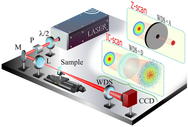
Experimental setup used to characterize the NL response of transparent and turbid media by Z-scan and IC-scan techniques. P: polarizer; M: mirror; L: lens; WDS: wavefront distortion sensor; A: aperture; D: diffuser.
The choice of WDS defines the technique that is used to measure the NL optical response of different media. For instance, when the WDS is an iris, the experimental setup corresponds to the well-established CA Z-scan technique43. On the other hand, when the WDS is removed, the integration of the intensity profile on the CCD, at each step of the NL medium, gives rise to the open aperture (OA) Z-scan technique43, while the D4σ technique is accessed when the transverse irradiance moments are analyzed45. Various types of WDS are reported in the literature as adaptations to the Z-scan technique for measuring NL refractive index (see46 and reference therein).
The IC-scan technique, proposed in this work, uses transparent light diffusers as WDS to generate speckle patterns, whose intensity correlation function is highly sensitive to wavefront changes induced by self-focusing (or self-defocusing) effects. Figure 2a illustrates a speckle pattern captured by the CCD (1024 × 1280 pixels), when the quartz (sample) cell is empty. The 2D spatial intensity self-correlation function , shown in Fig. 2b, exhibits the shape expected for a speckle pattern with Gaussian intensity distribution, varying from to 22, where the angular brackets denote averaging over many realizations. Fifty consecutive images were used to calculate the 2D spatial intensity correlation function at each position of the NL sample. For each image, the laser beam illuminates a different zone of the light diffusor, which is rotated from one image to the next. The width of gives the mean size of the speckles, which was measured to be ~ 20 pixels. In addition, the normal distribution is corroborated by the intensity-probability density function, , since it behaves as a function close to a straight line, on a semilogarithmic scale (see Fig. 2c). IC-scan curves are constructed by plotting the maximum value of as a function of the sample position around the focal plane of the lens L, as shown in the numerical simulation section.
Figure 2.
(a) Speckle pattern obtained by IC-scan technique and their (b) 2D spatial intensity correlation and (c) intensity-probability density function. The intensity profile was collected for an empty cell.
It is important to highlight that the experimental setup of the IC-scan technique is very similar to the well-known Z-scan and D4σ techniques. This is because in the three techniques used, the NL phase shift caused in the laser beam that interacts with the NL medium is the result of the refractive index modulation induced by the intensity beam profile, which causes the self-focusing (or self-defocusing) effect. However, the difference between the techniques lies in the detection methodology that varies depending on the type of WDS used. In the IC-scan technique, the analysis of the beam phase is carried out by means of an interferometric measurement introduced by the presence of the diffuser, in the far-field, which gives rises to speckle patterns with different intensity distributions. Thus, the IC-scan technique emerges as an application of speckle metrology dedicated to the measurement of the NL refractive index through the phase deformations monitored by the analysis of the intensity correlation function, which has a solid theoretical basis in several studies15,47. We emphasized that the diffusers act only as WDS, i.e., the speckles do not experience the self-focusing (self-defocusing) that usually occurs when they propagate in media with a positive (negative) NL refractive index, as reported in48.
Nonlinear media
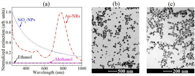
Four NL media, with and without scattering particles, were used to demonstrate the potential of IC-scan to measure the NL refractive indices compared to Z-scan and D4σ techniques. Two of them are typical NL solvents, viz. pure ethanol (≥ 99.9%) and methanol (≥ 99.9%), purchased from Sigma-Aldrich, used as transparent (without scatterers) NL media, as shown in Fig. 3a. To represent NL media with different Rayleigh scattering contributions, two colloids containing silica (SiO2) NPs suspended in ethanol were prepared, following the procedure described in49, with volume fractions of 8.2 × 10⁻³ and 4.1 × 10⁻². Its transmission electron microscopy (TEM) image (Fig. 3b) reveals spherical particles with an average diameter of ~ 120 nm, and their extinction spectrum is characteristic of Rayleigh scattering because it depends on , where is the incident light wavelength.
Figure 3.
(a) Normalized extinction spectra and (b,c) TEM images for scattering media represented by spherical silica (SiO2) NPs and gold nanorods (Au-NRs) suspended in ethanol and water, respectively. The extinction spectra of the solvents were plotted to demonstrate their transparency.
Colloidal gold nanorods (Au-NRs) were also used as scattering media with strong NL optical behavior. For this, Au-NRs were chemically synthesized by a seed-mediated growth method50 to exhibit an average cross-sectional diameter of 15.0 ± 0.6 nm and aspect ratio (AR) equal to 3.4 (see Fig. 3c). The NR’s dimensions were chosen so that its longitudinal localized surface plasmon (l-LSP) resonance, which is known to exhibit a significant scattering contribution51, is centralized near the laser wavelength (788 nm), as shown in Fig. 3a. For the IC-scan measurements, the Au-NR colloids were diluted in deionized water to present volume fractions of 2.5 × 10⁻⁵, 5.0 × 10⁻⁵ and 7.5 × 10⁻⁵.
Numerical simulations
Nonlocal NL response: Z-scan and D4σ
Due to the high repetition rate of the excitation laser, the media studied in this work exhibit an optical nonlinearity dominated by thermal effects. On this origin, the nonlocal model proposed in52 was used to simulate the experimental results obtained by the Z-scan, D4σ and IC-scan techniques, because it presented better results than the thermal lens and aberrant thermal lens models. By expressing the optical field in the amplitude-phase form, , the nonlocal model defines that the evolution equations, along the beam propagation axis (z-axis), for the phase and intensity of an incident Gaussian beam passing through a thin NL sample can be written as53:
| 1 |
| 2 |
where for media exhibiting a third-order refractive index, , and an intensity-dependent extinction coefficient, . is the nonlocal intensity that can be expressed as the product of the maximum on-axis value, , and a Gaussian nonlocal profile, , with , , Gaussian beam waist: , wavelength: , and Rayleigh length: . Notice that the nonlocality factor, , is introduced as a constant that affects the radius of the Gaussian beam in the NL sample52,53. For instance, for the NL phase shift extends (compresses) beyond the incident intensity distribution, while for the NL response of the medium is considered as local43. It is worth mentioning that the values measured in this work for are related to the thermo-optic coefficients that tend to induce self-defocusing effects in an equivalent way to the third-order NL refractive indices for the Kerr effect.
To find the amplitude and phase of the optical field in the detection plane, the Fast Fourier beam propagation method (BPM) was used54. Numerically, the NL medium of thickness was divided into parts of size , with the optical field at the end of each step given by . The operator represents the linear propagation through distance , in a homogeneous medium with linear refractive index and transverse derivative in the Fourier domain. The linear and NL intensity losses along propagation are considered through the operator , while the operator incorporates the NL phase-shift. Note that represents the propagation depth in the NL medium and the sample position around the focus plane. The last two operators, and conclude the beam propagation in a step , where the resulting field will be used as an initial condition for the next step. After sequentially performing N iterations, the far-field beam patterns for the different position of the NL sample, around the focal plane, were obtained by numerical simulations on the free-field propagation using the Huygens–Fresnel formalism, following the method described in54. Approximate analytical expressions for the nonlocal model were also reported in53 using the Gaussian decomposition method.
It is worth mentioning that due to the strong contributions of nonlocal nonlinearities, which induce large NL phase variations (), the BPM is important to allow experimental curve fitting considering the thin-sample approximation. That is, in the NL regime, the medium is regarded as “thin”, if the sample length is small enough that changes in the beam diameter within the sample due to nonlinear refraction can be neglected43. Since in the BPM method Eqs. (1) and (2) are solved iteratively for sample lengths of a step size, , the thin-sample criterion implies that , which is easily obeyed since large values of N were used. In the numerical simulations, N was chosen as the minimum value such that when it is duplicated the Z-scan, IC-scan and D4σ curves present the same results. In this work, the minimum value used for N was 1000.
From the simulated far-field beam patterns, , the Z-scan and D4σ curves were fitted by calculating the normalized transmittance, , and normalized second-order moments, , respectively. For Z-scan, the normalized transmittance was calculated by using the expression , where is the radius of a circular aperture and is the far-field beam pattern when the absorption and refraction NL contributions are zero. To reproduce the experimental conditions of the CA Z-scan scheme, the transmittance was calculated over the area of a circular aperture with a radius of 335 µm ( 50 pixels), centered at . The D4σ curves were calculated using the total beam area through the expression: with , and also normalized by , calculated when the nonlinearities are zero.
Light transmission by a diffuser: IC-scan
Since the IC-scan technique uses a light diffuser as an element sensitive to wavefront distortions, the experimental curves were modeled by transmitting far-field beam patterns through a rough surface using the optical transfer function55. Briefly, the random fields were generated by multiplying , obtained in the previous section, by , where corresponds to a random matrix generated from a uniform distribution in the range of . Thus, the random phase field is given by . However, since rough media randomize the field’s phase based on its degree of scattering, a spatial frequency filter was added to reproduce the experimentally obtained speckles patterns. In the spatial frequency domain, we use: , where the first term denotes the 2D Fourier transform of the and is a spatial frequency filter consisting of a 4-f imaging system containing a mask in the Fourier plane, which allows the transmission of photons whose spatial frequencies pass through a circular aperture of radius ρ56. Since the spatial frequencies are mapped in the Fourier plane at the points , where k corresponds to the wavevector and f to the focal length of the lenses used in the 4-f system, we have that has the same behavior as a pupil function in real space, given by: for coordinates inside a circle of radius ρ and for otherwise. Thus, filters out photons with spatial frequencies (cut-off frequency) lower than . Finally, the speckle patterns were obtained by calculating the field in the spatial domain through the inverse Fourier transform, , followed by free-space propagation to the detection plane, via the split-step BPM57.
In this work, we use a circular aperture of radius ρ = 2.2 mm to properly simulate the speckle patterns, in the linear regime, as shown by the solid curves in Fig. 2b,c. These values were kept constant for the analysis of the IC-scan curves in the NL regime.
Results and discussions
NL refractive optical characterization of transparent media
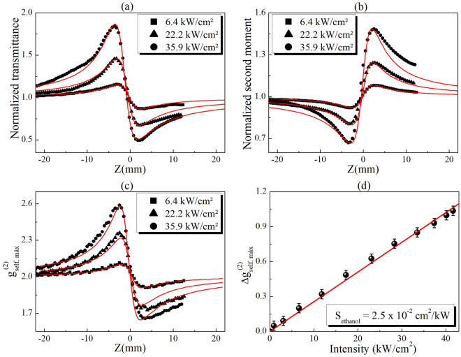
Figure 4 shows the experimental results obtained by the CA Z-scan, D4σ and IC-scan techniques performed to characterize the refractive NL response of pure ethanol. A good signal-to-noise ratio is observed in the curves because scattering particles (or defects) are not present in the NL medium. In Fig. 4a,b, by fitting the experimental results using the nonlocal model (with ) described in the Numerical simulation section, the NL refractive indices (for Z-scan) and (for D4σ), were obtained. The difference between the values measured by CA Z-scan and D4σ is probably due to the asymmetry or imperfection in the Gaussian beam profile that causes alterations when analyzing the transmittance through a small aperture or the transverse irradiance (second-order) moments, respectively. Nevertheless, reports in the literature using the local NL model to fit the experimental curves showed values lower than those obtained by us (see for example58). However, because the thermal contribution is dominant in continuous or quasi-continuous excitation, the nonlocal NL model presents better results when compared to the experimental curves, as shown in Fig. 4. Comparisons between the local and nonlocal models to fit the experimental curves are described in the Supplementary Material.
Figure 4.
(a) Z-scan, (b) D4σ and (c) IC-scan curves obtained for ethanol at different intensities. The solid lines in (a) and (b) represent the best curve fits using the nonlocal model (section 3.A), while in (c) represent the numerical simulation using the procedure described in section 3.B. (d) Peak-to-valley variation of IC-scan curves for ethanol as a function of incident intensity.
The IC-scan curves, shown in Fig. 4c, were obtained by calculating the maximum value of , i.e. , as a function of the sample position, when a light diffuser is used as WDS in the far field (5 cm before the CCD). Profiles similar to those of Z-scan, with a peak-valley structure, are observed in the IC-scan curves, starting with in the linear regime and increasing (or decreasing) due to beam divergence angle variations caused by the NL phase shift at high intensities. The variations of can be understood from the different intensity distributions that the speckle patterns exhibit when a light diffuser is illuminated with different spot sizes. For instance, it was reported in59 that the speckle size increases as the illuminated area decreases, regardless of the complex structure embedded in the phase and amplitude distribution of the beam illuminating the diffuser. Thus, if the CCD analysis window is considered to have a fixed area, increasing the speckle size results in a pattern with fewer speckles, but exhibiting a higher contrast . Conversely, large illumination diffuser areas lead to the construction of a pattern with a large number of speckles, with smaller sizes, resulting in a more homogeneous intensity distribution, i.e., lower intensity contrast . For this reason, the IC-scan curves present a peak-to-valley structure opposite to those of D4σ, which directly measure the beam size in the detection plane.
To validate the experimental IC-scan results, numerical simulations were performed using the and , which coincides with the values obtained by D4σ (also being close to those of Z-scan). The solid lines in Fig. 4c reveal the good agreement between the experimental results and the numerical simulation of well-characterized transparent NL media.
In a simpler way, the NL refractive index in IC-scan can also be obtained by using an external reference method, provided that the NL parameters ( and ) of a reference material are known. For instance, Fig. 4d shows the evolution of the peak-to-valley variations of , i.e. , as a function of the incident intensity for pure ethanol. A linear behavior with slope is obtained for intensities up to 42 kW/cm2. Assuming that is known for pure ethanol (reference material), similar to the Z-scan and D4σ techniques, where the peak-to-valley transmittance variations: and the peak-to-valley second moment variations: , respectively, in IC-scan we propose that . Thus, the NL refractive index for a different material can be obtained by using the relationship: , where the subscripts ref and j represent the reference and the new NL media, respectively. This proposal was applied to the study of the NL refraction of pure methanol, which also exhibits a linear dependence of on the incident intensity (see Fig. 2S). By using , calculated from the linear fit of the experimental results, and the measured by IC-scan, it is possible to find , which is very close to the values obtained by Z-scan and D4σ , demonstrating the reliability of the IC-scan technique. The calculated by the external reference method coincides with the value obtained from the numerical fit described in the Numerical simulation section (see Fig. 2S of the supplementary material).
It is important to mention that the expression to calculate , using the external reference method, is modified by a multiplicative factor when the reference and new NL material do not present the same nonlocality factor, m. In this work, the dependence of on m can be calculated numerically, as was done in previous works for Z-scan52,53. However, new studies are being developed to analytically describe IC-scan curves.
NL refractive optical characterization of turbid media
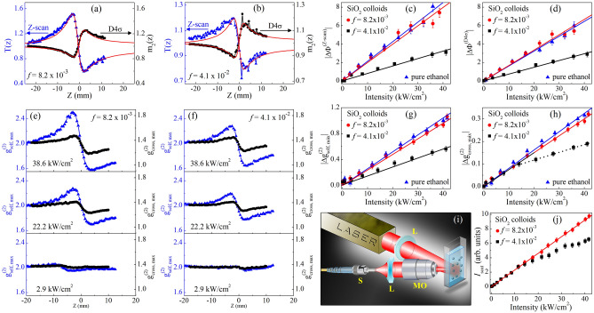
Although IC-scan can be used to characterize the NL refractive response of transparent media, the advantages of IC-scan over other techniques become relevant when measuring the NL response of scattering media. First, colloids containing SiO2 NPs suspended in ethanol were prepared as described in the NL media section. Since Rayleigh scattering is more predominant in the blue spectral region, large NP’s volume fractions were needed to induce from weak to moderate scattering at 788 nm. For instance, Fig. 5a illustrates a good signal-to-noise ratio in the CA Z-scan and D4σ curves for f = 8.2 × 10⁻³ and I = 22.2 kW/cm2. The experimental curves were fitted using the nonlocal nonlinearity model, obtaining as result a NL refractive index that coincides with that of pure ethanol . The analysis indicates that, under the excitation conditions used here, SiO2 NPs play the role of light scatterers with negligible nonlinearity, i.e. .
Figure 5.
CA Z-scan and D4σ curves for colloids containing SiO2 NPs suspended in ethanol with (a) f = 8.2 × 10⁻³ and (b) f = 4.1 × 10⁻², for an intensity of 22.2 kW/cm2. The solid curves in (a,b) were obtained by using the nonlocal nonlinearity model. (c, d) Intensity dependence of NL phase shift obtained from (c) CA Z-scan and (d) D4σ measurements. (e,f) IC-scan curves for SiO2 colloids obtained from 2D spatial intensity self-correlation (blue curves) and cross-correlation (black curves) functions and (g,h) their respective versus incident intensity. (i) Experimental scheme and (j) result of the measurement of the scattered light intensity (Iscat) as a function of the incident intensity for SiO2 colloids.
In addition, the experimental curves show that the higher the NPs concentration (f = 4.1 × 10⁻²), the lower the signal-to-noise ratio and, consequently, the greater the deviation against the theoretical (without dispersion) model, as shown in Fig. 5b. A compilation of the effective NL phase shift, , measured by CA Z-scan and D4σ for pure ethanol and colloids containing SiO2 NPs can be seen in Fig. 5c,d, respectively. Notice, in both techniques, that for pure ethanol and the colloid with f = 8.2 × 10⁻³ are very close to each other, in agreement with Fig. 5a, for intensities up to 40 kW/cm2. Nevertheless, for f = 4.1 × 10⁻², the slope of the curve as a function of for CA Z-scan (D4σ) is 2.6 (2.2) times less than that of pure ethanol, contradicting what is expected through . In the latter case, it is evident that the scattering is responsible for causing distortions in the profile of the CA Z-scan and D4σ curves, leading to inadequate measurements of the NL refractive index in turbid media. Table 1 shows the obtained for both techniques using the nonlocal nonlinearity model.
Table 1.
NL refractive indices measured for ethanol and SiO2 colloids, with different volume fractions (f), using Z-scan, D4σ and IC-scan techniques.
| NL optical technique | (cm2/W) | (cm2/W) | ||
|---|---|---|---|---|
| f = 8.2 × 10⁻³ | 4.1 × 10⁻³ | |||
| Z-scan | −2.8 × 10⁻⁸ | −2.6 × 10⁻⁸ | −1.1 × 10⁻⁸ | |
| D4σ | −2.2 × 10⁻⁸ | −2.2 × 10⁻⁸ | −1.0 × 10⁻⁸ | |
| IC-scan | Self-correlation | −2.2 × 10⁻⁸ | −2.1 × 10⁻⁸ | −1.3 × 10⁻⁸ |
| Cross-correlation | −2.1 × 10⁻⁸ | −2.1 × 10⁻⁸† | ||
†NL refractive index measured for intensities up to 15 kW/cm2.
On the other hand, the IC-scan curves show greater robustness against the scattering caused by SiO2 NPs, even for larger concentrations, as shown in Fig. 5e,f. For visualization purposes, all IC-scan curves were set to start at , although the curves show a decrease in the maxima of the correlation functions due to contrast decrease caused by particle-induced light scattering. The vertical shift does not modify the results obtained since the measurement of by using the external reference method is based on the analysis of the intensity dependence of . Figure 5g shows that the slope of the curves for the different concentrations of SiO2 NPs behaves in a similar way to that observed in for Z-scan and D4σ. As expected, similar values of were found for pure ethanol and the colloid with f = 8.2 × 10⁻³. Meanwhile, for the colloid with f = 4.1 × 10⁻², the was 1.7 times lower than that of pure ethanol, as reported in Table 1. Although the obtained by IC-scan for the largest concentrations differs from the expected theoretical value , its accuracy is higher than that of the Z-scan and D4σ techniques.
An adaptation in the analysis methodology of the speckle patterns captured by the CCD, in the IC-scan technique, allows to achieve more exact measurements of the NL refractive index in strong-scattering media. For this purpose, instead of the self-correlation function, it is proposed to analyze the 2D spatial intensity cross-correlation function between the far-field intensity transverse profiles (I1 and I2) induced in two different regimes. The first regime is excited at low incident intensities (I = 0.1 kW/cm2), where linear scattering effects exist but refractive nonlinearities are negligible. Meanwhile, in the second regime, the incident intensities (I > 1.0 kW/cm2) are high enough to excite both linear and NL effects. Therefore, the cross-correlation function allows to analyze the statistical properties of the speckle patterns that were modified only by NL refraction effects.
The black curves in Fig. 5e,f show the new IC-scan profiles for SiO2 colloids obtained by analyzing the maximum values of the as a function of the sample position. It is important to mention that the 2D spatial intensity cross-correlation function was also calculated from 50 consecutive images captured for I1 (linear regime) and I2 (NL regime). Notice from Fig. 5h that the between the peak and the valley for pure ethanol and the SiO2 colloid with f = 8.2 × 10⁻³ evolve in a similar way with the increase of the incident intensity, in accordance with the other techniques. Even more interesting is that for intensities up to 15 kW/cm2, the IC-scan technique using the cross-correlation function is the only methodology that, as expected, shows that the slope of the curve for the colloid with f = 4.1 × 10⁻² is close to that of pure ethanol. As a result, is obtained for the most concentrated SiO2-colloid, corresponding to ~ 96% of the value obtained for pure ethanol, as indicated in Table 1. The results reveal the potential of the IC-scan technique to remove the contribution of linear scattering in the analysis of intensity cross-correlations, allowing a correct measurement of the NL refractive index in turbid media.
For I > 15 kW/cm2, it is observed that for the colloid with f = 4.1 × 10⁻², also deviates significantly from the values found for pure ethanol, indicating the contribution of some new NL phenomenon that influences the characterization of the NL refractive behavior. To understand the origin of the change in the slope of the versus I curve, experiments to characterize the behavior of the scattered light intensity with the increase of the laser intensity were performed. In these experiments, a cell with 1.0 mm thickness, containing SiO2 colloids, was located in the focus of a 10 cm lens, identical to that used in the Z-scan, D4σ and IC-scan experiments. The scattered light was collected in a direction nearly perpendicular to the propagation direction of the incident laser beam by using a microscope objective, a plano-convex lens and a photodetector, as schematized in Fig. 5i.
Figure 5j shows the dependence of the scattered light intensity (at 788 nm) with the incident laser intensity for the SiO2 colloids. Notice that for f = 8.2 × 10⁻³, the scattered light intensity (Iscat) presents a linear behavior (red line) versus the incident intensity that extends up to ~ 40 kW/cm2. However, similar to Fig. 5h for f = 4.1 × 10⁻², Iscat exhibits a significant deviation from the linear behavior for I > 15 kW/cm2. This NL scattering contributions can be understood from the Rayleigh-Gans model60, by expressing the scattering coefficient as: , where represents the difference between the effective refractive indices of the NP and the host medium, and is an intensity-independent parameter, but depends on the size, shape and concentration of the NPs and the optical wavelength. By considering the NL refractive behavior of the colloids , it is possible to find expressions for the linear and NL scattering coefficients, with . Since the NL contribution of the SiO2 NPs was considered small compared to the solvent, corresponds mainly to the NL refractive index of ethanol, which became significant for higher intensities. Thus, as shown in Table 1, , decreasing the linear scattering coefficient for high intensities and corroborating the results of Fig. 5h,j. Therefore, in addition to the IC-scan technique allowing scattering-free NL refraction measurements, it also has the ability to distinguish linear and NL scattering contributions.
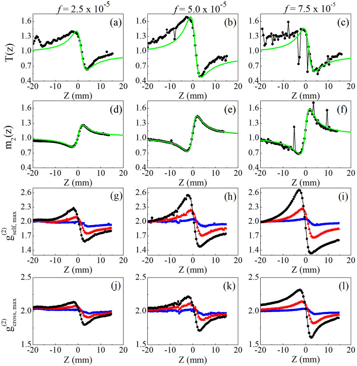
A similar study was performed with Au-NRs colloids, where the nonlinearity is dominated by the thermal response of the nanoparticles. However, due to the NR’s dimensions, a relevant contribution from linear scattering is present in the l-LSP band61, which makes the Au-NRs behave as both scatterers and NL particles. This dual behavior of the Au-NRs makes its NL characterization, using the Z-scan technique, suffer from scattering-induced wavefront distortions that cause erroneous NL refractive index measurements. In fact, Fig. 6a–c exhibit CA Z-scan curves whose signal-to-noise ratio decreases drastically with increasing volume fraction. An even more critical result is that for the Au-NRs colloid with f = 7.5 × 10⁻⁵ is less than for the more dilute colloids. By fitting the experimental curves using the nonlocal nonlinearity model (green solid lines), NL refractive indices that do not obey monotonic growth as a function of Au-NR’s concentration are obtained, as shown in Table 2. Thus, the NL characterization of these scattering media, by using the Z-scan technique, contradicts the expected optical behavior in effective media theories whose nonlinearity is dominated by the NP’s response. A clear example is the Maxwell–Garnett theory50,62, where the effective third-order susceptibility is the result of the contributions of the host medium and the NPs susceptibilities, weighted through the volume fraction. Figures 6d–f show the D4σ curves for Au-NR colloids where, despite exhibiting a better signal-to-noise ratio, also does not grow proportionally to the increase in Au-NR concentration.
Figure 6.
Normalized (a–c) CA Z-scan, (d–f) D4σ and (g-l) IC-scan curves for Au-NR colloids obtained from 2D spatial intensity (g–i) self-correlation and (j–l) cross-correlation functions, for Au-NRs colloids with volume fractions of (a,d,g,j) 2.5 × 10⁻⁵, (b,e,h,k) 5.0 × 10⁻⁵, (c,f,i,l) 7.5 × 10⁻⁵ and incident intensities of 0.1 kW/cm2 (blue squares), 0.5 kW/cm2 (red triangles) and 1.1 kW/cm2 (black circles). The solid (green) lines in (a-f) represent the best fit using the nonlocal nonlinearity model. For presentation purposes, all IC-scan curves have been shifted vertically to start at .
Table 2.
NL refractive indices measured for Au-NR colloids using Z-scan, D4σ and IC-scan techniques.
| NL optical technique | (× 10–6 cm2/W) | |||
|---|---|---|---|---|
| f = 2.5 × 10⁻⁵ | 5.0 × 10⁻⁵ | 7.5 × 10⁻⁵ | ||
| Z-scan | −0.56 | −0.86 | −0.75 | |
| D4σ | −0.27 | −0.45 | −0.46 | |
| IC-scan | Self-correlation | −0.56 | −0.88 | −1.04 |
| Cross-correlation | −0.86 | −1.26 | −1.73 | |
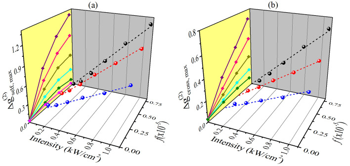
In contrast, the IC-scan curves, obtained through the analysis of the self- (Fig. 6g–i) and cross-correlation (Fig. 6j–l) functions, show excellent signal-to-noise ratios for all concentrations explored in this work. Furthermore, Fig. 7 exhibits a monotonic linear increase of with incident intensity, as expected for both IC-scan configurations. Regarding the concentration dependence of the NL refractive index, measurements using the CA Z-scan (Fig. 6a–c) or self-correlation IC-scan (Fig. 7a) show that for larger volume fractions, decreases or saturates, respectively, due to strong scattering. Nevertheless, when the IC-scan technique is applied using the cross-correlation functions (Fig. 7b), presents a linear behavior with the NP’s volumetric fraction, preserving the validity of models such as the Maxwell–Garnett one to study the NL response of composite media. Therefore, the studies with Au-NR’s colloids reinforce the potential of the cross-correlation IC-scan technique to measure the NL refractive indices of turbid media.
Figure 7.
Intensity and volume fraction dependence of peak-to-valley variation of IC-scan curves obtained for Au-NRs colloids using the 2D spatial intensity (a) self-correlation and (b) cross-correlation functions. In the yellow planes, the curves were built from the projection of the values of and versus f, for the different intensities. From top to bottom: I = 1.1, 0.8, 0.5, 0.3, 0.2 and 0.04 kW/cm2, the latter corresponding to the linear regime (used as a reference in the cross-correlation functions).
Conclusions
In summary, the present experiments demonstrate the potential of the IC-scan technique to adequately characterize the NL optical response of heterogeneous media, which can exhibit such strong (multiple) scattering that conventional NL spectroscopy techniques fail. The ability for NL characterization of turbid media with IC-scan lies in the analysis of the wavefront changes induced by the self-focusing and self-defocusing effects through the statistical properties of the speckle patterns that are formed in media with a high degree of scattering (or passing through an external light diffuser). Therefore, elastic scattering, which is detrimental to Z-scan and D4σ techniques, is the fundamental phenomenon that gives rise to the IC-scan technique. As a proof of principle, the NL refractive indices of colloids containing highly concentrated silica nanospheres in ethanol and gold nanorods in water were measured with Z-scan, D4σ and IC-scan. The results reveal that the IC-scan experiments present curves with a better signal-to-noise ratio, resulting in more precise measurements, as well as NL refractive index that are in agreement with the expected theoretical values. The accuracy of IC-scan measurements in highly scattering media is achieved by analyzing the cross-correlation functions between linear and NL regimes, which measure the influence of NL refraction on the generated speckle patterns, without the interference effects caused by linear scattering. In this way, IC-scan is presented as a powerful tool to characterize the NL refractive index of media with significant linear (elastic) scattering, such as inhomogeneous vitreous materials, biological media and liquid crystals, maintaining a simple experimental setup compared to other techniques. In addition, the IC-scan technique presented the ability to identify regions where NL scattering effects are relevant, which is a topic of considerable fundamental interest, and its study is currently in progress. New studies are also being carried out to characterize the NL response of liquid crystals, which have the ability to generate speckle patterns by themselves, configuring an adaptation to the IC-scan technique in which the light diffuser is dispensable.
Supplementary Information
Acknowledgements
A S. Reyna thanks Dr. Cid B. de Araújo for the infrastructure provided for the development of this work.
Author contributions
A.S. Reyna conceived and supervised the project. M.J.B. Crispim and C.C.S. Pereira conducted the experimental work, including the assembly of the experimental setups, measurements, and analysis of the results. N.T.C. Oliveira synthesized and characterized the nanomaterials. All authors contributed to the scientific discussions and the preparation of the manuscript.
Funding
Conselho Nacional de Desenvolvimento Científico e Tecnológico (CNPq) - Universal 408016/2018-3; Fundação de Amparo à Ciência e Tecnologia do Estado de Pernambuco (FACEPE) - APQ-0962-1.05/21 and APQ-1006-1.05/21; Coordenação de Aperfeiçoamento de Pessoal de Nível Superior (CAPES).
Data availability
Data underlying the results presented in this paper are not publicly available at this time but may be obtained from the authors upon reasonable request.
Competing interests
The authors declare no competing interests.
Footnotes
The original online version of this Article was revised: The original version of this Article contained an error in the Funding section. Now reads: “Conselho Nacional de Desenvolvimento Científico e Tecnológico (CNPq) - Universal 408016/2018-3; Fundação de Amparo à Ciência eTecnologia do Estado de Pernambuco (FACEPE) - APQ-0962-1.05/21 and APQ-1006-1.05/21; Coordenação de Aperfeiçoamento de Pessoal de Nível Superior (CAPES).”
Publisher's note
Springer Nature remains neutral with regard to jurisdictional claims in published maps and institutional affiliations.
Change history
5/17/2023
A Correction to this paper has been published: 10.1038/s41598-023-34978-z
Supplementary Information
The online version contains supplementary material available at 10.1038/s41598-023-34486-0.
References
- 1.Carminati R, Schotland JC. Principles of Scattering and Transport of Light. Cambridge University Press, Cambridge; 2021. [Google Scholar]
- 2.Carvalho PM, Felício MR, Santos NC, Gonçalves S, Domingues MM. Application of light scattering techniques to nanoparticle characterization and development. Front. Chem. 2018;6:237. doi: 10.3389/fchem.2018.00237. [DOI] [PMC free article] [PubMed] [Google Scholar]
- 3.Szarvas, T., Molnár, G., Nádudvari, Gy., Tóth, Sz., Almásy, O., Spindler, Sz., Erdei, D., Pothorszky, Sz., Kretschmer, R. & Bauer, S. Bulk micro-defect detection with low-angle illumination. Rev. Sci. Instrum.92, 043701, DOI: 10.1063/5.0027147 (2021). [DOI] [PubMed]
- 4.Yun SH, Kwok SJJ. Light in diagnosis, therapy and surgery. Nat. Biomed. Eng. 2017;1:0008. doi: 10.1038/s41551-016-0008. [DOI] [PMC free article] [PubMed] [Google Scholar]
- 5.Mosk AP, Lagendijk A, Lerosey G, Fink M. Controlling waves in space and time for imaging and focusing in complex media. Nat. Photon. 2012;6:283–292. doi: 10.1038/nphoton.2012.88. [DOI] [Google Scholar]
- 6.Yu H, Lee K, Park J, Park Y. Ultrahigh-definition dynamic 3D holographic display by active control of volume speckle fields. Nat. Photon. 2017;11:186–192. doi: 10.1038/nphoton.2016.272. [DOI] [Google Scholar]
- 7.Pappu R, Recht B, Taylor J, Gershenfeld N. Physical one-way functions. Science. 2002;297:2026–2030. doi: 10.1126/science.1074376. [DOI] [PubMed] [Google Scholar]
- 8.Wiersma DS. The physics and applications of random lasers. Nat. Phys. 2008;4:359–367. doi: 10.1038/nphys971. [DOI] [Google Scholar]
- 9.Ghofraniha N, Viola I, Di Maria F, Barbarella G, Gigli G, Leuzzi L, Conti C. Experimental evidence of replica symmetry breaking in random lasers. Nat. Comm. 2015;6:6058. doi: 10.1038/ncomms7058. [DOI] [PubMed] [Google Scholar]
- 10.Raposo EP, González IRR, Macêdo AMS, Lima BC, Kashyap R, de Menezes LS, Gomes ASL. Evidence of a floquet phase in a photonic system. Phys. Rev. Lett. 2019;122:143903. doi: 10.1103/PhysRevLett.122.143903. [DOI] [PubMed] [Google Scholar]
- 11.Segev M, Silberberg Y, Christodoulides DN. Anderson localization of light. Nat. Photon. 2013;7:197–204. doi: 10.1038/nphoton.2013.30. [DOI] [Google Scholar]
- 12.Bashkansky M, Reintjes J. Statistics and reduction of speckle in optical coherence tomography. Opt. Lett. 2000;25:545–547. doi: 10.1364/OL.25.000545. [DOI] [PubMed] [Google Scholar]
- 13.Chaigneau E, Wright AJ, Poland SP, Girkin JM, Silver RA. Impact of wavefront distortion and scattering on 2-photon microscopy in mammalian brain tissue. Opt. Express. 2011;19:22755–22774. doi: 10.1364/OE.19.022755. [DOI] [PMC free article] [PubMed] [Google Scholar]
- 14.Strudley T, Bruck R, Mills B, Muskens OL. An ultrafast reconfigurable nanophotonic switch using wavefront shaping of light in a nonlinear nanomaterial. Light Sci. Appl. 2014;3:207. doi: 10.1038/lsa.2014.88. [DOI] [Google Scholar]
- 15.Dainty JC. Laser Speckle and Related Phenomena. Springer, Berlin; 1975. [Google Scholar]
- 16.Liu Y, Zhang C, Wang LV. Effects of light scattering on optical-resolution photoacoustic microscopy. J. Biomed. Opt. 2012;17:126014. doi: 10.1117/1.JBO.17.12.126014. [DOI] [PMC free article] [PubMed] [Google Scholar]
- 17.He, P., Perlin, M. A., Muleady, S. R., Lewis-Swan, R. J., Hutson, R. B., Ye, J. & Rey, A. M. Engineering spin squeezing in a 3D optical lattice with interacting spin-orbit-coupled fermions. Phys. Rev. Res.1, 033075, DOI: 10.1103/PhysRevResearch.1.033075 (2019).
- 18.Steponavicius R, Thennadil SN. Extraction of chemical information of suspensions using radiative transfer theory to remove multiple scattering effects: Application to a model multicomponent system. Anal. Chem. 2011;83:1931–1937. doi: 10.1021/ac1024073. [DOI] [PubMed] [Google Scholar]
- 19.Zhan P, Si J, Tan W, Liu X, Wu B, Xu S, Chen F, Hou X. The influence of turbid medium properties on object visibility in optical Kerr gated imaging. Laser Phys. 2014;24:015401. doi: 10.1088/1054-660X/24/1/015401. [DOI] [Google Scholar]
- 20.Meitav N, Ribak EN, Shoham S. Point spread function estimation from projected speckle illumination. Light Sci. Appl. 2016;5:e16048. doi: 10.1038/lsa.2016.48. [DOI] [PMC free article] [PubMed] [Google Scholar]
- 21.Liu Y, Wang Z, Huang J. Recent progress on aberration compensation and coherent noise suppression in digital holography. Appl. Sci. 2018;8:444. doi: 10.3390/app8030444. [DOI] [Google Scholar]
- 22.Steinberg S, Yan L-Q. Rendering of subjective speckle formed by rough statistical surfaces. ACM Trans. Graph. 2022;41:2. doi: 10.1145/3472293. [DOI] [Google Scholar]
- 23.Goodman, J. W. Statistical Properties of Laser Speckle Pattern. In Laser Speckle and Related Phenomena, vol. 9 in series Topics in Applied Physics, (ed. Dainty, J. C.), 9–75 (Springer-Verlag, Berlin, Heidelberg New York Tokyo, 1984).
- 24.Scaddan RJ, Walker JG. Statistics of stellar speckle patterns. Appl. Opt. 1978;17:3779–3784. doi: 10.1364/AO.17.003779. [DOI] [PubMed] [Google Scholar]
- 25.Soest, G. V., Poelwijk, F. J. & Lagendijk, A. Speckle experiments in random lasers. Phys. Rev. E65, 04660, DOI: 10.1103/PhysRevE.65.046603 (2002). [DOI] [PubMed]
- 26.Oliveira, N. T. C., Vieira, A. M., de Araújo, C. B., Martins, W. S., de Oliveira, R. A., & Reyna, A. S. Light disorder as a degree of randomness to improve the performance of random lasers. Phys. Rev. Appl.15, 064062. 10.1103/PhysRevApplied.15.064062 (2021).
- 27.Vieira AM, Oliveira NTC, de Araújo CB, Martins WS, de Oliveira RA, Reyna AS. Influence of the excitation light disorder on the spatial coherence in the stimulated raman scattering and random lasing coupled regime. J. Phys. Chem. C. 2021;125:5919–5926. doi: 10.1021/acs.jpcc.1c00266. [DOI] [Google Scholar]
- 28.Bender N, Sun M, Yılmaz H, Bewersdorf J, Cao H. Circumventing the optical diffraction limit with customized speckles. Optica. 2021;8:122–129. doi: 10.1364/OPTICA.411007. [DOI] [Google Scholar]
- 29.Bender N, Yılmaz H, Bromberg Y, Cao H. Customizing speckle intensity statistics. Optica. 2018;5:595–600. doi: 10.1364/OPTICA.5.000595. [DOI] [Google Scholar]
- 30.Chu TC, Ranson WF, Sutton MA. Applications of digital-image-correlation techniques to experimental mechanics. Exp. Mech. 1985;25:232–244. doi: 10.1007/BF02325092. [DOI] [Google Scholar]
- 31.Valent E, Silberberg Y. Scatterer recognition via analysis of speckle patterns. Optica. 2018;5:204–207. doi: 10.1364/OPTICA.5.000204. [DOI] [Google Scholar]
- 32.Heeman, W., Steenbergen, W., van Dam, G. M. & Boerma, E. C. Clinical applications of laser speckle contrast imaging: A review. J. Biomed. Opt.24, 080901, DOI: 10.1117/1.JBO.24.8.080901 (2019). [DOI] [PMC free article] [PubMed]
- 33.Samineni P, Perret Z, Warren WS, Fischer MC. Measurements of nonlinear refractive index in scattering media. Opt. Express. 2010;18:12727–12735. doi: 10.1364/OE.18.012727. [DOI] [PMC free article] [PubMed] [Google Scholar]
- 34.Jorge, K. C., García, H. A., Amaral, A. M., Reyna, A. S., Menezes, L. de S. & de Araújo, C. B. Measurements of the nonlinear refractive index in scattering media using the Scattered Light Imaging Method – SLIM. Opt. Express23, 19512–19521. 10.1364/OE.23.019512 (2015). [DOI] [PubMed]
- 35.Liu, W.-W., Zeng, X., Lin, L., Qi, P.-F. & Zhang, N. Spectral domain Z-scan technique. Opt. Lasers Eng.146, 106693. 10.1016/j.optlaseng.2021.106693 (2021).
- 36.Purwar H, Idlahcen S, Rozé C, Sedarsky D, Blaisot J-B. Collinear, two-color optical Kerr effect shutter for ultrafast time-resolved imaging. Opt. Express. 2014;22:15778–15790. doi: 10.1364/OE.22.015778. [DOI] [PubMed] [Google Scholar]
- 37.Rockwell BA, Roach WP, Rogers ME, Mayo MW, Toth CA, Cain CP, Noojin GD. Nonlinear refraction in vitreous humor. Opt. Lett. 1993;18:1792–1794. doi: 10.1364/OL.18.001792. [DOI] [PubMed] [Google Scholar]
- 38.Joshi JH, Kalainathan S, Kanchan DK, Joshi MJ, Parikh KD. Effect of l-threonine on growth and properties of ammonium dihydrogen phosphate crystal. Arab. J. Chem. 2020;13:1532–1550. doi: 10.1016/j.arabjc.2017.12.005. [DOI] [Google Scholar]
- 39.Kim, G. Y. & Kwak, C. H. Simple Optical Methods for Measuring Optical Nonlinearities and Rotational Viscosity in Nematic Liquid Crystals. In New Developments in Liquid Crystals, (ed. Tkachenko, G.) (IntechOpen, 2009).
- 40.Rodríguez-Rosales, A. A., Ortega-Martínez, R. & Morales-Saavedra, O. G. Impact of dyes on the nonlinear optical response of liquid crystals implementing the Z-scan technique. J. Phys. Conf. Ser.274, 012138. 10.1088/1742-6596/274/1/012138 (2011).
- 41.Jorge, K. C., Amaral, A. M., Reyna, A. S., Menezes, L. de S. & de Araújo, C. B. Observation and analysis of nonlinear scattering using the Scattered Light Imaging Method (SLIM). Phys. Rev. A105, 063520. 10.1103/PhysRevA.105.063520 (2022).
- 42.Zeng X, Qi P, Li M, Cheng Z, Lin L, Liu W, Wang Y, Zhang N. Nonlinear optical properties of human cornea measured by spectral domain Z-scan method. Opt. Express. 2021;29:38870–38878. doi: 10.1364/OE.441731. [DOI] [PubMed] [Google Scholar]
- 43.Sheik-Bahae, M., Said, A. A., Wei, T. H., Hagan, D. J. & Van Stryland, E. W. Sensitive measurements of optical nonlinearities using a single beam. IEEE J. Quantum Electron. QE-26, 760–769. 10.1109/3.53394 (1990).
- 44.Boudebs G, Besse V, Cassagne C, Leblond H, de Araújo CB. Nonlinear characterization of materials using the D4σ method inside a Z-scan 4f-system. Opt. Lett. 2013;38:2206–2208. doi: 10.1364/OL.38.002206. [DOI] [PubMed] [Google Scholar]
- 45.Amaral AM, Mejía HA, Falcão-Filho EL, de Araújo CB. D4σ curves described analytically through propagation analysis of transverse irradiance moments. Opt. Lett. 2016;41:2081–2084. doi: 10.1364/OL.41.002081. [DOI] [PubMed] [Google Scholar]
- 46.de Araújo, C. B., Gomes, A. S. L. & Boudebs, G. Techniques for nonlinear optical characterization of materials: a review. Rep. Prog. Phys.79, 036401. 10.1088/0034-4885/79/3/036401 (2016). [DOI] [PubMed]
- 47.Kim K-D, Yu H, Lee K, Park Y-K. Universal sensitivity of speckle intensity correlations to wavefront change in light diffusers. Sci. Rep. 2017;7:44435. doi: 10.1038/srep44435. [DOI] [PMC free article] [PubMed] [Google Scholar]
- 48.Bromberg Y, Lahini Y, Small E, Silberberg Y. Hanbury Brown and Twiss interferometry with interacting photons. Nat. Photon. 2010;4:721–726. doi: 10.1038/nphoton.2010.195. [DOI] [Google Scholar]
- 49.Brito-Silva AM, Sobral-Filho RG, Barbosa-Silva R, de Araújo CB, Galembeck A, Brolo AG. Improved synthesis of gold and silver nanoshells. Langmuir. 2013;29:4366–4372. doi: 10.1021/la3050626. [DOI] [PubMed] [Google Scholar]
- 50.Oliveira NTC, Reyna AS, Falcão EHL, de Araújo CB. Light scattering, absorption, and refraction due to high-order optical nonlinearities in colloidal gold nanorods. J. Phys. Chem. C. 2019;123(20):12997–13008. doi: 10.1021/acs.jpcc.9b01369. [DOI] [Google Scholar]
- 51.Huang X, El-Sayed MA. Gold nanoparticles: Optical properties and implementations in cancer diagnosis and photothermal therapy. J. Adv. Res. 2010;1:13–28. doi: 10.1016/j.jare.2010.02.002. [DOI] [Google Scholar]
- 52.Ramirez, E. V. G., Carrasco, M. L. A., Otero, M. M. M., Lara, E. R., Chavez-Cerda, S. & Castillo, M. D. I. Z-scan and spatial self-phase modulation of a Gaussian beam in a thin nonlocal nonlinear media. J. Opt.13, 085203. 10.1088/2040-8978/13/8/085203 (2011).
- 53.Ortega AB, Carrasco MLA, Otero MMM, Lara ER, Ramírez EVG, Castillo MDI. Analytical expressions for z-scan with arbitrary phase change in thin nonlocal nonlinear media. Opt. Express. 2014;22:27932–27941. doi: 10.1364/OE.22.027932. [DOI] [PubMed] [Google Scholar]
- 54.Hughes S, Burzler JM, Spruce G, Wherrett BS. Fast Fourier transform techniques for efficient simulation of Z-scan measurements. J. Opt. Soc. Am. B. 1995;12:1888–1893. doi: 10.1364/JOSAB.12.001888. [DOI] [Google Scholar]
- 55.Song L, Zhou Z, Wang X, Zhao X, Elson DS. Simulation of speckle patterns with pre-defined correlation distributions. Biomed. Opt. Express. 2016;7:798–809. doi: 10.1364/BOE.7.000798. [DOI] [PMC free article] [PubMed] [Google Scholar]
- 56.Saleh, B. E. A. & Teich, M. C. Fundamentals of Photonics. (John Wiley & Sons, 2019).
- 57.Poon, T.-C. & Kim, T. Engineering Optics with MATLAB, 2nd ed. (World Scientific Publishing Company, 2017).
- 58.Bautista JEQ, da Silva-Neto ML, Campos CLAV, Maldonado M, de Araújo CB, Gomes ASL. Thermal and non-thermal intensity dependent optical nonlinearities in ethanol at 800 nm, 1480 nm, and 1560 nm. J. Opt. Soc. Am. B. 2021;38:1104–1111. doi: 10.1364/JOSAB.418635. [DOI] [Google Scholar]
- 59.Hu X-B, Dong M-X, Zhu Z-H, Gao W, Rosales-Guzmán C. Does the structure of light influence the speckle size? Sci. Rep. 2020;10:199. doi: 10.1038/s41598-019-56964-0. [DOI] [PMC free article] [PubMed] [Google Scholar]
- 60.Liberman, V., Sworin, M., Kingsborough, R. P., Geurtsen, G. P., Rothschild, M. Nonlinear Bleaching, Absorption, and Scattering of 532-nm-Irradiated Plasmonic Nanoparticles. J. Appl. Phys.113, 053107. 10.1063/1.4790798 (2013).
- 61.Sburlan, S. E., Blanco, L. A. & Nieto-Vesperinas, M. Plasmon excitation in sets of nanoscale cylinders and spheres. Phys. Rev. B73, 035403. 10.1103/PhysRevB.73.035403 (2006).
- 62.Reyna, A. S. & de Araújo, C. B. High-order nonlinearities of metal–dielectric nanocomposites. In: Metal Nanostructures for Photonics (eds. Kassab, L. R. P. & de Araújo, C. B.) pp. 61–86. Ch. 4, (Elsevier, 2019).
Associated Data
This section collects any data citations, data availability statements, or supplementary materials included in this article.
Supplementary Materials
Data Availability Statement
Data underlying the results presented in this paper are not publicly available at this time but may be obtained from the authors upon reasonable request.


