Abstract
Background
Recent studies have investigated the features of breast cancer (BC) with low human epidermal growth factor receptor 2 (HER2) expression or HER2-0 expression. However, the results were inconsistent. In this study, we investigated the differences in the pathological complete response (pCR) rate and disease-free survival (DFS) between HER2-low and HER2-0 BC patients and between subgroups.
Methods
HER2-negative BC patients who received neoadjuvant chemotherapy between January 2013 and December 2019 in our hospital were retrospectively reviewed. First, the pCR rate and DFS were compared between HER2-low and HER2-0 patients and among different hormone receptor (HR) and HER2 statuses. Subsequently, DFS was compared between different HER2 status populations with or without pCR. Finally, a Cox regression model was used to identify the prognostic factors.
Results
Overall, 693 patients were selected: 561 were HER2-low, and 132 were HER2-0. Between the two groups, there were significant differences in N stage (P = 0.008) and HR status (P = 0.007). No significant difference in the pCR rate (12.12% vs 14.39%, P = 0.468) or DFS was observed, independent of HR status. HR+/HER2-low patients had a significantly worse pCR rate (P < 0.001) and longer DFS (P < 0.001) than HR-/HER2-low or HER2-0 patients. In addition, a longer DFS was found in HER2-low patients versus HER2-0 patients among those who did not achieve pCR. Cox regression showed that N stage and HR status were prognostic factors in the overall and HER2-low populations, while no prognostic factor was found in the HER2-0 group.
Conclusion
This study suggested that HER2 status is not associated with the pCR rate or DFS. Longer DFS was found only among patients who did not achieve pCR in the HER2-low versus HER2-0 population. We speculated that the interaction of HR and HER2 might have played a crucial role in this process.
Keywords: HER2-low, HER2-0, hormone receptor, pCR, DFS
Introduction
In recent decades, the number of breast cancer (BC) patients has increased rapidly, and BC is the most common malignant tumor in women around the world.1 Currently, according to the 2018 ASCO guidelines, BC subtypes are defined by the statuses of the estrogen receptor (ER), progesterone receptor (PR), human epidermal growth factor receptor 2 (HER2), and Ki67.2 HER2 status, which is evaluated by immunohistochemistry (IHC), historically is classified as positive (IHC score of 3+ or 2+ and amplification of the ERBB2 gene) or negative (IHC score of 0, 1+, or 2+ with nonamplification of the ERBB2 gene).3
However, in recent years, several clinical trials have reported the existence of a novel subtype among HER2-negative BC patients. The results showed that there was a significant difference in characteristics between HER2-0 (IHC score of 0) patients and HER2-low (IHC score of 1+ or 2+ without detecting ERBB2 gene amplification) patients.4–6 Novel antibody-drug conjugates (ADCs) targeting HER2 could significantly improve the prognosis of BC patients with low HER2 expression.7,8 Subsequent studies have been conducted to explore the differences in response to neoadjuvant chemotherapy (NAC), as well as in DFS of BC patients with HER2-low or HER2-0 status.9–16 However, these results have been inconsistent and even conflicting.
Therefore, in this study, we retrospectively reviewed data from our hospital. This study aimed to compare the pathological complete response (pCR) rate and disease-free survival (DFS) between HER2-low and HER2-0 BC patients, as well as between subgroups.
Methods
Patient Selection and Study Design
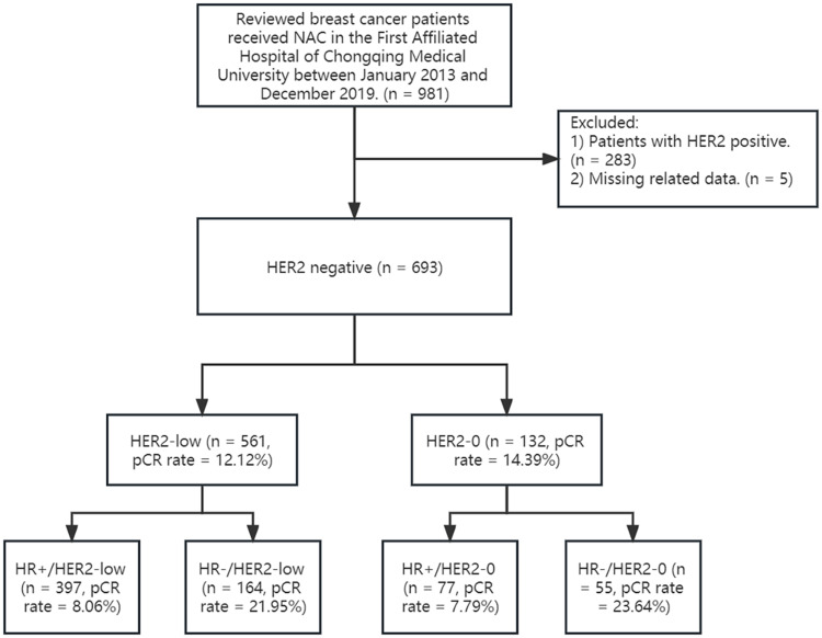
This retrospective study was approved by the ethics committee of the First Affiliated Hospital of Chongqing Medical University (ID: No. 2020–59) and was performed according to the Declaration of Helsinki. We retrospectively reviewed BC patients at the First Affiliated Hospital of Chongqing Medical University from January 2013 to December 2019. Patients who received NAC were selected for the subsequent analysis. We compared the pCR rate and DFS between the HER2-low group and the HER2-0 group. Next, the pCR rate and DFS were compared among the following 4 subgroups: 1) HR+/HER2-low patients; 2) HR-/HER2-low patients; 3) HR+/HER2-0 patients; and 4) HR-/HER2-0 patients. DFS was further compared among patients with different HER2 statuses and with different responses to NAC. Finally, we identified prognostic factors using a Cox regression model. The flowchart of this study is shown in Figure 1.
Figure 1.
The flowchart of this study.
Treatment Protocol
NAC was performed according to the National Comprehensive Cancer Network (NCCN) and the Chinese Society of Clinical Oncology (CSCO) guidelines.17,18 Once the patients were diagnosed as needing to receive NAC, treatment started within a week. The treatment protocol was TEC (docetaxel 75 mg/m2, epirubicin 75 mg/m2, and cyclophosphamide 500 mg/m2) or EC (epirubicin 75 mg/m2 and cyclophosphamide 500 mg/m2), and drugs were administered at 21-day intervals. After 4–6 cycles of NAC, the response was evaluated by both the clinicians and the pathologists. Mastectomy or breast-conserving surgery plus axillary lymphadenectomy was subsequently performed.
Pathological Evaluation
The IHC index was assessed based on the core needle biopsy of the tumor. Hormone receptor (HR) positivity was defined as an estrogen receptor (ER)- or progesterone receptor (PR)-expressing cell percentage > 10% by IHC. pCR was defined as no residual invasive tumor in either the breast or axillary lymph nodes (ypT0ypN0).19 HER2 was evaluated by IHC and FISH. The possible IHC scores were as follows: 1) 0: no staining is observed or membrane staining that is incomplete and faint/barely perceptible in ≤ 10% of tumor cells; 2) 1+: incomplete membrane staining that is faint/barely perceptible in > 10% of tumor cells; 3) 2+: weak to moderate complete membrane staining observed in > 10% of tumor cells; and 4) 3+: circumferential membrane staining that is complete, intense, and in > 10% of tumor cells. An IHC score of 0 was defined as HER2-0. An IHC core of 1+ or 2+ with negative FISH was defined as HER2-low. An IHC core of 3+ or 2+ with positive FISH was defined as HER2-positive.20 These results were evaluated by two pathologists independently and blindly.
Follow-Up
All patients were interviewed by telephone from discharge to February 1st, 2021. DFS was selected for assessing patient prognoses. DFS was defined as the period between surgery and either 1) the first relapse of the tumor locally, regionally, or distantly; 2) a diagnosis of secondary malignant tumor or contralateral BC; or 3) death due to any reason.
Statistical Analysis
We used IBM SPSS 23.0 (Chicago, IL, USA), Stata/SE 16.0 and RStudio 1.1.456 (R version 4.2.1) software for statistical analysis. For each patient, age; histological type of tumor; T stage; N stage; ER, PR, and HR status; HER2 score; Ki67 index; P53; and pCR were recorded. ER, PR, HR, and Ki67 were divided according to current guidelines. P53 was divided based on the ideal cutoff value, which was determined using receiver operating characteristic (ROC) curves. The chi-square test was used to compare categorical data, and the t-test was used to compare continuous data. Kaplan‒Meier curves were used to describe the survival probability, and the Log rank test was used for comparisons between groups. A Cox proportional hazard model was used to identify prognostic factors in the whole cohort, HER2-low cohort, and HER2-0 cohort. A p value < 0.05 was considered to indicate a significant difference.
Results
Patient Characteristics
Overall, a total of 693 HER2-negative BC patients were selected, and the median age was 48 years old (range 20–72). Of them, 132 patients had HER2-0 expression, and 561 patients had HER2-low expression. We first compared the characteristics and response to NAC between the HER2-low group and the HER2-0 group. There were significant differences in N stage and ER, PR, and HR status. Compared with HER2-0 patients, HER2-low patients were significantly associated with positive ER, PR, and HR and N1 stage. Moreover, HER2-low patients exhibited lower Ki67 and higher P53 levels, but the differences were not statistically significant. Table 1 displays the characteristics of the patients.
Table 1.
Characteristics of the Patients at Diagnosis
| Variable | HER2-Low (n = 561) | HER2-0 (n = 132) | Total | P value |
|---|---|---|---|---|
| Age | 48 (20–72) | 49 (26–70) | 48 (20–72) | 0.399 |
| Histologic type | 0.431 | |||
| Invasive ductal carcinoma | 543 (96.79%) | 126 (95.45%) | 669 | |
| Other | 18 (3.21%) | 6 (4.55%) | 24 | |
| T | 0.444 | |||
| T1 | 61 (10.87%) | 10 (7.58%) | 71 | |
| T2 | 385 (68.63%) | 97 (73.48%) | 482 | |
| T3 | 115 (20.50%) | 25 (18.94%) | 140 | |
| N | 0.008 | |||
| N0 | 205 (36.54%) | 58 (43.94%) | 263 | |
| N1 | 271 (48.31%) | 47 (35.61%) | 318 | |
| N2 | 74 (13.19%) | 19 (14.39%) | 93 | |
| N3 | 11 (1.96%) | 8 (6.06%) | 19 | |
| ER | 0.007 | |||
| ≤ 10 | 200 (45.65%) | 64 (48.48%) | 264 | |
| > 10 | 361 (64.35%) | 68 (51.52%) | 429 | |
| PR | 0.018 | |||
| ≤ 10 | 314 (55.97%) | 89 (67.42%) | 403 | |
| > 10 | 247 (44.03%) | 43 (32.58%) | 290 | |
| HR | 0.007 | |||
| HR+ | 397 (70.77%) | 77 (58.33) | 474 | |
| HR- | 164 (29.23%) | 55 (41.67%) | 219 | |
| Ki67 | 0.253 | |||
| ≤ 20 | 290 (51.69%) | 58 (43.94%) | 348 | |
| 21–30 | 128 (22.82%) | 33 (25.00%) | 161 | |
| > 30 | 143 (25.49%) | 41 (31.06%) | 184 | |
| p53 | 0.384 | |||
| ≤ 17.5 | 284 (50.62%) | 73 (55.30%) | 357 | |
| > 17.5 | 277 (49.38%) | 59 (44.70%) | 336 | |
| pCR | 0.468 | |||
| pCR | 68 (12.12%) | 19 (14.39%) | 87 | |
| Non-pCR | 493 (87.88) | 113 (85.61%) | 606 |
Comparison of pCR Rate
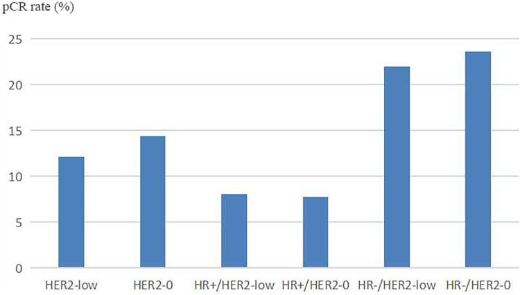
We analyzed the pCR rate among BC patients with HER2-low or HER2-0 status. Although the pCR rate of HER2-low patients was slightly lower than that of HER2-0 patients, no significant difference was observed (12.12% vs 14.39%, P = 0.468). Additionally, no significant difference was found between the HR+ and HR- populations (P = 0.937 and P = 0.795, respectively). Moreover, in line with consensus, the results showed that patients with HR-/HER2-low or HER2-0 status had a significantly higher pCR rate than patients with HR+/HER2-low or HER2-0 status (P < 0.001, Figure 2).
Figure 2.
The pCR rate of patients with HER2-low, HER2-0, HR+/HER2-low, HR+/HER2-0, HR-/HER2-low, and HR-/HER2-0. No significant difference was observed in the whole, HR+ and HR- populations (P = 0.468, P = 0.937 and P = 0.795, respectively).
Comparison of DFS
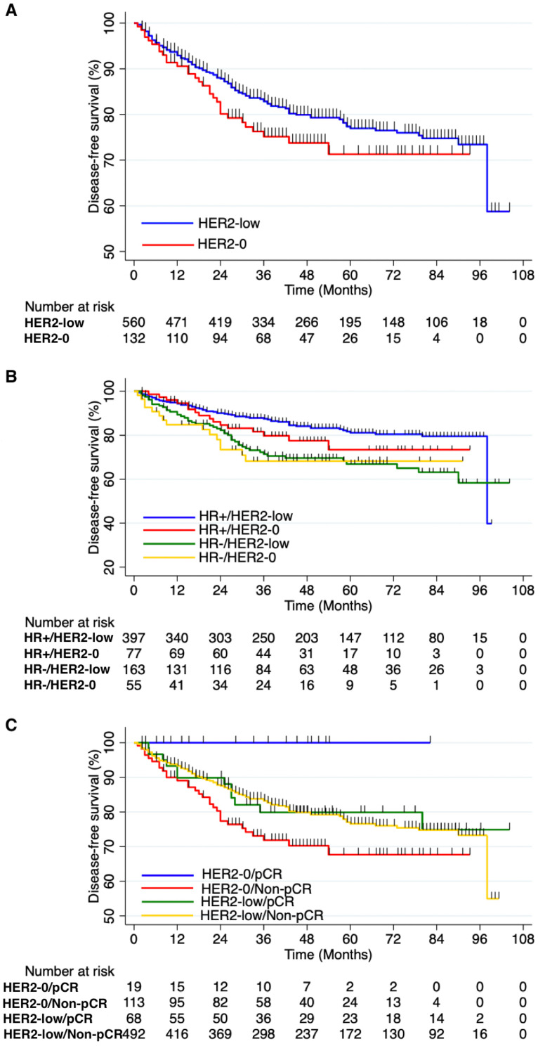
The median follow-up time was 43 months (IQR: 23–68). In total, 140 events were detected (109 cases in the HER2-low group and 31 cases in the HER2-0 group). The 3-year DFS rates for HER2-low and HER2-0 patients were 85.0% and 77.9%, respectively. The 5-year DFS rates for HER2-low and HER2-0 patients were 81.6% and 76.3%, respectively. Although we observed that HER2-low BC patients had a longer DFS than HER2-0 BC patients, there was no significant difference (P = 0.156, Figure 3A).
Figure 3.
DFS analysis (A) Comparison of DFS between patients with HER2-low and patients with HER2-0. Although HER2-low BC patients exhibited a longer DFS compared to HER2-0 BC patients, the difference was not statistically significant (P = 0.156). (B) Comparison of DFS between patients with different HR and HER2 status. No statistically significant difference existed between the HER2-low and HER2-0 groups within both the HR+ and HR- populations (P = 0.229 and P = 0.535, respectively). HR+/HER2-low patients had significantly longer DFS than patients with HR-/HER2-low or HER2-0 status (P < 0.001 and P = 0.005, respectively). (C) Comparison of DFS between patients with different HER2 status and with or without pCR. Patients with HER2-0/non-pCR status demonstrated a significantly worse DFS when compared to both patients with HER2-0/pCR and patients with HER2-low/non-pCR status. (P = 0.027 and P = 0.036, respectively).
Next, we compared DFS between BC patients with different HR and HER2 statuses. No significant difference existed between the HER2-low and HER2-0 groups in either the HR+ or HR- populations (P = 0.229 and P = 0.535, respectively). However, HR+/HER2-low patients had significantly longer DFS than patients with HR-/HER2-low or HER2-0 status (P < 0.001 and P = 0.005, respectively). Moreover, we observed that HR+/HER2-0 patients had longer DFS than patients with HR-/HER2-low or HER2-0 status, although the difference was not significant (P = 0.179 and P = 0.106, respectively) (Figure 3B).
Additionally, we compared DFS between BC patients with different HER2 statuses with or without pCR to NAC. The results showed that patients with HER2-0/non-pCR had significantly worse DFS than both patients with HER2-0/pCR and patients with HER2-low/non-pCR (P = 0.027 and P = 0.036, respectively, Figure 3C).
We further investigated the predictive factors in the overall patient group, HER2-low patient group, and HER2-0 patient group by using Cox regression analysis. Both univariate and multivariate analyses identified that N stage and HR status were prognostic factors in the whole population and in the HER2-low population, while no factor was associated with DFS in the HER2-0 population (Table 2).
Table 2.
Univariate and Multivariate Analyses for DFS
| The Whole Patients’ Group | |||||||
|---|---|---|---|---|---|---|---|
| Univariate Analysis | Multivariate Analysis | ||||||
| p | Hazard Ratio | 95% CI | p | Hazard Ratio | 95% CI | ||
| T1+T2 vs T3 | 0.188 | 0.767 | 0.517–1.138 | T1+T2 vs T3 | 0.301 | 0.811 | 0.545–1.206 |
| N0+N1 vs N2+N3 | 0.008 | 0.587 | 0.396–0.872 | N0+N1 vs N2+N3 | 0.011 | 0.598 | 0.401–0.891 |
| HR- vs HR+ | < 0.001 | 1.942 | 1.39–2.713 | HR- vs HR+ | < 0.001 | 2.063 | 1.453–2.929 |
| HER2-low vs HER2-0 | 0.159 | 0.75 | 0.502–1.119 | HER2-low vs HER2-0 | 0.334 | 0.819 | 0.546–1.228 |
| Ki67 | 0.72 | 1.002 | 0.993–1.01 | Ki67 | 0.768 | 0.999 | 0.99–1.008 |
| Non-pCR vs pCR | 0.217 | 1.205 | 0.896–1.62 | Non-pCR vs pCR | 0.118 | 1.627 | 0.884–2.995 |
| The HER2-low group | |||||||
| Univariate analysis | Multivariate analysis | ||||||
| p | Hazard Ratio | 95% CI | p | Hazard Ratio | 95% CI | ||
| T1+T2 vs T3 | 0.225 | 0.758 | 0.485–1.185 | T1+T2 vs T3 | 0.302 | 0.788 | 0.502–1.238 |
| N0+N1 vs N2+N3 | 0.048 | 0.628 | 0.396–0.996 | N0+N1 vs N2+N3 | 0.039 | 0.613 | 0.284–0.977 |
| HR- vs HR+ | < 0.001 | 2.029 | 1.388–2.967 | HR- vs HR+ | < 0.001 | 2.125 | 1.431–3.158 |
| Ki67 | 0.505 | 1.003 | 0.994–1.013 | Ki67 | 0.505 | 1 | 0.994–1.011 |
| Non-pCR vs pCR | 0.846 | 1.061 | 0.582–1.934 | Non-pCR vs pCR | 0.846 | 1.201 | 0.646–2.231 |
| The HER2-0 group | |||||||
| Univariate analysis | Multivariate analysis | ||||||
| p | Hazard Ratio | 95% CI | p | Hazard Ratio | 95% CI | ||
| T1+T2 vs T3 | 0.608 | 0.802 | 0.345–1.862 | T1+T2 vs T3 | 0.527 | 0.755 | 0.316–1.803 |
| N0+N1 vs N2+N3 | 0.071 | 0.489 | 0.225–1.064 | N0+N1 vs N2+N3 | 0.126 | 0.537 | 0.242–1.19 |
| HR- vs HR+ | 0.21 | 1.569 | 0.775–3.176 | HR- vs HR+ | 0.118 | 1.821 | 0.86–3.859 |
| Ki67 | 0.502 | 0.994 | 0.978–1.011 | Ki67 | 0.702 | 0.996 | 0.978–1.015 |
| Non-pCR vs pCR | 0.113 | 1.677 | 0.884–3.180 | Non-pCR vs pCR | 0.969 | / | / |
Discussion
In recent decades, BC was classified according to the evaluation of ER, PR, Ki67, and HER2 status. HER2 status is traditionally divided into positive and negative, and only HER2-positive patients can benefit from anti-HER2 targeted therapy.19,21 However, with novel evidence emerging, researchers have observed that patients with HER2 1+ or 2+ scores without ERBB2 gene overexpression could also have better prognoses after receiving anti-HER2 therapy, and these patients were defined as having HER2-low expression.22,23 Recently, studies have debated whether a significant biological and prognostic difference exists between HER2-low patients and HER2-0 patients.
Zhou et al indicated that there was no difference in the pCR rate between HER2-low and HER2-0 patients.10,14 In contrast, a pooled analysis by Denkert et al showed that the pCR rate was significantly lower in HER2-low patients than in HER2-0 patients, and the same trend was also found in the hormone receptor-positive subgroup. Similarly, controversy has also arisen in the study of prognosis. Denkert et al have argued that HER2-low patients had significantly longer OS and DFS than HER2-0 patients,24 while Alves observed that no differences existed.14 Moreover, two other large cohort studies concluded that HER2-low patients had a better prognosis than HER2-0 patients.25,26 A recent meta-analysis concluded that HER2-low status is associated with enhanced DFS and OS in early-stage BC, regardless of HR status.27 Furthermore, almost all of these studies demonstrated that HR+ was strongly positively associated with HER2-low expression and lower tumor stage. Denkert et al further indicated that HER2-low expression was correlated with lower Ki67 and lower TP53 levels. He suggested that these might result in HER2-low patients having lower pCR rates and better survival outcomes.24 Most importantly, a Phase 3 trial by Modi et al revealed that trastuzumab deruxtecan could significantly improve the survival time of patients with HER2-low status.7
In this retrospective study, we first compared the characteristics between the two groups, and the results were mostly consistent with those of previous works. Then, we analyzed whether the pCR rate and DFS were different between the two groups and subgroups. However, no significant differences were observed in the whole, HR+ and HR- groups. Subgroup analysis indicated that compared with patients with HR-/HER2-low or HER2-0 status, patients with HR+/HER2-low status had significantly lower pCR rates and better prognoses. Interestingly, among patients who did not achieve pCR, HER2-low patients had significantly longer DFS than HER2-0 patients. Furthermore, the Cox regression model confirmed that HR was a prognostic factor for DFS in both the overall patient group and the HER2-low group. Additionally, we noticed that HR+ was strongly associated with HER2-low status.
Zhang et al indicated that mediator subunit 1 (MED1), a biomarker that coamplified with HER2 and a coactivator of ER, played a key role in tamoxifen resistance.28 Yang et al revealed that there was a significantly positive correlation between MED1 and insulin-like growth factor 1 (IGF-1), and IGF-1 pathways were of vital importance in mediating MED1 functions during tumorigenesis.29 Meanwhile, Groot et al reported that BC patients with decreased expression of IGF-1 receptor after NAC could have a better prognosis than those with normal expression.30 In addition, an earlier finding by Bellacosa et al demonstrated that MLH1, the most abundantly expressed mismatch repair protein, forms a complex and interacts with MED1.31,32 The loss of mismatch repair could develop during chemotherapy and lead to chemotherapy resistance.33 Recently, Wang et al discovered that the MLH1-positive rate was lower in the HR+ group than in the HR- group. Based on these findings, we speculated that the reason for HER2-low BC patients having a worse pCR rate and better prognosis in several studies may lie in HR, IGF-1, MED1 and MLH1. Previous studies had inconsistent conclusions, which might be explained by the different expression rates of IGF-1, MED1 and MLH1 in each center.
To the best of our knowledge, this is the first study to compare the pCR rate and DFS between BC patients with different HR and HER2 statuses. However, there were limitations in our work. First, this was a single-center retrospective study, which might lead to bias in the results. Second, we have not performed studies using animal or cell models to validate our hypothesis. Regardless, we hope our work will have significance for clinical procedures and future research. BC patients with HER2-low expression could benefit from these studies.
In summary, our findings indicated that there was no difference in the pCR rate or DFS between HER2-low and HER2-0 BC patients in the overall, HR+ and HR- populations. DFS was significantly longer in HER2-low patients than in HER2-0 patients only when pCR was not achieved. Future studies should focus on the interaction between HR and HER2, especially changes in the expression levels of IGF-1, MED1 and MLH1 among HER2-low or HER2-0 patients pre- and post-NAC.
Acknowledgments
The authors thank Dr. Yang Peng for his kindly help in the statistical analysis.
Funding Statement
This work was supported by the Key Research and Development Project of Chongqing’s Technology Innovation and Application Development Special Big Health Field (Grant no. CSTC2021jscx-gksb-N0027), and the First-class Discipline Construction Project of Clinical Medicine in the First Clinical College of Chongqing Medical University (Grant no. 472020320220007).
Data Sharing Statement
The data are available and can be requested from the corresponding author.
Ethics Approval and Informed Consent
This retrospective study was approved by the ethics committee of the First Affiliated Hospital of Chongqing Medical University (ID: No. 2020–59) and was performed according to the Declaration of Helsinki. And informed consent has been obtained from the study participants prior to study commencement. We declared that patient data was maintained with confidentiality.
Author Contributions
All authors made a significant contribution to the work reported, whether in the conception, study design, execution, acquisition of data, analysis and interpretation, or in all these areas; took part in drafting, revising or critically reviewing the article; gave final approval of the version to be published; have agreed on the journal to which the article has been submitted; and agree to be accountable for all aspects of the work.
Disclosure
The authors declare that the research was conducted in the absence of any commercial or financial relationships that could be construed as a potential conflict of interest.
References
- 1.Sung H, Ferlay J, Siegel RL, et al. Global cancer statistics 2020: GLOBOCAN estimates of incidence and mortality worldwide for 36 cancers in 185 countries. CA Cancer J Clin. 2021;71(3):209–249. doi: 10.3322/caac.21660 [DOI] [PubMed] [Google Scholar]
- 2.Cardoso F, Kyriakides S, Ohno S, et al. Early breast cancer: ESMO Clinical Practice Guidelines for diagnosis, treatment and follow-up. Ann Oncol. 2019;30(8):1194–1220. doi: 10.1093/annonc/mdz173 [DOI] [PubMed] [Google Scholar]
- 3.Wolff AC, Hammond M, Allison KH, Harvey BE, McShane LM, Dowsett M. HER2 testing in breast cancer: American Society of Clinical Oncology/College of American pathologists clinical practice guideline focused update summary. J Oncol Pract. 2018;14(7):437–441. doi: 10.1200/JOP.18.00206 [DOI] [PubMed] [Google Scholar]
- 4.Cherifi F, Da Silva A, Johnson A, et al. HELENA: HER2-Low as a prEdictive factor of response to Neoadjuvant chemotherapy in eArly breast cancer. BMC Cancer. 2022;22(1):1081. doi: 10.1186/s12885-022-10163-9 [DOI] [PMC free article] [PubMed] [Google Scholar]
- 5.Zhang H, Katerji H, Turner BM, Audeh W, Hicks DG. HER2-low breast cancers: incidence, HER2 staining patterns, clinicopathologic features, MammaPrint and BluePrint genomic profiles. Mod Pathol. 2022;35(8):1075–1082. doi: 10.1038/s41379-022-01019-5 [DOI] [PubMed] [Google Scholar]
- 6.Tarantino P, Hamilton E, Tolaney SM, et al. HER2-low breast cancer: pathological and clinical landscape. J Clin Oncol. 2020;38(17):1951–1962. doi: 10.1200/JCO.19.02488 [DOI] [PubMed] [Google Scholar]
- 7.Modi S, Jacot W, Yamashita T, et al. Trastuzumab Deruxtecan in Previously Treated HER2-Low Advanced Breast Cancer. N Engl J Med. 2022;387(1):9–20. doi: 10.1056/NEJMoa2203690 [DOI] [PMC free article] [PubMed] [Google Scholar]
- 8.Modi S, Park H, Murthy RK, et al. Antitumor activity and safety of trastuzumab deruxtecan in patients with HER2-low-expressing advanced breast cancer: results from a phase ib study. J Clin Oncol. 2020;38(17):1887–1896. doi: 10.1200/JCO.19.02318 [DOI] [PMC free article] [PubMed] [Google Scholar]
- 9.Xu H, Han Y, Wu Y, et al. Clinicopathological characteristics and prognosis of HER2-low early-stage breast cancer: a single-institution experience. Front Oncol. 2022;12:906011. doi: 10.3389/fonc.2022.906011 [DOI] [PMC free article] [PubMed] [Google Scholar]
- 10.Zhou S, Liu T, Kuang X, et al. Comparison of clinicopathological characteristics and response to neoadjuvant chemotherapy between HER2-low and HER2-zero breast cancer. Breast. 2022;67:1–7. doi: 10.1016/j.breast.2022.12.006 [DOI] [PMC free article] [PubMed] [Google Scholar]
- 11.Tang L, Li Z, Jiang L, Shu X, Xu Y, Liu S. Efficacy evaluation of neoadjuvant chemotherapy in patients with HER2-low expression breast cancer: a real-world retrospective study. Front Oncol. 2022;12:999716. doi: 10.3389/fonc.2022.999716 [DOI] [PMC free article] [PubMed] [Google Scholar]
- 12.de Moura Leite L, Cesca MG, Tavares MC, et al. HER2-low status and response to neoadjuvant chemotherapy in HER2 negative early breast cancer. Breast Cancer Res Treat. 2021;190(1):155–163. doi: 10.1007/s10549-021-06365-7 [DOI] [PubMed] [Google Scholar]
- 13.Li JJ, Yu Y, Ge J. HER2-low-positive and response to NACT and prognosis in HER2-negative non-metastatic BC. Breast Cancer. 2023;30(3):364–378. doi: 10.1007/s12282-022-01431-4 [DOI] [PubMed] [Google Scholar]
- 14.Alves FR, Gil L, Vasconcelos de Matos L, et al. Impact of human epidermal growth factor receptor 2 (HER2) low status in response to neoadjuvant chemotherapy in early breast cancer. Cureus. 2022;14(2):e22330. [DOI] [PMC free article] [PubMed] [Google Scholar]
- 15.Kang S, Lee SH, Lee HJ, et al. Pathological complete response, long-term outcomes, and recurrence patterns in HER2-low versus HER2-zero breast cancer after neoadjuvant chemotherapy. Eur J Cancer. 2022;176:30–40. doi: 10.1016/j.ejca.2022.08.031 [DOI] [PubMed] [Google Scholar]
- 16.Wang W, Zhu T, Chen H, Yao Y. The impact of HER2-low status on response to neoadjuvant chemotherapy in clinically HER2-negative breast cancer. Clin Transl Oncol. 2022. doi: 10.1007/s12094-022-03062-9 [DOI] [PMC free article] [PubMed] [Google Scholar]
- 17.Gradishar WJ, Moran MS, Abraham J, et al. Breast cancer, version 3.2022, NCCN clinical practice guidelines in oncology. J Natl Compr Canc Netw. 2022;20(6):691–722. doi: 10.6004/jnccn.2022.0030 [DOI] [PubMed] [Google Scholar]
- 18.Li JB, Jiang ZF. 李健斌, 江泽飞. 2021年中国临床肿瘤学会乳腺癌诊疗指南更新要点解读 [Chinese society of clinical oncology breast cancer guideline version 2021: updates and interpretations]. Zhonghua Yi Xue Za Zhi. 2021;101(24):1835–1838. Chinese. doi: 10.3760/cma.j.cn112137-20210421-00954 [DOI] [PubMed] [Google Scholar]
- 19.Korde LA, Somerfield MR, Carey LA, et al. Neoadjuvant Chemotherapy, Endocrine Therapy, and Targeted Therapy for Breast Cancer: ASCO Guideline. J Clin Oncol. 2021;39(13):1485–1505. doi: 10.1200/JCO.20.03399 [DOI] [PMC free article] [PubMed] [Google Scholar]
- 20.Wolff AC, Hammond M, Allison KH, et al. Human epidermal growth factor receptor 2 testing in breast cancer: American Society of Clinical Oncology/College of American Pathologists Clinical Practice guideline focused update. J Clin Oncol. 2018;36(20):2105–2122. doi: 10.1200/JCO.2018.77.8738 [DOI] [PubMed] [Google Scholar]
- 21.Wolff AC, Hammond M, Allison KH, et al. Human epidermal growth factor receptor 2 testing in breast cancer: American Society of Clinical Oncology/College of American Pathologists Clinical Practice Guideline Focused Update. Arch Pathol Lab Med. 2018;142(11):1364–1382. doi: 10.5858/arpa.2018-0902-SA [DOI] [PubMed] [Google Scholar]
- 22.Banerji U, van Herpen C, Saura C, et al. Trastuzumab duocarmazine in locally advanced and metastatic solid tumours and HER2-expressing breast cancer: a Phase 1 dose-escalation and dose-expansion study. Lancet Oncol. 2019;20(8):1124–1135. doi: 10.1016/S1470-2045(19)30328-6 [DOI] [PubMed] [Google Scholar]
- 23.Fehrenbacher L, Cecchini RS, Geyer CE, et al. NSABP B-47/NRG oncology phase III randomized trial comparing adjuvant chemotherapy with or without trastuzumab in high-risk invasive breast cancer negative for HER2 by FISH and with IHC 1+ or 2. J Clin Oncol. 2020;38(5):444–453. doi: 10.1200/JCO.19.01455 [DOI] [PMC free article] [PubMed] [Google Scholar]
- 24.Denkert C, Seither F, Schneeweiss A, et al. Clinical and molecular characteristics of HER2-low-positive breast cancer: pooled analysis of individual patient data from four prospective, neoadjuvant clinical trials. Lancet Oncol. 2021;22(8):1151–1161. doi: 10.1016/S1470-2045(21)00301-6 [DOI] [PubMed] [Google Scholar]
- 25.Won HS, Ahn J, Kim Y, et al. Clinical significance of HER2-low expression in early breast cancer: a nationwide study from the Korean Breast Cancer Society. Breast Cancer Res. 2022;24(1):22. doi: 10.1186/s13058-022-01519-x [DOI] [PMC free article] [PubMed] [Google Scholar]
- 26.Tan R, Ong WS, Lee KH, et al. HER2 expression, copy number variation and survival outcomes in HER2-low non-metastatic breast cancer: an international multicentre cohort study and TCGA-METABRIC analysis. BMC Med. 2022;20(1):105. doi: 10.1186/s12916-022-02284-6 [DOI] [PMC free article] [PubMed] [Google Scholar]
- 27.Ergun Y, Ucar G, Akagunduz B. Comparison of HER2-zero and HER2-low in terms of clinicopathological factors and survival in early-stage breast cancer: a systematic review and meta-analysis. Cancer Treat Rev. 2023;115:102538. doi: 10.1016/j.ctrv.2023.102538 [DOI] [PubMed] [Google Scholar]
- 28.Zhang Y, Leonard M, Shu Y, et al. Overcoming tamoxifen resistance of human breast cancer by targeted gene silencing using multifunctional pRNA nanoparticles. ACS Nano. 2017;11(1):335–346. doi: 10.1021/acsnano.6b05910 [DOI] [PMC free article] [PubMed] [Google Scholar]
- 29.Yang Y, Leonard M, Zhang Y, et al. HER2-driven breast tumorigenesis relies upon interactions of the estrogen receptor with coactivator MED1. Cancer Res. 2018;78(2):422–435. doi: 10.1158/0008-5472.CAN-17-1533 [DOI] [PMC free article] [PubMed] [Google Scholar]
- 30.de Groot S, Charehbili A, van Laarhoven HW, et al. Insulin-like growth factor 1 receptor expression and IGF1R 3129G > T polymorphism are associated with response to neoadjuvant chemotherapy in breast cancer patients: results from the NEOZOTAC trial (BOOG 2010-01). Breast Cancer Res. 2016;18(1):3. doi: 10.1186/s13058-015-0663-3 [DOI] [PMC free article] [PubMed] [Google Scholar]
- 31.Cortellino S, Turner D, Masciullo V, et al. The base excision repair enzyme MED1 mediates DNA damage response to antitumor drugs and is associated with mismatch repair system integrity. Proc Natl Acad Sci USA. 2003;100(25):15071–15076. doi: 10.1073/pnas.2334585100 [DOI] [PMC free article] [PubMed] [Google Scholar]
- 32.Bellacosa A, Cicchillitti L, Schepis F, et al. MED1, a novel human methyl-CpG-binding endonuclease, interacts with DNA mismatch repair protein MLH1. Proc Natl Acad Sci USA. 1999;96(7):3969–3974. doi: 10.1073/pnas.96.7.3969 [DOI] [PMC free article] [PubMed] [Google Scholar]
- 33.Topping RP, Wilkinson JC, Scarpinato KD. Mismatch repair protein deficiency compromises cisplatin-induced apoptotic signaling. J Biol Chem. 2009;284(21):14029–14039. doi: 10.1074/jbc.M809303200 [DOI] [PMC free article] [PubMed] [Google Scholar]


