Abstract
OBJECTIVE
To investigate the effect of aqueous extract of Astragalus membranaceus on cognitive ability of rats living at high altitude.
METHODS
Rats were exposed to a simulated high-altitude hypobaric hypoxia chamber. The behavior of rats was tested by eight-arm maze. The contents of malondialdehyde (MDA), glutathione (GSH), reactive oxygen species (ROS) and activity of total superoxide dismutase (T-SOD) in hippocampus were measured. The expressions of mammalian target of rapamycin (mTOR) and cleaved capase-3 in hippocampus were determined by reverse transcription-polymerase chain reaction and Western blot.
RESULTS
The behavioral cognitive ability of the hypoxic control group was significantly lower than that of the normoxic control group. Under hypoxic environment, after the administration of aqueous extract of Astragalus membranaceus, the behavioral cognitive ability of rats was significantly improved. In hippocampal tissue, the content of MDA and ROS were significantly decreased, while the content of GSH and activity of T-SOD in hippocampus were significantly increased. The mRNA expression of mTOR and P70S6K and the protein expression of p-mTOR were significantly increased; the mRNA expression of 4E-binding protein 1 (4E-BP1) and the protein expression of phosphorylated-4E-BP1 (p-4E-BP1) and cleaved capase-3 were significantly decreased.
CONCLUSION
When the rats are exposed to high altitude hypoxia, the behavioral cognitive ability could be significantly reduced. Aqueous extract of Astragalus membranaceus can significantly improve cognitive function in rats under hypoxia. The potential mechanism is related to improving oxidative stress, reducing the accumulation of free radicals and metabolites, and activating mTOR signaling pathway.
Keywords: hypoxia, cognition, Astragalus propinquus, oxidative stress, TOR serine-threonine kinases
1. INTRODUCTION
Areas with altitudes above 3000 m account for about a quarter of Chinese area, and hypoxia is considered to be the main threat to mammals living in high altitude areas.1,2 The air pressure decreases exponentially with the increase of altitude, and the oxygen concentration in the atmosphere will also decrease. This will lead to insufficient oxygen intake of the body tissues, inhibit the ability to use oxygen, damage the body and cause tissue structure and a series of changes in physiological function.3, 4 Among them, the brain tissue is particularly sensitive to oxygen.5 When the body is exposed to the high altitude environment, the brain metabolism and function would be affected, including degenerative lesions of brain tissue, prolonged reaction time, decreased mental and motor alertness, continued attention loss, and impaired learning and memory ability. Eventually, it will cause the body's behavioral cognitive ability to decrease.6-8 However, the exact mechanism of cognitive impairment induced by hypoxia is still unclear, and the drug for the treatment of cognitive impairment is limited.
Huangqi (Radix Astragali Mongolici), a dried root of the legume family Astragalus membranaceus, is one of the largest flowering plant genera of legumes.9 Astragalus is the first-class herbal medicine recorded in the Shennong Materia Medica. As a traditional tonic medicine, it has a long history of medication. It is a supplementing/ tonifying Qi medicine in the classification of Traditional Chinese Medicine (TCM). It is valued for its ability to treat patients with a deficiency in vitality, which symptomatically presents as fatigue, anorexia, chronic diarrhea, and so on.10 Astragalus has also been used as a health food supplement in many traditional soups and dietary supplements.11 Modern pharmacological studies showed that the main active components of Astragalus arepolysaccharides, flavonoids, Astragalosides and so on.9 Among them, the pharmacological effects of polysaccharides are mainly shown to anti-oxidation, immune repair and regulation, anti-aging and inhibit tumor growth.12-14 Flavones mainly showed antioxidant, anti-inflammatory and anti-tumor activities.11 Astragalus saponin tend to have immune regulation, heart-strengthening, cholesterol-lowering and anti-dep-ressant.15, 16
In this experiment, water was used as a solvent to extract most of the hydrophilic active ingredients in Astragalus, such as polysaccharides, saponins, etc., to obtain a water extract of Astragalus with various components. Besides, the water extract of Astragalus is consistent with the traditional use of herbal medicine which is often decoction. Combined with the simulated low-pressure hypoxia experimental cabin and the eight-arm labyrinth experimental analysis system, investigated was the effect of aqueous extract of Astragalus membranaceus on the cognitive ability of rats living at high altitude.
2. MAERIALS AND METHODS
2.1. Preparation of aqueous extract
Huangqi (Radix Astragali Mongolici) used in this study came from dried root of Astragalus Mongholicus Bunge planted in Inner Mongolia. They were purchased from Lanzhou Huanghe medicine market and were identified by an expert in TCM. 1 kg of Huangqi (Radix Astragali Mongolici) decoction pieces and 8 times the amount of ultrapure water were put into a multi-functional dynamic extraction and concentration unit. The extraction temperature was set at 100 ℃, and the boiler temperature was 120 ℃. This process was repeated for three times. After extraction, all filtrates were transferred to a multi-functional dynamic concentration tank. The con- centration boiler temperature was set at 100 ℃, and the concentration material temperature was at 65 ℃. Filtrates were concentrated to 1000 mL and then they were concentrated to pastes at 94 ℃ in evaporation dish. Finally, the pastes were freeze-dried. The yield of aqueous extract was 34.0%.
2.2. Experimental animal
Totally 120 specified pathogen free Wistar male rats weighing 180-220 g were purchased from the Animal Experiment Department of the 940th Hospital (Lanzhou, China). Rats were housed in a 12 h light/dark cycle room with (25 ± 2) ℃ temperature and 40%-70% humidity. All experiments were conducted according to the guidelines of the committee on Care and Use of Laboratory Animals of the 940th Hospital (Approval No. 2018KYLL004).
2.3. Experimental reagent
Assay kits for total superoxide dismutase (T-SOD), malondialdehyde (MDA), glutathione (GSH), reactive oxygen species (ROS) were purchased from Beijing Annuolun Biotechnology Co., Ltd. (Beijing, China). Rabbit Cleaved Caspase-3 polyclonal antibody, rabbit phosphorylated 4E-binding protein 1 (p-4E-BP1) polyclonal antibody, rabbit phosphorylated mammalian target of rapamycin (p-mTOR) polyclonal antibody and rabbit p-P70S6K polyclonal antibody were purchased from Cell Signaling Technology Co., Ltd. (Shanghai, China). Polyvinylidene fluoride (PVDF) membrane column Total RNA extraction kit (9767), reverse transcription kit dye method fluorescence quantitative kit (RR820A) and reverse transcription-polymerase chain reaction (RT-PCR) primers were purchased from Takara Biotechnology (Beijing) Co., Ltd. (Beijing, China).
2.4. Eight-arm maze training
The rats were trained for eight-arm maze system, which is a widely used experimental device for studying the animal cognitive function, and the training refers to our previous research.17,18 The following indicators were recorded: WME: working memory errors; RME: reference memory errors; TE: total errors; TT: total reaction times. The rats were trained continuously for 48 h. The training is considered successful when TE ≤ 1 and WME = 0 for five consecutive tests.
2.5. Experimental design
Sixty successfully trained Wistar rats were randomly divided into six group (n = 10): normoxic control group (NCG), hypoxia control group (HCG), positive group (PG, intragastric administration with L-leucine (1.35 g/kg), Astragalus membranaceus high dose group (H-AMG, 0.35 g/kg), middle dose group (M-AMG, 0.15 g/kg), and low dose group (L-AMG, 0.05 g/kg). The hypoxia model was created according to our previous study with slight modification.17 At the end of the 3rd day of administration, rats in NCG was placed in breeding chamber [altitude: 1500 m, atmospheric pressure: 86.3 kPa, oxygen partial pressure: 17.59 kPa, temperature: (25 ± 2) ℃], while rats in other groups were placed in simulated low-press hypobaric hypoxic breeding chamber [altitude: 7500 m, atmospheric pressure 35.9 kPa, oxygen partial pressure 8.0 kPa, temperature: (25 ± 2) ℃] for 7 d. At 1 h after the last administration, WME, RME, TE and TT of each group of rats were recorded by the eight-arm maze analysis system.
2.6. Hematoxylin-eosin (HE) staining and oxidative stress detecting
After the eight-arm maze experiment, the rats were anesthetized were perfused with 4% paraformaldehyde. The brains were removed and embedded in paraffin. Then they were cut into 5 μm-thick sections. The sections were stained with HE, and the CA1 areas in hippocampus were observed under an optical microscopy. After the eight-arm maze experiment, brain and hippocampal tissues were quickly taken. The contents of ROS, MDA, and GSH and activity of T-SOD were determined according to the corresponding instructions.
2.7. mRNA expression analysis
The total RNA was extracted according to the instructions of Takara Mini BEST Universal RNA Extraction Kit. 10 μL reaction system was used for reverse transcription of cDNA. The conditions were set as follows: 37 ℃ (15 min) and 85 ℃ (5 s). A 20 μL reaction system was used for the RT-PCR reaction. The conditions were 95 (30 s), 95 (5 s), and 60 ℃ (34 s). This cycle was repeated 40 times. GAPDH was used as the internal reference. All experiments were performed in triplicate. After the measurement, the ΔCt of mTOR, P70S6K and 4E-BP1 in the hippocampus of each group of rats was calculated. The relative expression of the target gene was analyzed and calculated with 2–ΔΔCt. The sequence of primer base list is shown in Table 1.
Table 1.
GAPDH, m-TOR, P70S6K and 4E-BP1 primer sequences
| Primer name | Base sequence |
|---|---|
| GAPDH | Upstream: 5'-GGCACAGTCAAGGCTGAGAATG-3' |
| Downstream: 5'-ATGGTGGTGAAGACGCCAGTA-3' | |
| m-TOR | Upstream: 5'-CGTGCTGTTGGGTGAGAGAG-3' |
| Downstream: 5'-TTCGTGTCCATCTTCTTGTCG-3' | |
| P70S6K | Upstream: 5'-GCCTCCCTACCTCACACAAGA-3' |
| Downstream: 5'-CCACCTTCCGAGCCAAAA-3' | |
| 4E-BP1 | Upstream: 5'-GGAGAGCCACAGCAGTCAGG-3' |
| Downstream: 5'-TCAACAGAGGCACAAGGAGGTAT-3' |
Notes: GAPDH: glyceraldehyde-3-phosphate dehydrogenase; m-TOR: mammalian target of rapamycin; 4E-BP1: 4E-binding protein 1.
2.8. Protein expression analysis
The total proteins were extracted from rat hippocampus tissue by RIPA lysate. Protein concentration was determined by the BCA method. Equal amounts of proteins were loaded on sodium dodecyl sulfate polyacrylamide gel electrohoresis, followed by transferred to PVDF membrane. After blocking in 5% non-fat milk for 2 h at room temperature, membranes were incubated overnight at 4 ℃ with corresponding primary antibodies: p-mTOR (1∶750), p-P70S6K (1∶1000), p-4E-BP1 (1∶1000), cleaved capase-3 (1∶750), β-actin (1∶1000). Then the membrane was washed and incubated with the secondary antibody. The membrane was coated with ECL luminescent liquid and was exposed in a dark room. Image-Po plus 6.0 (Media Cybernetics, Maryland, USA) software was used to measure the gray scale of the exposed images.
2.9. Statistical analysis
All experimental data were processed with SPSS 20.0 (IBM, New York, USA) statistical software. Measurement data were presented as mean ± standard deviation ( ± s). One-way analysis of variance followed by Tukey’s multiple-comparison test was is used for comparison between each group of data. P < 0.05 was considered as statistical significance.
3. RESULTS
3.1. Eight-arm maze test
Compared with NCG, the results of WME, RME and TE of the remaining 5 groups of rats increase significantly (P < 0.05, < 0.01), while the TT of HCG and L-AMG was significantly increased (P < 0.01). Compared with HCG, the RME and TT of PG, H-AMG, M-AMG and L-AMG were significantly reduced (P < 0.05, < 0.01), while the TE of H-AMG, M-AMG and L-AMG was significantly decreased (P < 0.05, < 0.01). The test results of the eight-arm maze experiment are shown in Table 2.
Table 2.
Results of the eight-arm maze experimental ($\bar{x}$ ± s)
| Group | Dose | n | WME (times) | RME (times) | TE (times) | TT (s) |
|---|---|---|---|---|---|---|
| NCG | 0.1 mL/10 g | 10 | 0.5±0.4 | 0.5±0.5 | 1.0±0.6 | 112.1±31.0 |
| HCG | 0.1 mL/10 g | 10 | 1.8±1.0b | 3.2±1.2b | 5.0±1.3b | 224.2±42.9b |
| PG | 0.35 g/kg | 10 | 1.3±1.0a | 1.8±0.8bd | 3.1±1.9b | 154.5±57.6d |
| H-AMG | 0.35 g/kg | 10 | 1.4±0.6a | 1.9±0.8bc | 3.2±0.7bd | 133.3±47.5d |
| M-AMG | 0.15 g/kg | 10 | 1.2±0.7a | 1.9±0.8bc | 3.1±1.0bd | 152.1±49.0d |
| L-AMG | 0.05 g/kg | 10 | 1.3±0.9a | 2.2±0.9bc | 3.5±1.2bc | 174.3±50.9bc |
Notes: NCG: normoxic control group; HCG: hypoxia control group; PG: positive group (1.35 g/kg L-leucine); H-AMG: high dose group (0.35 g/kg Astragalus membranaceus); M-AMG: middle dose group (0.15 g/kg Astragalus membranaceus); L-AMG: low dose group (0.05 g/kg Astragalus membranaceus). Rats were administered by gavage once a day for 7 d. WME: working memory errors; RME: reference memory errors; TE: total errors; TT: total reaction times. Compared with NCG, aP < 0.05, bP < 0.01; compare with HCG, cP < 0.05, dP < 0.01.
3.2. Pathological damages in hippocampus
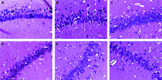
The results showed that the structure of CA1 area in hippocampus of NCG group was orderly and complete, the cell morphology was normal, and the boundary between nucleus and cytoplasm was obvious. While cells in hippocampal CA1 area of HCG group were markedly distorted, and there was a large area of edema. Treatment with different doses of AMG could ameliorate pathological damages, and the high dosage group exhibited fewer pathological lesions. The pathological images have been supplied in the revised manuscript (Figure 1).
Figure 1. Pathological damages in hippocampus of each group of rats (n = 8, ×100).

A: NCG: normoxic control group; B: HCG: hypoxia control group; C: PG: positive group (1.35 g/kg L-leucine); D: H-AMG: high dose group (0.35 g/kg Astragalus membranaceus); E: M-AMG: middle dose group (0.15 g/kg Astragalus membranaceus); F: L-AMG: low dose group (0.05 g/kg Astragalus membranaceus). Rats were administered by gavage once a day for 7 d.
3.3. MDA, GSH, ROS content and T-SOD activity in hippocampus
Compared with NCG, the content of MDA in the hippocampus of HEG and L-AMG increase significantly (P < 0.01, < 0.05), while the GSH content decrease significantly (P < 0.01, < 0.05). Compared with HCG, the content of MDA in hippocampus of PG, H-AMG, M-AMG and L-AMG decrease significantly (P < 0.01), while the content of GSH in hippocampus of PG, H-AMG and M-AMG increase significantly (P < 0.05) (Table 3).
Table 3.
Contents of MDA and GSH in hippocampus of each group of rats ($\bar{x}$ ± s)
| Group | Dose | n | MDA (nmol/mgprot) | GSH (mg/gprot) |
|---|---|---|---|---|
| NCG | 0.1 mL/10 g | 8 | 1.6±0.3 | 21.3±2.7 |
| HCG | 0.1 mL/10 g | 8 | 2.8±0.5a | 16.6±1.0a |
| PG | 0.35 g/kg | 8 | 1.7±0.5b | 19.4±2.5d |
| H-AMG | 0.35 g/kg | 8 | 1.8±0.6b | 19.1±2.6d |
| M-AMG | 0.15 g/kg | 8 | 1.9±0.4b | 19.0±2.3d |
| L-AMG | 0.05 g/kg | 8 | 2.0±0.4bc | 17.6±2.6cd |
Notes: NCG: normoxic control group; HCG: hypoxia control group; PG: positive group (1.35 g/kg L-leucine); H-AMG: high dose group (0.35 g/kg Astragalus membranaceus); M-AMG: middle dose group (0.15 g/kg Astragalus membranaceus); L-AMG: low dose group (0.05 g/kg Astragalus membranaceus). MDA: malondialdehyde, GSH: glutathione. Rats were administered by gavage once a day for 7 d. Compared with NCG, aP < 0.01, cP < 0.05; compared with HCG group, bP < 0.01, dP < 0.05.
Compared with NCG, the activity of T-SOD in hippocampal tissue of HCG and L-AMG was obviously decreased (P < 0.01), while the content of ROS was obviously increased (P < 0.01, < 0.05). There was no significant change in the content of T-SOD and ROS in hippocampus of H-AMG and M-AMG (P > 0.05). Compared with HCG, the activity of T-SOD in hippocampus of PG, H-AMG and M-AMG was significantly increased (P < 0.05), and the content of ROS was obviously decreased (P < 0.05), while the activity of T-SOD and the content of ROS in hippocampus of L-AMG had no significant change (P > 0.05). The results are shown in Table 4.
Table 4.
T-SOD activity and ROS content in hippocampus of each group of rats ($\bar{x}$ ± s)
| Group | Dose | n | T-SOD (U/mgprot) | ROS (FU/mgprot) |
|---|---|---|---|---|
| NCG | 0.1 mL/10 g | 8 | 303.2±34.7 | 1.4±0.3 (107) |
| HCG | 0.1 mL/10 g | 8 | 244.3±39.1a | 2.0±0.3a (107) |
| PG | 0.35 g/kg | 8 | 286.0±34.0b | 1.4±0.2c (107) |
| H-AMG | 0.35 g/kg | 8 | 285.0±25.0b | 1.5±0.2c (107) |
| M-AMG | 0.15 g/kg | 8 | 280.4±26.6b | 1.6±0.3c (107) |
| L-AMG | 0.05 g/kg | 8 | 249.6 ±19.2a | 1.8±0.3d (107) |
Notes: NCG: normoxic control group; HCG: hypoxia control group; PG: positive group (1.35 g/kg L-leucine); H-AMG: high dose group (0.35 g/kg Astragalus membranaceus); M-AMG: middle dose group (0.15 g/kg Astragalus membranaceus); L-AMG: low dose group (0.05g/kg Astragalus membranaceus). Rats were administered by gavage once a day for 7 d. T-SOD: total superoxide dismutase; ROS: reactive oxygen species. Compared with NCG, aP < 0.01, dP < 0.05; compared with HCG group, cP < 0.01, bP < 0.05.
3.4. Expression of mTOR, P70S6K, 4E-BP1 mRNA in hippocampus
Compared with NCG, the mTOR mRNA and P70S6K mRNA expression in HCG hippocampus were significantly reduced (P < 0.01), while 4E-BP1 mRNA expression was significantly increased (P < 0.05). Compared with HCG, mTOR mRNA expression was significantly increased in PG, H-AMG, M-AMG and L-AMG hippocampus (P < 0.05, < 0.01), the expression of P70S6K mRNA was significantly increased in PG, H-AMG hippocampus (P < 0.05, < 0.01), while the expression of 4E-BP1 mRNA in hippocampus of PG, H-AMG and M-AMG was significantly decreased (P < 0.05, < 0.01). The results are shown in Table 5.
Table 5.
Effect of aqueous extract of Astragalus membranaceus on target gene expression ($\bar{x}$ ± s)
| Group | Dose | n | mTOR mRNA | P70S6K mRNA | 4E-BP1 mRNA |
|---|---|---|---|---|---|
| NCG | 0.1 mL/10 g | 8 | 1.0±0.1 | 1.0±0.1 | 1.0±0.0 |
| HCG | 0.1 mL/10 g | 8 | 0.5±0.1a | 0.7±0.1a | 1.5±0.2d |
| PG | 0.35 g/kg | 8 | 0.9±0.1b | 1.0±0.1b | 0.8±0.1c |
| H-AMG | 0.35 g/kg | 8 | 0.9±0.2b | 0.9±0.2c | 0.8±0.1c |
| M-AMG | 0.15 g/kg | 8 | 0.9±0.1b | 0.8±0.1 | 0.9±0.1c |
| L-AMG | 0.05 g/kg | 8 | 0.7±0.1c | 0.8±0.1 | 1.26± 0.3 |
Notes: NCG: normoxic control group; HCG: hypoxia control group; PG: positive group (1.35 g/kg L-leucine); H-AMG: high dose group (0.35 g/kg Astragalus membranaceus); M-AMG: middle dose group (0.15 g/kg Astragalus membranaceus); L-AMG: low dose group (0.05 g/kg Astragalus membranaceus). Rats were administered by gavage once a day for 7 d. mTOR: mammalian target of rapamycin; 4E-BP1: 4E-binding protein 1. Compared with NCG, aP < 0.01, dP < 0.05; compared with HCG group, bP < 0.01, cP < 0.05.
3.5. Proteins expression of mTOR, p-P70S6K and p-4E-BP1 in hippocampus
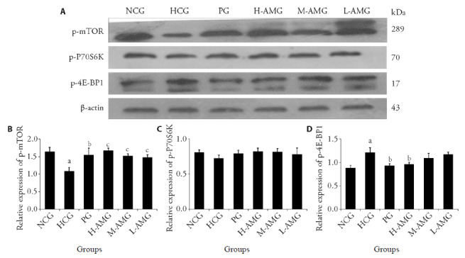
Compared with NCG, the protein expression of mTOR in hippocampal tissue of HCG rats was significantly decreased (P < 0.01), while the protein expression of p-4E-BP1 was significantly increased (P < 0.05) while p-P70S6K decreased insignificantly (P > 0.05). Compared with HCG, mTOR protein expression in hippocampus of PG, H-AMG, M-AMG and L-AMG rats was significantly increased (P < 0.05, < 0.01), While in hippocampus of PG and H-AMG rats, the protein expression of p-4E-BP1 was significantly reduced (P < 0.05), and the protein expression of p-P70S6K was not statistically significant (P > 0.05) (Figure 2).
Figure 2. Effect of aqueous extract of Astragalus membranaceus on target gene expression ($\bar{x}$± s, n = 3).

A: Western blot banding of protein expression; B: quantitative analysis of p-mTOR; C: quantitative analysis of p-P70S6K; D: quantitative analysis of p-4E-BP1. NCG: normoxic control group; HCG: hypoxia control group; PG: positive group (1.35 g/kg L-leucine); H-AMG: high dose group (0.35 g/kg Astragalus membranaceus); M-AMG: middle dose group (0.15 g/kg Astragalus membranaceus); L-AMG: low dose group (0.05 g/kg Astragalus membranaceus). Rats were administered by gavage once a day for 7 d. mTOR: mammalian target of rapamycin; 4E-BP1: 4E-binding protein 1. Compared with NCG, aP < 0.01; compared with HCG group, bP < 0.05, cP < 0.01.
3.6. Proteins expression of cleaved capase-3 protein in hippocampus
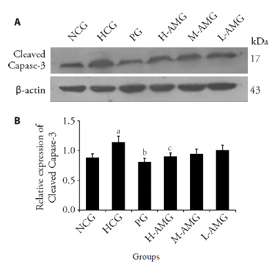
Compared with NCG, the protein expression of Cleaved Capase-3 in hippocampus of HCG rats was significantly increased (P < 0.05). Compared with the HCG group, the expression of Cleaved Capase-3 protein in the hippocampus of PG and H-AMG rats was significantly reduced (P < 0.05), while the expression of protein in M-AMG and L-AMG rats did not decrease significantly (P > 0.05) (Figure 3).
Figure 3. Effect of aqueous extract of Astragalus membranaceus on cleaved capase-3 protein expression ($\bar{x}$ ± s, n = 3).

A: Western blot banding of protein expression; B: quantitative analysis of cleaved capase-3. NCG: normoxic control group; HCG: hypoxia control group; PG: positive group (1.35 g/kg L-leucine); H-AMG: high dose group (0.35 g/kg Astragalus membranaceus); M-AMG: middle dose group (0.15 g/kg Astragalus membranaceus); L-AMG: low dose group (0.05 g/kg Astragalus membranaceus). Rats were administered by gavage once a day for 7 d. Compared with NCG, aP < 0.05; compared with HCG group, bP < 0.01, cP < 0.05.
4. DISCUSSION
At present, the decline of cognitive function caused by high altitude hypoxia has attracted more and more people's attention. Many researchers have shifted the focus of high altitude disease research to improving the high altitude cognitive ability.19,20 Early experiments found that a variety of TCMs such as Phenylethanoid Glycosides in Cistanche deserticola, Pedicularis Kansuensis and Tibetan medicine have shown good pharmacological activity to improve memory damage in the plateau.17,21
When the body is under low pressure and low oxygen, the nerve conduction velocity will decrease. Moreover, the functions of the visual and auditory systems and the central nervous system could be also affected. Structural changes in rat hippocampal synapses can lead to changes in high-level brain functions, such as learning, memory and thinking, which directly affect the cognitive function of the human brain. The cognitive function is the most important psychological condition for people to complete learning and memory.22 Exposure to hypoxia at high altitude can induce oxidative stress in cortex, hippocampus and striatum, leading to the imbalance of antioxidant enzyme-oxidase system in vivo, and oxidative damage of lipid, protein and DNA. Hippocampal tissue is related to learning and memory, and damage to this area will affect learning and cognitive function. Memory is the ability of the brain's nervous system to store past experience. It is the accumulation of a person's impression of previous activities, feelings and experiences.23 Davranche study the effects of hypoxia on cognitive function in high altitude population. The results showed that exposure to high altitude would increase reaction time and damage memory coding and retention, resulting in memory and behavioral cognition impairment.24 In this study, the values of WME, RWE, TE and TT of HCG rats were significantly higher than those of NCG, indicating that the spatial memory ability of rats was indeed impaired when exposed to hypobaric and hypoxic environment. After intervention with Astragalus membranaceus water extract, WME, RWE, TE and TT of rats in each group were significantly lower than those of HCG, indicating that Astragalus water extract can improve the behavioral cognitive function of rats in hypobaric hypoxia environment, prevent and treat of memory impairment caused by hypobaric hypoxia.
In addition, the activities of GSH, T-SOD and other enzymes were decreased significantly when exposure to hypoxia at high altitude, while the concentration of ROS increased, resulting in neuro-degeneration and memory impairment, hippocampal dendritic atrophy and decreased hippocampal synaptic plasticity.25 When exposed to hypoxia, ROS are produced in the brain, and ROS can induce oxidative damage, which may also be one of the mechanisms of neurobehavioral damage.26 In this experiment, the contents of ROS, MDA, GSH and T-SOD activity in the hippocampus of rats were measured. The results showed that hypoxia-induced oxidative stress is also one of the causes of neuronal damage and memory damage. Intervention of aqueous extract of Astragalus membranaceus in rats can significantly increase T-SOD activity and GSH content, reduce ROS and MDA content, indicating that aqueous extract of Astragalus membranaceus can improve memory impairment by alleviating oxidative stress induced by hypoxia.
The mTOR serves as an "integrator" of multiple signaling pathways, and its main role is to regulate gene transcription, protein translation and apoptosis. The function of mTOR is mainly exerted by mTORC1, mTORC2 and its multiple downstream substrates [such as eukaryotic initiation factor 4E-BP1, p70 ribosomal S6 kinase (P70S6K), etc.]. Among them, mTORC1 is mainly involved in cytoskeleton construction, and regulates mRNA translation, cell proliferation, autophagy and apoptosis through phosphorylation of downstream factors 4E-BP1 and P70S6K.27 Hypoxia induced oxidative stress could inhibit the mTOR signaling pathway in certain pathway, leading to hypo phosphorylation of mTOR and its downstream 4E-BP1 and P70S6K.28 The results showed that the mTOR signal pathway in hippocampus was indeed inhibited when rats were exposed to high altitude hypobaric hypoxia. After intervention with aqueous extract of Astragalus membranaceus in rats, the relative expression of mTOR, P70S6K mRNA and the expression of p-mTOR, p-P70S6K and cleaved capase-3 protein were increased, and the expression of 4E-BP1 mRNA and p-4E-BP1 protein were significantly reduced. This indicates that aqueous extract of Astragalus membranaceus can counteract the inhibition of mTOR signaling pathway caused by hypobaric and hypoxia, improve memory impairment at high altitude, and protect the behavioral and cognitive ability of rats.
Caspase-3 is the main terminal splicing protease in the process of apoptosis, and plays a vital role in the occurrence and development of apoptosis. Cleaved caspase-3 is produced by the lytic activation of caspase-3. It cannot be detected in cells that have not undergone apoptosis or have been necrotic.29 In this experiment, the expression of cleaved capase-3 protein in the hippocampus tissue of HCG rats increased significantly. After intragastric administration of aqueous extract of Astragalus membranaceus in rats, the expression of cleaved capase-3 protein was significantly decreased, indicating that aqueous extract of Astragalus membranaceus can improve memory impairment by inhibiting cell apoptosis. Undeniably, there was limitation of the present study that should be considered. Although behavioral results are direct evidence for cognitive function, the neurotransmitters in the blood should also be tested. This would be analyzed in the further study.
In conclusion, our findings suggest that high altitude hypoxia could cause severe cognitive impairment. Aqueous extract of Astragalus membranaceus significantly improved the condition of the impairment induced by hypoxia. The underlying mechanism may be related to relieving the oxidative stress, reducing the accumulation of free radicals and metabolites, and activating the mTOR signaling pathway. Aqueous extract of Astragalus membranaceus could be considered as a promising candidate for the treatment of altitude hypoxia induced cognitive impairment.
REFERENCES
- [1]. Zhao J, Zhang R, Yu Q, et al. Characteristics of EEG activity during high altitude hypoxia and lowland reoxygenation. Brain Res 2016;1648:243-9. [DOI] [PubMed] [Google Scholar]
- [2]. Wei D, Wei L, Li X, et al. Effect of hypoxia on Ldh-c expression in somatic cells of plateau pika. Int J Env Res Pub He 2016;13:773. [DOI] [PMC free article] [PubMed] [Google Scholar]
- [3]. Vargas M, Becerril-Ángeles M, Medina-Reyes I, et al. Altitude above 1500 m is a major determinant of asthma incidence. An ecological study. Resp Med 2018;135:1-7. [DOI] [PubMed] [Google Scholar]
- [4]. Luks A, Auerbach P, Freer L, et al. Wilderness medical society clinical practice guidelines for the prevention and treatment of acute altitude illness: 2019 update. Wild Environ Med 2019;30:S3-S18. [DOI] [PubMed] [Google Scholar]
- [5]. Bärtsch P, Swenson E. Acute high-altitude illnesses. NEJM 2013;368:2294-302. [DOI] [PubMed] [Google Scholar]
- [6]. Asmaro D, Mayall J, Ferguson S. Cognition at altitude: impairment in executive and memory processes under hypoxic conditions. Aviat Space Environ Med 2013;84:1159-65. [DOI] [PubMed] [Google Scholar]
- [7]. Yan X. Cognitive impairments at high altitudes and adaptation. High Alt Med Biol 2014;15:141-5. [DOI] [PubMed] [Google Scholar]
- [8]. Wilson M, Newman S, Imray C. The cerebral effects of ascent to high altitudes. Lancet Neurol 2009;8:175-91. [DOI] [PubMed] [Google Scholar]
- [9]. Movafeghi A, Djozan D, Razeghi J, et al. Identification of volatile organic compounds in leaves, roots and gum of Astragalus compactus Lam. using solid phase microextraction followed by GC-MS analysis. Nat Prod Res 2010;24:703-9. [DOI] [PubMed] [Google Scholar]
- [10]. Auyeung KK, Han Q, Ko JK. Astragalus membranaceus: a review of its protection against inflammation and gastrointestinal cancers. Am J Chin Med 2016;44:1-22. [DOI] [PubMed] [Google Scholar]
- [11]. Li X, Qu L, Dong Y, et al. A review of recent research progress on the astragalus genus. Molecules 2014;19:18850-80. [DOI] [PMC free article] [PubMed] [Google Scholar]
- [12]. Wang Y, Liu L, Ma Y, et al. Chemical discrimination of astragalus mongholicus and astragalus membranaceus based on metabolomics using UHPLC-ESI-Q-TOF-MS/MS approach. molecules 2019;24:4064. [DOI] [PMC free article] [PubMed] [Google Scholar]
- [13]. Liu M, Wu K, Mao X, et al. Astragalus polysaccharide improves insulin sensitivity in KKAy mice: regulation of PKB/GLUT4 signaling in skeletal muscle. J Ethnopharmacol 2010;127:32-37. [DOI] [PubMed] [Google Scholar]
- [14]. Du X, Chen X, Zhao B, et al. Astragalus polysaccharides enhance the humoral and cellular immune responses of hepatitis B surface antigen vaccination through inhibiting the expression of transforming growth factor β and the frequency of regulatory T cells. FEMS Immunol Med Microbiol 2011;63:228-35. [DOI] [PubMed] [Google Scholar]
- [15]. Nalbantsoy A, Nesil T, Erden S, et al. Adjuvant effects of Astragalus saponins macrophyllosaponin B and astragaloside VII. J Ethnopharmacol 2011;134:897-903. [DOI] [PubMed] [Google Scholar]
- [16]. Linnek J, Mitaine-Offer A, Miyamoto T, et al. Cycloartane-type glycosides from two species of Astragalus (Fabaceae). Nat Prod Commun 2009;4:477-78. [PubMed] [Google Scholar]
- [17]. Li M, Zhu Y, Li J, et al. Effect and mechanism of verbascoside on hypoxic memory injury in plateau. Phytother Res 2019;33:2692-701. [DOI] [PubMed] [Google Scholar]
- [18]. Xu H, Baracskay P, O'Neill J, et al. Assembly responses of hippocampal CA1 place cells predict learned behavior in goal-directed spatial tasks on the radial eight-arm maze. Neuron 2019;101:119-32. [DOI] [PubMed] [Google Scholar]
- [19]. Sharma V, Das S, Dhar P, et al. Domain specific changes in cognition at high altitude and its correlation with hyperhomocysteinemia. Plos One 2014;9:e101448. [DOI] [PMC free article] [PubMed] [Google Scholar]
- [20]. Rimoldi S, Rexhaj E, Duplain H, et al. Acute and chronic altitude-induced cognitive dysfunction in children and adolescents. J Pediatr 2016;169:238-43. [DOI] [PubMed] [Google Scholar]
- [21]. Zhou B, Li M, Cao X, et al. Phenylethanoid glycosides of Pedicularis muscicola Maxim ameliorate high altitude-induced memory impairment. Physiol Behav 2016;157:39-46. [DOI] [PubMed] [Google Scholar]
- [22]. Zhang G, Zhou S, Yuan C, et al. The effects of short-term and long-term exposure to a high altitude hypoxic environment on neurobehavioral function. High Alt Med Biol 2013;14:338-41. [DOI] [PubMed] [Google Scholar]
- [23]. Joyce K, Lucas S, Imray C, et al. Advances in the available non-biological pharmacotherapy prevention and treatment of acute mountain sickness and high altitude cerebral and pulmonary oedema. Expert Opin Pharmaco 2018;19:1891-902. [DOI] [PubMed] [Google Scholar]
- [24]. Davranche K, Casini L, Arnal P, et al. Cognitive functions and cerebral oxygenation changes during acute and prolonged hypoxic exposure. Physiol Behav 2016;164:189-97. [DOI] [PubMed] [Google Scholar]
- [25]. Ma H, Li X, Liu M, et al. Mental Rotation Effect on Adult Immigrants with Long-term Exposure to High Altitude in Tibet: An ERP Study. Neuroscience 2018;386:339-50. [DOI] [PubMed] [Google Scholar]
- [26]. Fuhrmann D, Brüne B. Mitochondrial composition and function under the control of hypoxia. Redox Biol 2017;12:208-15. [DOI] [PMC free article] [PubMed] [Google Scholar]
- [27]. Zeng H. mTOR signaling in immune cells and its implications for cancer immunotherapy. Cancer Lett 2017;408:182-89. [DOI] [PubMed] [Google Scholar]
- [28]. Woillard J, Kamar N, Rousseau A, et al. Association of sirolimus adverse effects with mTOR, p70S6K or Raptor polymorphisms in kidney transplant recipients. Pharmacogenet Genomics 2012;22:725-32. [DOI] [PMC free article] [PubMed] [Google Scholar]
- [29]. Fu S, Imai K, Sawasaki T, et al. ScreenCap3: Improving prediction of caspase-3 cleavage sites using experimentally verified noncleavage sites. Proteomics 2014;14:2042-6. [DOI] [PMC free article] [PubMed] [Google Scholar]


