Abstract
Background
To report the occurrence of abdominal symptoms in patients who presented with prolonged heterogeneous liver enhancement (PHLE) after injecting contrast agent SonoVue®.
Methods
A total of 105 patients who indicated to have contrast-enhanced ultrasound (CEUS) examinations were consecutively observed. The liver scanning under ultrasound was performed before and after the contrast agent injection. Patients’ basic information, clinical manifestations, and ultrasound images under B-mode and CEUS mode were respectively recorded. For patients exhibiting abdominal symptoms, the occurrence and last time of symptoms were recorded in detail. We subsequently compared the difference in clinical characteristics between patients with and without the PHLE phenomenon.
Results
In 20 patients with the PHLE phenomenon, 13 showed abdominal symptoms. Eight patients (61.5%) appeared to have mild defecation sensation, and 5 (38.5%) showed apparent abdominal pain. The PHLE phenomenon began to appear within 15 minutes to 1.5 hours after the intravenous injection of SonoVue®. This phenomenon lasted for 30 minutes to 5 hours in ultrasound. Patients with severe abdominal symptoms showed large-area and diffuse PHLE patterns. Only sparse hyperechoic spots in the liver were detected in patients with mild discomfort. Abdominal discomfort resolved spontaneously in all patients. Meanwhile, the PHLE gradually disappeared without any medical treatment. In the PHLE-positive group, the proportion of patients with a history of gastrointestinal disease was significantly higher (P=0.02).
Conclusions
Patients with the PHLE phenomenon can exhibit abdominal symptoms. We suggest gastrointestinal disorders may contribute to PHLE, which can be considered a harmless phenomenon that does not affect the safety profile of SonoVue®.
Keywords: Contrast agent, ultrasound, liver enhancement, abdominal symptom
Introduction
Contrast-enhanced ultrasound (CEUS) is a widely-used, safe, and effective imaging technique that visualizes micro-vascularization using specific contrast agents (1-3). The contrast agents are lipid-encapsulated microbubbles with a diameter of about 1–10 µm. SonoVue® (Bracco, Milan, Italy) is the second-generation ultrasound contrast agent widely used in clinical practice, which is comprised of phospholipid-stabilized microbubbles filled with sulfur hexafluoride (4,5). The contrast agents enter the blood circulation through intravenous injection to generate contrast reflections for imaging and can be eliminated through the lung within 15–20 minutes. Previous studies have shown that severe adverse event associated with SonoVue® is rare, reflecting its good safety in clinical applications (6,7).
Prolonged heterogeneous liver enhancement (PHLE) is a rare post-contrast manifestation (8). It is characterized by the appearance of diffuse and unevenly distributed gas-like hyperechoic staining in the liver parenchyma under after contrast agent injection. Even increasing the mechanical index (MI) cannot clear hyperechoic spots. At present, the reason for the appearance of PHLE still needs to be determined. Several hypotheses have been put forward in previous reports, all reasonable but also contain limitations (1,8-13). Meanwhile, the existing studies are primarily based on European and Japanese populations, and no study of Chinese populations has been reported. Through consecutive observation of the liver performance after CEUS examinations, we found that patients with the PHLE phenomenon were accompanied by abdominal symptoms, which lasted for a short time and could resolve spontaneously.
This study aimed to summarize the characteristics of abdominal symptoms in patients who presented with PHLE in ultrasound. Furthermore, we tried to figure out clinical factors associated with this unique phenomenon. We present the following article in accordance with the STROBE reporting checklist (available at https://qims.amegroups.com/article/view/10.21037/qims-22-1035/rc).
Methods
Study participants
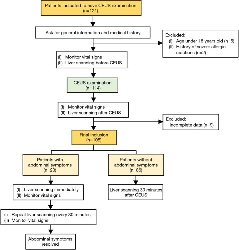
This cross-sectional study included all patients indicated to have CEUS examination from March to May 2022 in the outpatient clinic of China-Japan Friendship Hospital. This study was conducted following the Declaration of Helsinki (as revised in 2013). The ethics committee of China-Japan Friendship Hospital approved this study. The inclusion criteria of this study were all patients who had confirmed to have CEUS examination. The exclusion criteria were as follows: (I) age <18 years old; (II) patients with history of severe allergic reactions; (III) incomplete data (Figure 1). Written informed consent was obtained from all patients. Before the CEUS examination, we asked patients for related medical history, including the history of allergic reactions, gastrointestinal disorders, and hepatobiliary disease. The patient’s age, gender, height, weight, examination item, total injection dose of SonoVue®, and the number of injections were respectively recorded.
Figure 1.
The flow chart of study design. CEUS, contrast-enhanced ultrasound.
Ultrasound technique and parameters
The following ultrasound techniques were used in this study: EPIQ Elite (Philips, Netherlands), Resona R9 (Mindray, China), and ACUSON Sequoia (Siemens, Germany). Conventional B-mode and color Doppler ultrasound examinations were performed to scan the organ. All the patients underwent liver scanning in B-mode ultrasound before CEUS examination. The equipment parameters for liver scanning were set to abdominal setting with MI 1.1 to 1.3 both in B-mode and CEUS mode. A low MI level ranging from 0.06–0.07 was used in CEUS examination. The focal point at the lesion’s deepest level or the liver’s deepest level was set to guarantee optimal conditions for CEUS. Conventional ultrasound and CEUS examinations were performed by radiologists in the ultrasound department. The operators had more than four years of experience in ultrasound. All the radiologists had been carried out for standardized training of reading images and they did not know about the study design.
CEUS examination
The patients in this study only underwent one type of CEUS examination. For thyroid CEUS and lymph node CEUS, 0.5–1.0 mL SonoVue® was usually administered for imaging. Breast CEUS was usually performed using 2.5–3.0 mL of SonoVue®. If the patient had multiple nodules, the usage of SonoVue® was increased accordingly. SonoVue® was administered as a bolus using an intravenous catheter followed by 10 mL of a 0.9% saline bolus. Directly after the SonoVue® injection, scanning was performed in real-time for at least two minutes. The equipment settings for the contrast imaging were set to contrast harmonic imaging mode, frequency of 2.0–2.5 MHz. In this study, microbubble destruction was not performed at the end of the CEUS examination.
Most patients received only one bolus injection. However, depending on the indication (for example, multiple thyroid lesions), some patients received multiple bolus injections of SonoVue®. Before and after the CEUS examination, the vital signs were respectively monitored for patients, including blood pressure (mmHg), heart rate (beats per minute), respiratory rate (breaths per minute), and oxygen saturation (%). All patients were observed for at least 40 minutes after the CEUS examination.
Liver scanning
Before CEUS examination, all the patients underwent liver scanning under B-mode ultrasound with MI 1.1 to 1.3. At the end of CEUS, all the patients conducted liver examinations, which observed under B-mode and CEUS settings (MI 1.1 to 1.3). MI of 1.1 to 1.3 was defined as high MI under CEUS mode. During observation time, if the patient did not experience discomfort, we performed liver scans 30 minutes after the CEUS examination and recorded the images under B-mode and CEUS settings. If the patient appeared to have abdominal discomfort, we immediately performed the liver examination and recorded the images under B-mode and CEUS settings. We did the abdominal scanning every 30 minutes until the patient’s abdominal discomfort was relieved. The flow chart of the study design was illustrated in Figure 1.
Statistical analysis
Continuous variables were described by using the median (range, minimum to maximum). Categorical variables were reported by absolute frequencies and percentages. Differences between groups were assessed accordingly using the Wilcoxon rank sum test, Fisher’s precision probability test, Chi-square (χ2) test and Yates’ correction. All the statistical analyses were performed using SPSS 22.0 (IBM Corp., Armonk, NY, USA), and a P value less than 0.05 indicated statistical significance.
Results
Basic information
Of 105 patients who had CEUS examination, 20 showed PHLE phenomenon in ultrasound. Thirteen of the 20 patients presented with abdominal symptoms, while the other 7 showed no discomfort. These 13 patients included 11 females and two males. The mean age was 42.2 (range, 27 to 73) years old. Eleven patients underwent CEUS to diagnose thyroid nodules, one for breast lesions and one for abnormal lymph nodes. All these 13 patients received at least a 1.0 mL injection of SonoVue®. The demographic and clinical information of patients with PHLE and abdominal symptoms are summarized in Table 1.
Table 1. Demographic and clinical information of 13 patients with PHLE and abdominal symptoms.
| Case number | Gender | Age (years) | Total dose (mL) | Number of injections | Examination item |
|---|---|---|---|---|---|
| 1 | Female | 28 | 1.0 | 1 | Thyroid CEUS |
| 2 | Female | 73 | 2.0 | 2 | Thyroid CEUS |
| 3 | Female | 43 | 2.0 | 2 | Thyroid CEUS |
| 4 | Female | 40 | 5.0 | 1 | Breast CEUS |
| 5 | Female | 35 | 3.0 | 3 | Thyroid CEUS |
| 6 | Female | 27 | 2.0 | 2 | Thyroid CEUS |
| 7 | Female | 59 | 5.0 | 5 | Thyroid CEUS |
| 8 | Female | 42 | 2.0 | 2 | Thyroid CEUS |
| 9 | Male | 42 | 3.0 | 3 | Thyroid CEUS |
| 10 | Female | 58 | 1.5 | 1 | Thyroid CEUS |
| 11 | Female | 39 | 1.0 | 1 | Thyroid CEUS |
| 12 | Male | 31 | 2.5 | 5 | Thyroid CEUS |
| 13 | Female | 31 | 2.0 | 2 | Lymph node CEUS |
PHLE, prolonged heterogeneous liver enhancement; CEUS, contrast-enhanced ultrasound.
Ultrasound features in the liver
After intravenously administered contrast-enhanced agent, the hyperechoic hepatic enhancement pattern was observed along portal branches. Using the conventional B-mode and switching to the CEUS mode did not influence the appearance of hyperechoic spots. The PHLE phenomenon began to appear within 30 minutes to 1.5 hours after the intravenous injection of SonoVue®, and lasted for 30 minutes to 5 hours. Conventional B-mode ultrasound showed no sign of hepatic hyperechogenicity in two patients who underwent a second CEUS examination approximately 24 hours after the first examination. Abdominal symptoms were not observed the day after the occurrence of hyperechoic staining. We summarized the characteristics of hyperechoic spots in the liver as follows:
There were no hyperechoic spots in the liver before the CEUS examination.
The PHLE pattern manifested as diffusely distributed hyperechoic spots in the liver parenchyma, located along the portal vein.
The PHLE phenomenon was mainly focused on the right lobe of the liver. The contrast effect in the left lobe was slightly weaker than in the right lobe.
Flowing hyperechoic spots were detected in the portal vein.
For all the participants, hyperechoic staining was visible on both conventional B-mode and contrast-enhanced mode.
The PHLE appearance persisted stably even at high MI under CEUS mode (1.1 to 1.3).
Abdominal symptoms
All 13 patients with PHLE patterns experienced varying degrees of abdominal discomfort (Table 2). Eight patients (61.5%) appeared to have noticeable defecation sensation, and five (38.5%) showed abdominal pain—three patients presented with fatigue, nausea, and paleness on the face separately. An abdominal red rash was observed in one patient. Those abdominal symptoms appeared approximately 15–30 minutes after the CEUS examination. All patients’ abdominal symptoms resolved spontaneously. None of these patients experienced deleterious effects related to SonoVue® administration, and no changes in vital signs were observed (Table 3).
Table 2. Ultrasound features on liver and abdominal symptoms for the 13 patients with PHLE and abdominal symptoms.
| Case | Ultrasound features on liver | Time of PHLE appearance after CEUS | Clinical symptoms |
|---|---|---|---|
| 1 | Diffuse hyperechoic staining | 15 minutes | Persistent abdominal pain, defecate reaction, paleness |
| 2 | Diffuse hyperechoic staining | 1.5 hours | Defecate reaction |
| 3 | Diffuse hyperechoic staining | 30 minutes | Defecate reaction |
| 4 | Diffuse hyperechoic staining | 47 minutes | Mild abdominal discomfort |
| 5 | Diffuse hyperechoic staining | 1 hour 27 minutes | Defecate reaction |
| 6 | Diffuse hyperechoic staining | 3 hours | Abdominal distention and pain, defecate reaction |
| 7 | Tiny hyperechoic spots | 1 hour 14 minutes | Defecate reaction |
| 8 | Diffuse hyperechoic staining | 30 minutes | Nausea, abdominal distention and pain, red rash on the skin of the abdomen |
| 9 | Tiny hyperechoic spots | 32 minutes | Defecate reaction |
| 10 | Diffuse hyperechoic staining | 29 minutes | Abdominal pain, distention, nausea, dizziness, fatigue |
| 11 | Diffuse hyperechoic staining | 30 minutes | Defecate reaction |
| 12 | Diffuse hyperechoic staining | 30 minutes | Nausea, dizziness, fatigue |
| 13 | Diffuse hyperechoic staining | 15 minutes | Abdominal pain, defecate reaction |
PHLE, prolonged heterogeneous liver enhancement; CEUS, contrast-enhanced ultrasound.
Table 3. Vital signs after CEUS examination of the 13 patients with PHLE and abdominal symptoms.
| Case number | Blood pressure (mmHg) | Heart rate | Respiration | Oxygen saturation (%) |
|---|---|---|---|---|
| 1 | 122/80 | 87 | 18 | 98 |
| 2 | 125/98 | 91 | 18 | 99 |
| 3 | 102/90 | 80 | 17 | 99 |
| 4 | 106/95 | 82 | 17 | 99 |
| 5 | 110/79 | 79 | 17 | 99 |
| 6 | 96/75 | 70 | 22 | 100 |
| 7 | 138/92 | 64 | 16 | 98 |
| 8 | 120/94 | 76 | 18 | 99 |
| 9 | 118/78 | 63 | 17 | 99 |
| 10 | 101/67 | 54 | 17 | 99 |
| 11 | 132/79 | 58 | 18 | 99 |
| 12 | 104/64 | 60 | 18 | 99 |
| 13 | 112/74 | 72 | 17 | 99 |
CEUS, contrast-enhanced ultrasound; PHLE, prolonged heterogeneous liver enhancement.
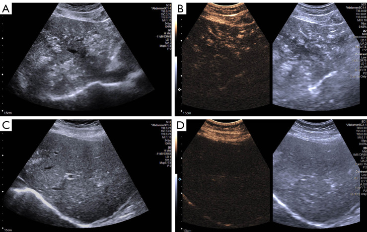
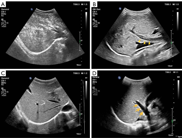
When the patient presented with abdominal symptoms, abdominal scanning revealed hyperechoic spots in the liver. Among 11 patients who manifested diffuse hyper-echoic staining in ultrasound, five patients experienced serious abdominal symptoms, such as abdominal pain, nausea, and even a red rash on the skin. The other six patients only showed mild abdominal discomfort, like defecation sensation or bowel movement (Figure 2). Only two patients exhibited tiny hyper-echoic spots in ultrasound. Regarding abdominal symptoms, the two patients merely presented with defecate reactions (Figure 3).
Figure 2.
A 42-year-old female patient was suspected of thyroid carcinoma. Thirty minutes after the injection of 2.0 mL SonoVue®, the patient appeared to have a stomach ache, nausea, and a red rash on the skin of the abdomen. Diffuse and large-area hyperechoic staining in the liver was detected in (A) B-mode and (B) CEUS mode after 30 minutes of SonoVue® injection. One hour later, the hyper-echoic area decreased both in (C) B-mode and (D) CEUS mode, and the patient’s abdominal symptoms resolved spontaneously. CEUS, contrast-enhanced ultrasound.
Figure 3.
A 42-year-old male patient was intravenously injected with 3.0 mL of SonoVue® to discriminate malignant thyroid nodules. The patient presented with mild abdominal discomfort after 32 minutes of CEUS. (A) At the same time, scattered tiny hyperechoic spots were detected in the liver. (B) Flowing hyperechoic spots were observed in the portal vein (arrows) (C) After 2 hours of SonoVue® injection, the patient’s abdominal discomfort was relieved, and the hyperechoic areas in the liver almost disappeared. (D) No hyperechoic spots were found in the portal vein (arrows). CEUS, contrast-enhanced ultrasound.
Of 13 patients who presented with abdominal symptoms, four patients showed abdominal symptoms varying degrees earlier than the PHLE phenomenon. We detected PHLE patterns in ultrasound approximately 27 minutes, one hour, 2.5 hours, and 1 hour after patients appeared with abdominal symptoms, respectively. The remaining nine patients exhibited PHLE immediately after having abdominal discomfort.
Comparison of characteristics between patients with and without PHLE phenomenon
We analyzed the difference in clinical characteristics between patients with (n=20) and without the PHLH phenomenon (n=85). Patients of the two groups did not differ in age (P=0.44), gender (P=0.76), height (P=0.22), weight (P=0.10), BMI (P=0.12), allergic history (P=0.90), hepatobiliary disease (P=0.52), number of injections (P=0.81) and the total injection dose (P=0.72). Notably, significant differences were observed in the history of gastrointestinal disease between the PHLE-positive and negative patients (P=0.02). In the PHLE-positive group, the proportion of patients with a history of the gastrointestinal disease was higher (Table 4). Of the three patients in the PHLE group who presented with gastrointestinal disease, one had a history of dyspepsia, one had a history of appendectomy, and one had a history of gastritis. The only patient in the PHLE-negative group with the gastrointestinal disease had a history of gastric reflux.
Table 4. Comparison of clinical characteristics between patients present and absent with PHLE phenomenon.
| Variables | Positive (n=20) | Negative (n=85) | P value |
|---|---|---|---|
| Age (years), median [range] | 35.5 [24, 73] | 38 [27, 69] | 0.44 |
| Gender, n (%) | 0.76 | ||
| Male | 3 (15.0) | 18 (21.2) | |
| Female | 17 (85.0) | 67 (78.8) | |
| Height (m), median [range] | 1.64 [1.55, 1.78] | 1.65 [1.53, 1.82] | 0.22 |
| Weight (kg), median [range] | 56.5 [43, 75] | 60 [45, 115] | 0.10 |
| BMI, median [range] | 21.5 [16.9, 25.0] | 22.0 [15.2, 37.0] | 0.12 |
| Allergic history, n (%) | 0.90 | ||
| Absent | 18 (90.0) | 73 (85.9) | |
| Present | 2 (10.0) | 12 (14.1) | |
| Gastrointestinal disease, n (%) | 0.02 | ||
| Absent | 17 (85.0) | 84 (98.8) | |
| Present | 3 (15.0) | 1 (1.2) | |
| Hepatobiliary disease, n (%) | 0.52 | ||
| Absent | 18 (90.0) | 68 (80.0) | |
| Present | 2 (10.0) | 17 (20.0) | |
| Number of injections, n (%) | 0.81 | ||
| 1 | 4 (20.0) | 14 (16.5) | |
| 2 | 7 (35.0) | 37 (43.5) | |
| 3 | 4 (20.0) | 21 (24.7) | |
| 4 | 3 (15.0) | 9 (10.6) | |
| 5 | 2 (10.0) | 4 (4.7) | |
| Injection dose (mL), median [range] | 2 [1, 5] | 2 [0.5, 10] | 0.72 |
PHLE, prolonged heterogeneous liver enhancement; BMI, body mass index.
Discussion
SonoVue® is the second-generation contrast agent characterized as a “blood-pool” agent, mimicing the behavior of red blood cells in the circulation (10). Adverse reactions of SonoVue® are rare (only 0.125%) (12). The safety of microbubbles is crucial for their wide application in clinical practice. In previous studies, the PHLE phenomenon has been reported in different medical centers (1,8-11). However, all the patients with abnormal performance were not accompanied by any clinical symptoms. Meanwhile, the hyperechoic staining pattern gradually resolved in all the patients by the following day without any treatment.
Our center’s study found that 13 of 105 patients (approximately 12.4%) developed both the PHLE phenomenon and abdominal discomfort after the CEUS examination. The incidence of this phenomenon is higher than that in previous reports (8,9). This high incidence might be because we included patients who underwent CEUS for multiple organs, including thyroid, breast and lymph nodes. Various types of contrast agents used in the investigation may also contribute to the different incidences of the PHLE phenomenon. For instance, in Okada et al.’s study, they utilized five different types of contrast agents, including EchoGen® (Sonus, Bothell, WA, USA), SonoVue® (Bracco, Milan, Italy), Sonazoid®, Optison® (Mallinckrodt Medical, St Louis, MO, USA), and Levovist®. Only SonoVue® was utilized as a contrast agent in our study. Furthermore, our study prospectively included 105 patients, and we performed close observation and liver scanning after CEUS, reducing the attrition of some symptomatic patients (6). In Caruso et al.’s study, hepatic examinations were performed in the CEUS arterial phase (20–30 s), portal phase (60 s), late phase (180 s), and the second late phase (240 s), respectively (7). It can be seen that the investigators observed up to 240 seconds after CEUS examinations. In comparison, we performed liver scanning 30 minutes after CEUS. Meanwhile, we added liver examinations if the patient experienced abdominal discomfort. Therefore, the incidence of PHLE was relatively higher, probably due to the long observation and the high frequency of liver examinations in our study. In our findings, the abdominal symptoms from patients gradually resolved spontaneously. These patients’ vital signs were all consistently stable, indicating that PHLE is harmless.
At present, the reason for the appearance of PHLE still needs to be determined. Several hypotheses have been reported in previous studies. One hypothesis suggests that the shell of microbubbles is destroyed initially. However, the microbubbles immediately combine to form large gas bubble conglomerates that are more stable (10). The higher dosage of injected SonoVue®, the larger the fused gas bubbles (8,9). Finally, these gas bubble conglomerates are trapped in the liver parenchyma, particularly along the portal vein branches. This hypothesis might explain the long time (more than 30 minutes) for which the hyperechoic spots remain in the liver, but it still has limitations in explaining some findings in our study. We found that patients with and without PHLE patterns did not differ in the injection dose of SonoVue®. The two patients showed tiny hyper-echoic spots with an injection dose of 3.0 and 5.0 mL, respectively. The remaining 11 patients showed diffuse hyper-echoic staining with an injection dose of 1.0 mL in two cases, 1.5 mL in one case, 2.0 mL in five cases, 2.5 mL in one case, and 3.0 mL in one case, and one case of 5.0 mL. Therefore, it seemed hard to find a correlation between the injection dose and the degree of PHLE in our study. Furthermore, SonoVue® is characterized as the “pure blood pool” contrast agent, so it is hardly taken up by Kupffer cells in the liver parenchyma.
Another hypothesis is that gas emboli from intestinal microcirculation are transported to the liver via the enter portal circulation (8,9). Hyperechoic staining might be the free gas caused by intestinal diseases, such as ischemia and necrotic enterocolitis (9,14). Nevertheless, this hypothesis cannot explain why the hyperechoic staining can be consistently detected two hours after CEUS examination when the microbubbles are supposed to dissipate from the circulation. Also, none of the 13 patients in our study had related intestinal disease to form free gas, but the hepatic enhancement still appeared.
Kupffer cells, the specialized macrophages in the liver, can remove foreign particles in the blood circulation by phagocytosis (8). In this context, one hypothesis indicates that phagocytosis of microbubbles by macrophages is the basis of the hepatic hyperechoic staining that last more than 5 minutes after SonoVue® injection (15). However, SonoVue® is characterized as a “pure blood pool agent” and is hardly taken up by Kupffer cells in the liver parenchyma. Meanwhile, this hypothesis cannot explain patients’ abdominal discomfort, accompanied by hepatic manifestations.
Our study found that 13 of 20 (65%) patients manifested hepatic hyperechoic spots after CEUS and were accompanied by abdominal symptoms. Meanwhile, we found that patients with PHLE manifestation had a higher proportion of containing a history of gastrointestinal disease. In this context, we speculated that it might be related to mild gastrointestinal allergic reactions. One patient (case 1) with abdominal symptoms concurred with paleness and fatigue. Another patient (case 8) showed a red rash on the skin of the abdomen, which is one manifestation of an allergic reaction. Based on the above performances, we hypothesize that SonoVue® causes a mild allergic reaction in the gastrointestinal tract, which increases the permeability of the capillaries on the surface of the intestinal wall. Thus, the gas in the intestinal tract is translated into the liver via the portal venous system. The dynamic hyperechoic spots were detected in the portal veins of most patients with obvious abdominal discomfort in our study (Figure 3B). The same performance in portal veins has also been previously reported (16,17). As the allergic reaction subsided, patients’ gastrointestinal symptoms were spontaneously relieved, and the hepatic hyperechoic staining gradually disappeared. This hypothesis was established based on some previous perspectives of delayed hepatic enhancement. We agree with Okada et al. (8) that the gas responsible for PHLE differs from the gas constituting the contrast agent microbubbles. The global volume of microbubbles was only a few milliliters, whereas the phenomenon observed was massive and continuous. Also, in Caruso et al.’s findings (9), marked hyperechogenicity was detected in the portal vein and superior mesenteric vein, confirming that the hepatic phenomenon originated from enter portal circulation.
This study is the first to report abdominal symptoms from patients with the PHLE phenomenon after the CEUS examination. This is also the first study based on the Chinese population to show this abnormal phenomenon’s incidence. Through consecutive observation and analysis, we figure out that these phenomena and symptoms are harmless and self-mitigating, which will not cause a negative impact on SonoVue®’s application in CEUS. Our study still contains some limitations. First of all, the sample size of this study is small. In future investigation, the relationship between PHLE and gastrointestinal reactions should be verified in a larger sample-size cohort. Secondly, in this study, we only evaluated the portal vein under B-mode ultrasound and CEUS. Other abdominal vessels, such as the superior mesenteric vein and the splenic vein, were not systematically examined when patients occurred abdominal symptoms. Thirdly, this study did not measure the allergic indexes for patients who presented with abdominal symptoms, so it cannot be directly concluded that gastrointestinal discomfort was related to allergic reactions. In addition, the presentation of the PHLE phenomenon in four patients lags behind the abdominal symptoms in this study. In the future investigation, the liver scanning should be considered to be extended to 1.5–2 hours after CEUS, regardless of whether the patients present with abdominal symptoms.
Conclusions
In our experience, patients who showed the PHLE phenomenon could appear abdominal symptoms. Having a history of gastrointestinal disorders may be one of the factors associated with PHLE, which considered a harmless phenomenon that does not affect the safety profile of SonoVue®.
Supplementary
The article’s supplementary files as
Acknowledgments
We thank Fang Zhou for editing the manuscript during revision.
Funding: This work was supported by the China-Japan Friendship Hospital Talent Introduction Project (No. 2019-RC-2).
Ethical Statement: The authors are accountable for all aspects of the work in ensuring that questions related to the accuracy or integrity of any part of the work are appropriately investigated and resolved. The study was conducted in accordance with the Declaration of Helsinki (as revised in 2013). This study was approved by the ethics committee of China-Japan Friendship Hospital and written informed consent was taken from all individual participants.
Footnotes
Reporting Checklist: The authors have completed the STROBE reporting checklist. Available at https://qims.amegroups.com/article/view/10.21037/qims-22-1035/rc
Conflicts of Interest: All authors have completed the ICMJE uniform disclosure form (available at https://qims.amegroups.com/article/view/10.21037/qims-22-1035/coif). The authors have no conflicts of interest to declare.
References
- 1.Tarnoki DL, Tarnoki AD, Sukosd H, Folhoffer A, Harkanyi Z. Delayed contrast enhancement of hepatic parenchyma after intravenous sonographic contrast agent: unusual phenomenon. Case report and review of literature. J Ultrasound 2021;24:3-9. 10.1007/s40477-020-00429-y [DOI] [PMC free article] [PubMed] [Google Scholar]
- 2.Liu H, Cao H, Chen L, Fang L, Liu Y, Zhan J, Diao X, Chen Y. The quantitative evaluation of contrast-enhanced ultrasound in the differentiation of small renal cell carcinoma subtypes and angiomyolipoma. Quant Imaging Med Surg 2022;12:106-18. 10.21037/qims-21-248 [DOI] [PMC free article] [PubMed] [Google Scholar]
- 3.Zhang P, Liu H, Yang X, Pang L, Gu F, Yuan J, Ding L, Zhang J, Luo W. Comparison of contrast-enhanced ultrasound characteristics of inflammatory thyroid nodules and papillary thyroid carcinomas using a quantitative time-intensity curve: a propensity score matching analysis. Quant Imaging Med Surg 2022;12:5209-21. 10.21037/qims-21-1208 [DOI] [PMC free article] [PubMed] [Google Scholar]
- 4.Westwood M, Joore M, Grutters J, Redekop K, Armstrong N, Lee K, Gloy V, Raatz H, Misso K, Severens J, Kleijnen J. Contrast-enhanced ultrasound using SonoVue® (sulphur hexafluoride microbubbles) compared with contrast-enhanced computed tomography and contrast-enhanced magnetic resonance imaging for the characterisation of focal liver lesions and detection of liver metastases: a systematic review and cost-effectiveness analysis. Health Technol Assess 2013;17:1-243. 10.3310/hta17090 [DOI] [PMC free article] [PubMed] [Google Scholar]
- 5.Pei XQ, Liu LZ, Xiong YH, Zou RH, Chen MS, Li AH, Cai MY. Quantitative analysis of contrast-enhanced ultrasonography: differentiating focal nodular hyperplasia from hepatocellular carcinoma. Br J Radiol 2013;86:20120536. 10.1259/bjr.20120536 [DOI] [PMC free article] [PubMed] [Google Scholar]
- 6.Henri M, Florence E, Aurore B, Denis H, Frederic P, Francois T, Lobna O. Contribution of contrast-enhanced ultrasound with Sonovue to describe the microvascularization of uterine fibroid tumors before and after uterine artery embolization. Eur J Obstet Gynecol Reprod Biol 2014;181:104-10. 10.1016/j.ejogrb.2014.07.030 [DOI] [PubMed] [Google Scholar]
- 7.Tang C, Fang K, Guo Y, Li R, Fan X, Chen P, Chen Z, Liu Q, Zou Y. Safety of Sulfur Hexafluoride Microbubbles in Sonography of Abdominal and Superficial Organs: Retrospective Analysis of 30,222 Cases. J Ultrasound Med 2017;36:531-8. 10.7863/ultra.15.11075 [DOI] [PubMed] [Google Scholar]
- 8.Okada M, Albrecht T, Blomley MJ, Heckemann RA, Cosgrove DO, Wolf KJ. Heterogeneous delayed enhancement of the liver after ultrasound contrast agent injection--a normal variant. Ultrasound Med Biol 2002;28:1089-92. 10.1016/S0301-5629(02)00553-7 [DOI] [PubMed] [Google Scholar]
- 9.Caruso G, Martegani A, Aiani L, Borghi C, Verderame F, Campisi A, Salvaggio G, Lagalla R, Cardinale AE. Heterogeneous delayed enhancement of hepatic parenchyma after intravenous infusion of sonographic contrast agent: a new hypothesis. Radiol Med 2007;112:56-63. 10.1007/s11547-007-0120-1 [DOI] [PubMed] [Google Scholar]
- 10.Cui XW, Ignee A, Hocke M, Seitz K, Schrade G, Dietrich CF. Prolonged heterogeneous liver enhancement on contrast-enhanced ultrasound. Ultraschall Med 2014;35:246-52. [DOI] [PubMed] [Google Scholar]
- 11.Shimada T, Maruyama H, Sekimoto T, Kamezaki H, Takahashi M, Yokosuka O. Heterogeneous staining in the liver parenchyma after the injection of perflubutane microbubble contrast agent. Ultrasound Med Biol 2012;38:1317-23. 10.1016/j.ultrasmedbio.2012.04.001 [DOI] [PubMed] [Google Scholar]
- 12.Lim AK, Patel N, Eckersley RJ, Taylor-Robinson SD, Cosgrove DO, Blomley MJ. Evidence for spleen-specific uptake of a microbubble contrast agent: a quantitative study in healthy volunteers. Radiology 2004;231:785-8. 10.1148/radiol.2313030544 [DOI] [PubMed] [Google Scholar]
- 13.Piscaglia F, Bolondi L, Italian Society for Ultrasound in Medicine and Biology (SIUMB) Study Group on Ultrasound Contrast Agents . The safety of Sonovue in abdominal applications: retrospective analysis of 23188 investigations. Ultrasound Med Biol 2006;32:1369-75. 10.1016/j.ultrasmedbio.2006.05.031 [DOI] [PubMed] [Google Scholar]
- 14.Abboud B, El Hachem J, Yazbeck T, Doumit C. Hepatic portal venous gas: physiopathology, etiology, prognosis and treatment. World J Gastroenterol 2009;15:3585-90. 10.3748/wjg.15.3585 [DOI] [PMC free article] [PubMed] [Google Scholar]
- 15.Yanagisawa K, Moriyasu F, Miyahara T, Yuki M, Iijima H. Phagocytosis of ultrasound contrast agent microbubbles by Kupffer cells. Ultrasound Med Biol 2007;33:318-25. 10.1016/j.ultrasmedbio.2006.08.008 [DOI] [PubMed] [Google Scholar]
- 16.Strohm WD, Römmele UE. The value of ultrasound in detection of collected gas in the portal vein or hepatic veins. Med Klin (Munich) 1994;89:538-42. [PubMed] [Google Scholar]
- 17.Metzler B, Blank W, Horn H, Schubert U, Braun B. Gas in the portal vein system of the liver. Value of ultrasound. Z Gastroenterol 1993;31:617-20. [PubMed] [Google Scholar]
Associated Data
This section collects any data citations, data availability statements, or supplementary materials included in this article.
Supplementary Materials
The article’s supplementary files as


