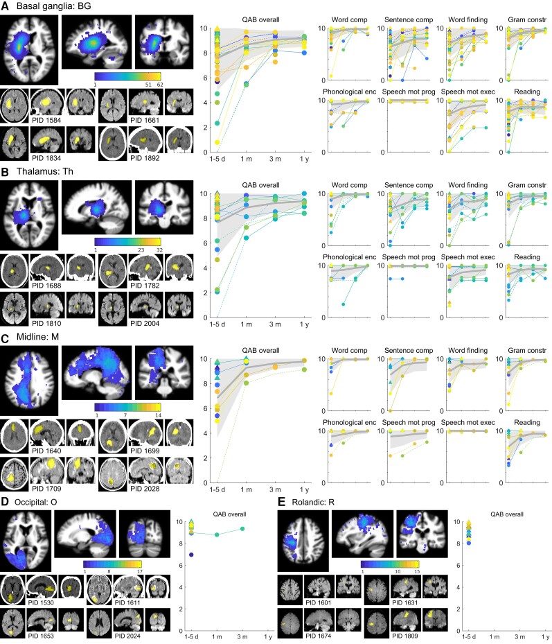
Figure 4.
Trajectories of recovery for patients with damage to the basal ganglia, thalamus, midline, occipital, and Rolandic regions. (A) BG group patients (n = 62). (B) Th group patients (n = 32). (C) M group patients (n = 14). (D) O group patients (n = 17). (E) R group patients (n = 15). See Fig. 2 legend for additional details.

