Abstract
Introduction:
Inborn errors of metabolism (IEM) are a rare cause of epilepsy in pediatric age group. Prompt diagnosis is essential, as some of these disorders are treatable.
Aim:
To determine the prevalence, clinical, and etiological profile of metabolic epilepsy in children.
Methods:
A prospective observational study of children with new onset seizures diagnosed as inherited metabolic disorder in a tertiary care hospital, South India.
Results:
Among 10,778 children with new onset seizures, 63 (0.58%) had metabolic epilepsy. The male female ratio was 1.3:1. Onset of the seizures were in neonatal period in 12 (19%), infancy in 35 (55.6%), and between one and 5 years of age in 16 (25.4%) children. Generalised seizures were seen in 46 (73%), followed by multiple seizure types (31.7%). The associated clinical features included developmental delay in 37 (58.7%), hyperactivity in 7 (11%), microcephaly in 13 (20.6%), optic atrophy in 12 (19%), sparse hair and/or seborrheic dermatitis in 10 (15.9%), movement disorder in 7 (11%), and focal deficit in 27 (42.9%) patients. Magnetic resonance imaging brain was abnormal in 44 (69.8%) and diagnostic in 28 (44.4%) patients. Causative metabolic errors included vitamin responsive errors in 20 (31.7%), disorders of complex molecules in 13 (20.6%), amino acidopathies in 12 (19%), organic acidemias in 10 (16%), disorders of energy metabolism in 6 (9.5%), and peroxisomal disorders in 2 (3.2%) patients. With specific treatment, seizure freedom could be achieved in 45 (71%) children. Five children lost to follow-up and two died. Among the remaining 56 patients, 11 (19.6%) had a good neurological outcome.
Conclusion:
Vitamin responsive epilepsies were the most frequent cause of metabolic epilepsy. Early diagnosis and prompt treatment is necessary as only one-fifth had a good neurological outcome.
Keywords: Inherited metabolic disorders, metabolic seizures, vitamin responsive epilepsy
INTRODUCTION
Inborn errors of metabolism (IEM) are an uncommon cause of epilepsy, but seizures and epilepsy often occur in children with IEM. Metabolic epilepsies are disorders related to inherited enzyme deficiencies with a resultant impact on metabolic/biochemical pathways.[1] International League Against Epilepsy recognizes the following conditions as metabolic epilepsy: Pyridoxine dependency/pyridoxal phosphate deficiency, Biotinidase/Holocarboxylase deficiency, Folinic acid-responsive seizures, Cerebral folate deficiency, Glucose transporter 1 deficiency, Creatine deficiency disorders, Mitochondrial disorders, and Peroxisomal disorders.[2,3] In addition, other metabolic disorders like nonketotic hyperglycinemia, molybdenum cofactor deficiency, organic acidemias, Menkes disease, and lysosomal storage disorders may present with seizures as the predominant feature in children.
Seizures due to inherited metabolic disorders are usually refractory to conventional antiseizure medications.[4] However, some of these disorders are amenable to specific treatments such as dietary therapy, vitamin and cofactor supplementation, and substrate reduction therapy. Hence, prompt diagnosis is essential for not only seizure control but a favorable outcome and better quality of life. Moreover, treatments of many inherited metabolic disorders have been optimized leading to an increasing number of patients who survive well into adulthood.[5] Hence, knowledge about these disorders is essential for every clinician serving patients of every age. There is not much literature from our country on metabolic epilepsy except individual case reports and review articles. Hence, an attempt was made to identify the prevalence of metabolic epilepsy among new-onset seizures in children, clinical presentation, imaging features, and outcomes.
METHODS
This prospective observational study was conducted at the Institute of Child Health and Hospital for Children, a tertiary care referral hospital in South India, between January 2015 and December 2021. All consecutive children aged less than 12 years attending the Department of Pediatric Neurology with new-onset seizures underwent evaluation. Children diagnosed with seizures due to an inherited metabolic disorder got included in the study after obtaining a written consent from their parents. Children with seizures due to acquired metabolic etiology such as dys-electrolytemia, hypocalcemia, hypomagnesemia, renal failure, and hypoglycemia due to non-IEM conditions were excluded.
We collected data regarding the age of the child, gender, parental consanguinity, age of onset of seizures, semiology, frequency, perinatal events, developmental milestones, and family history of similar illnesses. Detailed clinical examination was done looking for neurocutaneous markers, discolored hair, alopecia, seborrheic dermatitis, dysmorphism, micro/macrocephaly, organomegaly, and focal neurological deficits.
Children underwent investigations such as blood glucose, serum electrolytes, calcium, ammonia, lactate, urine ketones, arterial blood gas, tandem mass spectrometry (TMS), urine organic acids, magnetic resonance imaging (MRI) of the brain including magnetic resonance spectroscopy (MRS), and electroencephalogram (EEG). Special investigations such as serum copper, ceruloplasmin, uric acid, plasma homocysteine, cerebrospinal fluid analysis with corresponding blood sugar, Cerebrospinal fluid (CSF) lactate, serum glycine with CSF glycine, serum biotinidase levels, serum alpha amino adipic semialdehyde acid, urine sulfites, plasma very long chain fatty acid assay, serum enzyme assay for lysosomal storage disorders, and genetic analysis were done based on the clinical, imaging, and therapeutic trial clues accordingly. Detailed ophthalmologist opinion to look for optic atrophy, retinitis pigmentosa, macular cherry-red spot, lens dislocation, and cataract was obtained.
Children with alopecia and/or seborrheic dermatitis were treated with biotin pending serum biotinidase levels. Children with unexplained and refractory seizures received a therapeutic trial with pyridoxine, pyridoxal phosphate, folinic acid, and biotin successively. Those with treatable metabolic disorders were managed with dietary therapy, cofactor supplementation (pyridoxine in pyridoxine responsive epilepsy and hyperprolinemia, biotin in biotinidase deficiency, propionic acidemia and biotin responsive basal ganglia disease, folinic acid in folinic acid-responsive seizures and cerebral folate deficiency, thiamine in Maple syrup urine disease and biotin responsive basal ganglia disease, riboflavin in glutaric aciduria, and vitamin B12 in methylmalonic acidemia), mitochondrial cocktail (carnitine, cofactor Q, thiamine, riboflavin, biotin, and vitamin C) in mitochondrial disorders, substrate reduction therapy (sodium benzoate in conditions associated with hyperammonemia and hyperglycinemia), copper histidine in Menkes disease, and creatine monohydrate in Guanidino amino methyl transferase deficiency. Those who had refractory seizures despite dietary therapy, substrate reduction, and cofactor supplementation received antiseizure medications in addition. Children with untreatable metabolic disorders were managed with antiseizure medications as any other child with epilepsy. Sodium valproate as an antiseizure drug was avoided in mitochondrial disorders, hyperglycinemia, conditions associated with hyperammonemia, and phenobarbitone in glucose transporter deficiency. Children with developmental delay or regression underwent physiotherapy, occupational therapy, and speech therapy. Children without focal neurological deficits were considered as having good outcome and presence of any deficit was termed as poor outcome. All clinical information was recorded in a predesigned proforma and managed with a Microsoft Excel spreadsheet. Frequency was calculated as number (n) and percentage (%).
RESULTS
A total of 10,778 children were diagnosed with new-onset seizures during the study period, of which 63 (0.58%) had metabolic epilepsy based on the inclusion criteria. During the same period, 156 children were diagnosed to have IEM, of which 63 (40.4%) had seizures. The male-female ratio was 1.3:1. The mean age of onset was 11.83 ± 9.82 months (range 0.03-60 months). The onset of the seizures was during infancy in 47 (74.6%) children, of which 12 (19%) were in the neonatal period. The mean age at diagnosis was 23.43 ± 15.09 months (range 1-96 months). The commonest seizure type was generalized tonic-clonic noted in 46 (73%) children followed by focal seizures in 15 (23.8%) children. Twenty (31.7%) children had multiple seizure types [Table 1].
Table 1.
Clinical characteristics of children with metabolic epilepsy
| Variables | No. of patients (%) |
|---|---|
| Total no. of patients | 63 |
| Gender distribution | |
| Males | 36 (57.1) |
| Females | 27 (42.9) |
| Age of onset (months) | |
| 0-12 | 47 (74.6) |
| >12 | 16 (25.4) |
| Age at diagnosis (months) | |
| 0-12 | 32 (50.8) |
| 13-24 | 11 (17.5) |
| >24 | 20 (31.7) |
| H/O Consanguinity | 47 (74.6) |
| Seizure type | |
| Focal clonic seizures | 15 (23.8) |
| Generalised onset–motor | |
| Tonic clonic | 46 (73) |
| Myoclonic | 12 (19) |
| Epileptic spasms | 7 (11.1) |
| Multiple types | 20 (31.7) |
| Multiple episodes prior to specific therapy | 43 (68.3) |
| Status epilepticus | 7 (11.1) |
| Developmental delay | 37 (58.7) |
| Developmental regression | 20 (31.7) |
| Diagnosis confirmed by | |
| Biochemical including enzyme assay | 42 (66.7) |
| Genetic testing | 14 (22.2) |
| Therapeutic trial | 4 (6.3) |
| Skin biopsy | 3 (4.8) |
The associated clinical features noticed included developmental delay in 37 (58.7%) patients, lethargy and encephalopathy in nine (14.3%), hyperactivity in seven (11%), frequent hiccups in three (4.8%), autistic features in 2 (3.2%), abnormal startle in five (7.9%), and abnormal odor of urine in two (3.2%) children.
Examination revealed microcephaly in 13 (20.6%) patients, macrocephaly in three (4.8%), dysmorphic facies in six (9.5%), sparse hair or seborrheic dermatitis in 10 (15.9%), discolored hair in two (3.2%), hypotonia in four (6.3%), movement disorder in the form of choreoathetosis in four (6.3%), dystonia in three (4.8%), and focal neurological deficit in the form of spastic quadriparesis, diplegia, and ataxia in 27 (42.9%) patients. The ophthalmological evaluation revealed optic atrophy in 12 (19%), a macular cherry-red spot in three (4.8%), retinitis pigmentosa, and lens dislocation in one (1.6%) child each.
The mean time lag between the onset of symptoms and diagnosis was 12.44 ± 8.44 months. The mean number of antiepileptic drugs used before the diagnosis of a particular IEM was 2.28 ± 0.66.
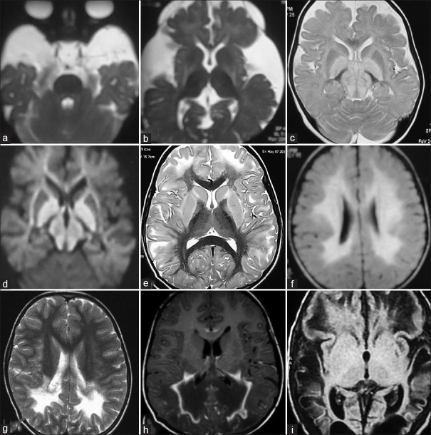
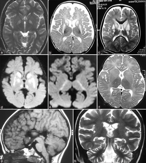
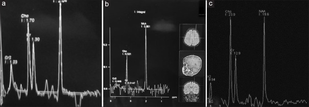
MRI brain was abnormal in 44 (69.8%) and was diagnostic in 28 (44.4%) patients. The imaging findings in individual patients are depicted in Figures 1 and 2. MRS was diagnostic in seven (11.1%). Figure 3 shows the abnormal peaks seen in MRS of our patients. EEG was normal in 29 (46%). The biochemical, imaging, electrophysiological, and genetic abnormalities in each group of metabolic epilepsies have been depicted in Table 2.
Figure 1.
(a and b) T2 axial Magnetic Resonance Imaging shows Bats wing appearance, widened sylvian fissure with poor operculisation in a child with Glutaric aciduria type 1. (c and d). T2 axial MR image showing hyperintense globus pallidi and midbrain with marked diffusion restriction in Maple Syrup Urine Disease. (e) T2 axial MR imaging showing subcortical white matter hyperintensity with sparing of periventricular white matter in a child with L2 hydroxy glutaric aciduria. (f) MR imaging axial FLAIR sequence showing symmetrical periventricular white matter hyperintensity typical of metachromatic leukodystrophy. (g) T2 axial MR image shows periventricular white matter hyperintensity in posterior regions in a child with Krabbe disease. (h) MR contrast image depicting peri ventricular white matter hyperintensity in the posterior region with contrast enhancement characteristic of adrenoleukodystrophy. (i) MR axial FLAIR image showing multicystic encephalomalacia in a child with molybdenum cofactor deficiency
Figure 2.
(a) T2 axial Magnetic Resonance Imaging shows bilateral globus pallidi involvement in methyl malonic acidemia. (b) T2 axial MR image showing bilateral caudate and putamen involvement in a child with propionic acidemia. (c) T2 axial MR image shows bilateral caudate and putamen involvement in a child with biotin responsive basal ganglia disease. (d) MR diffusion weighted imaging sequence showing diffusion restriction in bilateral basal ganglia and thalami. (e) MR diffusion weighted imaging sequence showing diffusion restriction in posterior limbs of both internal capsules. (f) T2 axial Magnetic Resonance Imaging shows hypomyelination in a child with gangliosidosis. (g) MR saggital FLAIR sequence depicting dysgenesis of corpus callosum in a child with pyridoxin dependency. (h) T2 coronal Magnetic Resonance Image showing cerebral and cerebellar atrophy in neuronal ceroid lipofuscinosis
Figure 3.
(a) Magnetic Resonance Spectroscopy showing lactate peak in a child with mitochondrial disorder. (b) MRS depicting absence of creatine peak in a child with Guanidino Acetate Methyl Transferase (GAMT) deficiency —a cerebral creatine deficiency disorder. (c) Normal MRS for comparison
Table 2.
Diagnostic work up of children with metabolic epilepsy
| Investigations | Disorders of vitamin & mineral metabolism (n=20) | Amino acido pathies (n=12) | Organic acide mias (n=10) | Disorders of energy meta bolism (n=6) | Storage disorders (n=13) | Peroxi somal disorders (n=2) | Total (n=63 | |
|---|---|---|---|---|---|---|---|---|
| 1 | MRI brain | |||||||
| Diagnostic | 0 | 6 | 10 | 5 | 5 | 2 | 28 | |
| Non specific | 6 | 4 | 0 | 0 | 6 | 0 | 16 | |
| Normal | 14 | 2 | 0 | 1 | 2 | 0 | 19 | |
| 2 | MRS | 0 | 3 | 0 | 4 | 0 | 0 | 7 |
| 3 | EEG abnormal | 8 | 9 | 3 | 3 | 10 | 1 | 34 |
| 4 | Biochem investig including enzyme levels | 14 | 12 | 7 | 1 | 6 | 2 | 42 |
| 5 | Axillary skin biopsy | 3 | 3 | |||||
| 6 | Genetic mutation studies | 5 (2*) | 3 | 3 (3*) | 5 (5*) | 5 (4*) | 2 | 23 (14*) |
| 7 | Therapeutic trial | 4 | 4 |
MRS – Magnetic Resonance Spectroscopy. (*) diagnosed only by genetic studies
Genetic studies were done in 23 (36.5%) children. Genetic testing confirmed the biochemical diagnosis in nine patients and provided the diagnosis in 14 patients. The biochemical, imaging, and genetic results of children with metabolic epilepsies who underwent genetic tests are described in Table 3.
Table 3.
Biochemical, imaging, and genetic results of children with metabolic epilepsies who underwent genetic tests
| Metabolic disorder | MRI brain | Biochemical tests | Genetic Result |
|---|---|---|---|
| Biotinidase deficiency | Hypomyelination | Low serum biotinidase | Homozygous for p.Q336X mutation |
| Biotinidase deficiency | Normal | Low serum biotinidase | c. 38_44delGCGG homozygous insertion mutation in BTD gene |
| Biotinidase deficiency | Normal | Low serum biotinidase | c. 1552C >T homozygous mutation in BTD gene |
| Pyridoxin Dependency | Thin corpus callosum | c. 1556G >A homozygous mutation in ALDH1A1gene | |
| Biotin thiamine responsive basal ganglia disease | Bilateral hyperintensities in striatum | c. 593T >C homozygous mutation in SLC19A3 gene | |
| Molybdenum cofactor deficiency | Multi cystic encephalomalacia | Low serum uric acid, high urine sulphites | c. 1762G >A homozygous mutation in MOCS1 gene |
| N- Acetyl Glutamate Synthase deficiency | Multi cystic encephalomalacia | Hyperammonemia, Low citrulline | c. 1535A >G homozygous mutation in NAGS gene |
| Non ketotic hyperglycinemia | diffusion restriction - internal capsule | Elevated CSF, serum glycine, ratio 0.09 | c. 2675C >T homozygous mutation in GLDC gene |
| L2-OH glutaric aciduria | subcortical WM involvement, peri ventricular sparing | c. 829 C >T homozygous mutation in L2HGDH gene | |
| L2-OH glutaric aciduria | Subcortical WM involvement, peri ventricular sparing | c. 829 C >T homozygous mutation in L2HGDH gene | |
| L2-OH glutaric aciduria | Subcortical WM involvement, peri ventricular sparing | homozygous deletion [chr: 14?50750588_?50750751_del] in L2HGDH gene | |
| GAMT deficiency | Globus pallidi HI Absent creatine peak in MRS | c. 164_171del-homozygous deletion in GAMT gene | |
| Fructose1,6 bis phosphatase def | Bilateral basal ganglia, midbrain | Hypoglycemia | c. 960_961insG homozygous insertion mutation in FBP1gene |
| Mitochondrial complex I | Bilateral basal ganglia, midbrain | Elevated serum lactate | c. 1156C >T homozygous mutation in NDUFV1 gene |
| Mitochondrial complex III deficiency | deep WM, diffusion restriction and rarefaction, lactate peak in MRS | Elevated serum lactate | c. 2T >C homozygous mutation in LYRM7 gene |
| Mitochondrial complex I def | Bilateral BG involvement | Elevated serum lactate | c. 1118T >C homozygous mutation in NDUFV1 gene |
| Metachromatic leuco dystrophy | Symmetrical periventricularWM | Low serum aryl sulfatase | c. 739G >A homozygous mutation in ARSA gene |
| Nieman Pick C disease | Normal | c. 436C >T homozygous mutation in NPC2 gene | |
| Neuronal ceroid lipofuscinosis-8 | Cerebral and cerebellar atrophy | c. 598_599delAThomozygous deletion in CLN 8 gene | |
| Neuronal ceroid lipofuscinosis-1 | Cerebral and cerebellar atrophy | c. 713C >T homozygous mutation in PPT1 gene | |
| Neuronal ceroid lipofuscinosis -2 | Cerebral and cerebellar atrophy, periventricularWM | c. 622 C >T homozygous mutation in TPP1 gene | |
| Zellweger syndrome | Perisylvian polymicrogyria | Elevated VLFA, low plasmalogen | C126+1G >T homozygous mutation in PEX12 gene |
| Adreno leukodystrophy | bilateral parieto occipital WM with contrast enhancement | Elevated VLFA | c. 1876G >A hemizygous missense mutation in ABCD1 gene |
HI, hyperintensities; WM, whitw matter; BG, basal ganglia; VLFA, very long chain fatty acids.
The causative metabolic errors included disorders of vitamin and metal metabolism in 20 (31.7%), amino acidopathies in 12 (19%), organic acidemias in 10 (15.9%), disorders of energy metabolism in six (9.5%), disorders of complex molecules in 13 (20.6%), and peroxisomal disorders in two (3.2%) children. The main cause of vitamin-responsive epilepsy was biotinidase deficiency in nine (14.3%) patients followed by pyridoxine dependency in six (9.5%). Treatable causes of metabolic epilepsy constituted 65.1% (41/63) of the cohort. The individual disorders under each group have been depicted in Table 4.
Table 4.
Etiological spectrum of metabolic epilepsies
| Metabolic disorder | Number (%) |
|---|---|
| Disorders of vitamin and metal metabolism* | 20 (31.7%) |
| Biotinidase deficiency | 9 |
| Pyridoxine dependency | 6 |
| Folinic acid responsive seizures | 1 |
| Hyperprolinemia | 1 |
| Cerebral folate deficiency | 1 |
| Biotin thiamine responsive basal ganglia disease | 1 |
| Menkes disease | 1 |
| Disorders of amino acid metabolism- Amino acidopathies | 12 (19%) |
| Non ketotic hyperglycinemia | 3 |
| Urea cycle disorders* | 3 |
| Molybdenum cofactor deficiency | 2 |
| Phenyl ketonuria* | 2 |
| Maple Syrup Urine Disease* | 2 |
| Organic acidemias* | 10 (15.9%) |
| Glutaric acidemia | 3 |
| L2 hydroxy glutaric aciduria | 3 |
| Propionic acidemia | 2 |
| Methyl malonic acidemia | 1 |
| Β keto-thiolase deficiency | 1 |
| Disorders of energy metabolism | 6 (9.5%) |
| Respiratory chain defects | 3 |
| Cerebral creatine deficiency* | 1 |
| Glucose transporter deficiency* | 1 |
| Fructose 1, 5 bisphosphatase deficiency* | 1 |
| Disorders of complex molecules | 13 (20.6%) |
| Neuronal ceroid lipofuscinosis | 6 |
| GM2 gangliosidosis | 3 |
| Metachromatic leukodystrophy | 2 |
| Krabbe disease | 1 |
| Niemann Pick C disease | 1 |
| Peroxisomal disorders | 2 (3.2%) |
| Zellweger syndrome | 1 |
| Adreno leukodystrophy* | 1 |
*Treatable metabolic epilepsy
With specific treatment, seizure freedom could be achieved in 45 (71%) children. Eighteen patients (29%) had refractory seizures. The prevalence of epilepsy and refractory epilepsy in each group of metabolic disorders is given in Table 5. The prevalence of epilepsy was higher in disorders of vitamin metabolism (95%), followed by amino acidopathies and disorders of energy metabolism (40%) each. Higher incidence of refractory seizures was noticed in disorders of complex molecules (69%) followed by disorders of energy metabolism and peroxisomal disorders (50%).
Table 5.
Prevalence of epilepsy and refractory epilepsy in various groups of metabolic epilepsies
| Etiological groups | Epilepsy prevalence No of children with epilepsy/Total no of children with IMD (63/156) | No. of patients with refractory epilepsy/No. of patients with epilepsy | Metabolic disorder with refractory cases |
|---|---|---|---|
| Disorders of vitamin and mineral metabolism | 20/21 (95.2%) | 0/20 | |
| Amino acidopathies | 12/30 (40%) | 5/12 (41.6%) | Nonketotic hyperglycinemia (3) Molybdenum cofactor deficiency (2) |
| Organic acidemias | 10/30 (33.3%) | 0/10 | |
| Disorders of energy metabolism | 6/15 (40%) | 3/6 (50%) | Mitochondrial disorders (3) |
| Disorders of complex molecules | 13/54 (24.1%) | 9/13 (69.2%) | Neuronal ceroid lipofuscinosis (6) GM2 gangliosidosis (2) Niemann Pick C disease (1) |
| Peroxisomal disorders | 2/6 (33.3%) | 1/2 (50%) | Zellweger syndrome (1) |
IMD, Inherited Metabolic Disorder
Complete seizure freedom was observed in all cases of vitamin-responsive epilepsies and organic acidemias. Antiseizure medications could be withdrawn in 28 (44.4%) patients. Five children were lost to follow-up and two died during follow-up. Among the remaining 56 patients, 11 (19.6%) had a good neurological outcome [Table 6].
Table 6.
Seizure control and outcome in various groups of metabolic epilepsies
| Variables | Disorders of vitamin & metal metabolism (n=20) | Amino acido pathies (n=12) | Organic acide mias (n=10) | Disorders of energy metabolism (n=6) | Storage disorders (n=13) | Peroxi somal disorders (n=2) | Total (n=63) (%) |
|---|---|---|---|---|---|---|---|
| Seizure control | 20/20 (100) | 7/12 (58.3) | 10/10 (100) | 3/6 (50) | 4/13 (30.8) | ½ (50) | 45 (71.4) |
| ASM withdrawn | 16/20 (80) | 6/12 (50) | 5/10 (50) | 1/6 (16.7) | 0 | 0 | 28 (44.4) |
| Outcome | |||||||
| Lost follow-up | 3 | 2 | 0 | 0 | 0 | 0 | 5 (7.9) |
| Expired | 0 | 0 | 0 | 0 | 2 | 0 | 2 (3.2) |
| Motor impairment | 7 | 7 | 1 | 2 | 13 | 2 | 32 (57.1) |
| Cognitive impairment | 10 | 8 | 2 | 5 | 8 | 1 | 34 (60.7) |
| Language disturbances | 4 | 2 | 2 | 3 | 6 | 1 | 18 (32.1) |
| Behavioral Problems | 1 | 2 | 2 | 3 | 0 | 0 | 8 (14.3) |
| Optic atrophy | 4 | 0 | 0 | 2 | 5 | 1 | 12 (21.4) |
| Hearing impairment | 2 | 0 | 0 | 0 | 0 | 0 | 2 (3.6) |
| Good Outcome | 6 | 0 | 5 | 0 | 0 | 0 | 11/56 (19.6) |
DISCUSSION
Inborn errors of metabolism are a rare cause of epilepsy in childhood. The precise prevalence of metabolic epilepsies is unknown, but they are likely to represent a small proportion of all patients with epilepsy.[6,7,8] The diagnostic yield from genetic testing in patients with epileptic encephalopathies is about 7% in a series and for treatable conditions about 4%.[9] Üstkoyuncu et al.[10] evaluated 268 patients with epilepsy and showed that 0.37% (1/268) of these patients had inherited metabolic disorders. We found that metabolic epilepsies accounted for 0.58% of children with new-onset seizures in our study.
On the other hand, seizures are a frequent symptom of many metabolic disorders. The prevalence of epilepsy among inherited metabolic disorders was found to be 40.4% (63/156) in our cohort, which is compatible with previous studies.[11,12] Youssef-Turki et al.[13] in a recent review have reported that 600 metabolic epilepsies have been identified, accounting for as much as 37% of all currently described inherited metabolic diseases.
No seizure semiology is specific to metabolic epilepsy. However, metabolic epilepsies are more frequently associated with generalized seizures.[12] We observed that generalized seizures were the dominant semiology that occurred in nearly three-fourth of our cases. Multiple seizure types were noticed in one-third of cases (20/63) which provided a clue to metabolic etiology and further workup. Similarly, multiple episodes of seizures not responding to antiseizure medications before specific therapy pointed toward a metabolic etiology. Nearly 70% (43/63) of the patients did not respond to antiseizure medications before specific therapy for the underlying metabolic disorder. When seizures are unexplained and refractory, vitamin-responsive epilepsies should always be considered and therapeutic trials using successively pyridoxine, pyridoxal phosphate, folinic acid, and biotin should be started.[14,15,16,17] Application of this protocol in the day-to-day practice resulted in the diagnosis of many cases of vitamin-responsive epilepsies in our cohort.
Parental consanguinity was noticed in three-fourth of the cases in our study, which is compatible with earlier studies.[11,18] As most of the neurometabolic disorders have an autosomal inheritance, presence of parental consanguinity provides a clue to the diagnosis of an inborn error of metabolism. Developmental delay, a clinical red flag to the diagnosis was noted in nearly 60% of our patients. Tumiene et al.[12] have reported that 66% of metabolic epilepsies are associated with developmental delay.
Vitamin-responsive epilepsies (31.7%) were found to be the commonest cause of metabolic epilepsies in our study followed by disorders of complex molecules (20.6%) and amino acidopathies (19%). Mohamed et al.[18] from Saudi Arabia have reported disorders of energy metabolism to be the most common (33%) aetiology, followed by aminoacidopathies (20.8%) and storage disorders (17.6%), whereas Karimzadeh et al.[11] from Iran noticed lysosomal storage disorders as the predominant disorder (64%) in their study. The etiological dissimilarities could be due to differences in ethnicity and geographical regions.
MRI brain including MRS should be performed in all children with suspected metabolic epilepsy as relatively specific; hence, diagnostic patterns may be seen in some IEM such as glutaric aciduria, maple syrup urine disease, L2 hydroxy glutaric aciduria, and leukodystrophies, while other diseases such as organic acidemias and mitochondrial cytopathies show imaging features that are more suggestive than specific.[4,19,20,21,22,23,24,25] MRI brain was diagnostic in nearly half of our patients. Proton MRS is of diagnostic value in some IEM such as nonketotic hyperglycinemia, mitochondriopathies, creatine deficiency disorders, and maple syrup urine disease.[19] MRS was abnormal in seven of our patients.
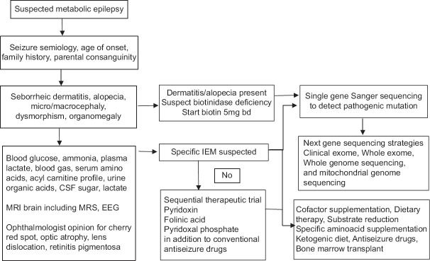
Tumiene et al.[12] have reported that in 70% of metabolic epilepsies, abnormalities could be identified by metabolic testing. Clinical clues, imaging findings, and a therapeutic trial (pyridoxin, folinic acid, and biotin) guided the biochemical investigations which were diagnostic in 66.7% (42/63) of the cases in our series, further confirmed by genetic studies in some (9/42, 21.4%). The diagnosis was established by genetic testing which showed pathogenic mutations in 14 cases (22.2%) - Fructose1,5 bisphosphate deficiency (1), biotin responsive basal ganglia disease (1) - earlier suspected to be mitochondrial disorders from neuroimaging, pyridoxin dependent epilepsy (1), N acetyl glutamate synthase deficiency (1), L2 hydroxy glutaric aciduria (3), Guanidinoacetate methyl transferase deficiency (1), mitochondrial disorders (3), and neuronal ceroid lipofuscinosis (3) and by axillary skin biopsy in three cases of neuronal ceroid lipofuscinosis. However, neuroimaging revealed distinct findings in L2 hydroxy glutaric aciduria, GAMT deficiency, and suggestive features in others. An approach to patients with suspected metabolic epilepsy has been summarized in Figure 4.
Figure 4.
An approach to children with suspected metabolic epilepsy
Nearly three-fourth of our patients achieved seizure freedom (45/63,71%) with treatment. When the individual etiological group was analyzed, with cofactor therapy (pyridoxine in pyridoxine-responsive epilepsy and hyperprolinemia, biotin in biotinidase deficiency, biotin, thiamine in biotin-responsive basal ganglia disease, folinic acid in folinic acid-responsive seizures, and cerebral folate deficiency), complete seizure control could be achieved in all patients (100%), and antiseizure medications could be withdrawn in 84% (16/19) of vitamin responsive epilepsies. Three children-profound biotinidase deficiency (1), biotin-responsive basal ganglia disease (1), and cerebral folate deficiency (1) required antiseizure medications in addition. Seizure freedom was also observed in all the children with organic acidemias (100%), in 58% (7/12) patients with amino acidopathies, 50% (3/6) with disorders of energy metabolism, and 31% (4/13) with lysosomal storage disorders. Seizures were refractory in nonketotic hyperglycinemia, molybdenum cofactor deficiency, storage disorders, and mitochondrial disorders. Mohamed et al.[18] have reported that one-third of patients in their study achieved seizure freedom and those who responded had treatable metabolic diseases such as citrullinemia and biotinidase deficiency. The higher percentage of children with seizure control was due to the higher prevalence of treatable metabolic disorders in our cohort.
Among our cohort of metabolic epilepsies, 41 (65.1%) disorders were treatable with specific cofactor administration, dietary therapy, and substrate reduction therapy. Antiseizure medications could be withdrawn in nearly two-thirds of them (26/41, 63.4%). This underscores the importance of proper diagnosis and prompt treatment, which can lead to better seizure control and withdrawal of unnecessary antiseizure medications. However, although two-thirds of the cohort (65.1%) were diagnosed as having treatable inherited metabolic disorders displaying excellent seizure control, only 27% (11/41) of them had a good neurological outcome without any neurological deficits. The remaining showed motor, cognitive, language, visual, and hearing impairments either single or in combination, reiterating the fact that an early diagnosis and specific treatment of the underlying metabolic disorder is crucial for a better neurological outcome.
In many countries, treatable metabolic disorders are now part of new-born screening programs.[26] Until new-born screening for inherited metabolic disorders becomes a routine practice, it is prudent to consider metabolic epilepsy in any child with refractory seizures, since early diagnosis and treatment can result in the prevention of a full-scale metabolic crisis and prevent neurological sequelae. With advances in genetic technology, next-generation sequencing using targeted gene panels for epileptic encephalopathy, whole exome sequencing, and whole genome sequencing would help in arriving at precise diagnosis enabling appropriate treatment.[4] It should also be noticed that even for those inherited metabolic disorders for which therapy is not available, the diagnosis is still important for genetic counselling.[27]
Limitations
Screening of metabolic disorders such as gamma-aminobutyric acid catabolism, purine and pyrimidine metabolism, and congenital disorders of glycosylation were not done for want of facilities. Similarly, genetic analysis was not carried out in all children with unexplained seizures, which would have identified more cases of metabolic epilepsies.
CONCLUSION
Although metabolic epilepsies constitute a small percentage of new-onset epilepsies, nearly two-thirds of them are treatable. The majority of metabolic epilepsies can be diagnosed by a good clinical examination, neuroimaging including MRS, and metabolic testing. A therapeutic trial with vitamins should be tried in any child with refractory seizures, as vitamin-responsive epilepsies are the common cause of metabolic epilepsies. A high index of suspicion is necessary as a timely diagnosis is crucial for good seizure control and better neurological outcome.
Financial support and sponsorship
Nil.
Conflicts of interest
There are no conflicts of interest.
Acknowledgments
We would like to thank Prof. Dr. Pramila, biochemistry professor of our institution, and her team for providing the necessary laboratory support in the evaluation of these children.
REFERENCES
- 1.Papetti L, Parisi P, Leuzzi V, Nardecchia F, Nicita F, Ursitti F, et al. Metabolic epilepsy:An update. Brain Dev. 2013;35:827–41. doi: 10.1016/j.braindev.2012.11.010. [DOI] [PubMed] [Google Scholar]
- 2.Berg AT, Berkovic SF, Brodie MJ, Buchhalter J, Cross JH, van Emde Boas W, et al. Revised terminology and concepts for organization of seizures and epilepsies:Report of the ILAE commission on classification and terminology, 2005–2009. Epilepsia. 2010;51:676–85. doi: 10.1111/j.1528-1167.2010.02522.x. [DOI] [PubMed] [Google Scholar]
- 3.Finsterer J, Mahjoub SZ. Presentation of adult mitochondrial epilepsy. Seizure. 2013;22:119–23. doi: 10.1016/j.seizure.2012.11.005. [DOI] [PubMed] [Google Scholar]
- 4.Sharma S, Prasad AN. Inborn errors of metabolism and epilepsy:Current understanding, diagnosis, and treatment approaches. Int J Mol Sci. 2017;18:1384. doi: 10.3390/ijms18071384. [DOI] [PMC free article] [PubMed] [Google Scholar]
- 5.Gariani K, Nascimento M, Superti-Furga A, Tran C. Clouds over IMD?Perspectives for inherited metabolic diseases in adults from a retrospective cohort study in two Swiss adult metabolic clinics. Orphanet J Rare Dis. 2020;15:210. doi: 10.1186/s13023-020-01471-z. [DOI] [PMC free article] [PubMed] [Google Scholar]
- 6.Scheffer IE, Berkovic S, Capovilla G, Connolly MB, French J, Guilhoto L, et al. ILAE classification of the epilepsies:Position paper of the ILAE Commission for Classification and Terminology. Epilepsia. 2017;58:512–21. doi: 10.1111/epi.13709. [DOI] [PMC free article] [PubMed] [Google Scholar]
- 7.Perucca P, Bahlo M, Berkovic SF. The genetics of epilepsy. Annu Rev Genom Hum Genet. 2020;21:205–30. doi: 10.1146/annurev-genom-120219-074937. [DOI] [PubMed] [Google Scholar]
- 8.Rahman S, Footitt EJ, Varadkar S, Clayton PT. Inborn errors of metabolism causing epilepsy. Dev Med Child Neurol. 2013;55:23–36. doi: 10.1111/j.1469-8749.2012.04406.x. [DOI] [PubMed] [Google Scholar]
- 9.Mercimek-Mahmutoglu S, Patel J, Cordeiro D, Hewson S, Callen D, Donner EJ, et al. Diagnostic yield of genetic testing in epileptic encephalopathy in childhood. Epilepsia. 2015;56:707–16. doi: 10.1111/epi.12954. [DOI] [PubMed] [Google Scholar]
- 10.Ustkoyuncu PS, Guven AS, Poyrazoglu HG, Gokay S, Kardas F, Kendirci M, et al. Screening inherited metabolic disorder in children with intellectual disability and Epilepsy. Turkish J Neurol. 2019;25:135–40. [Google Scholar]
- 11.Karimzadeh P, Habibi P. An approach to neurometabolic epilepsy in children with an underlying neurometabolic disorder. Iran J Child Neurol. 2020;14:79–86. [PMC free article] [PubMed] [Google Scholar]
- 12.Tumiene B, Ferreira CR, van Karnebeek CDM. Overview of Metabolic Epilepsies. Genes. 2022;13:508. doi: 10.3390/genes13030508. [DOI] [PMC free article] [PubMed] [Google Scholar]
- 13.Youssef-Turki IB, Kraoua I, Smirani S, Mariem K, Benrhouma H, Rouissi A, et al. Epilepsy aspects and EEG patterns in neuro-metabolic diseases. J Behav Brain Sci. 2011;1:69–74. [Google Scholar]
- 14.Baxter P, Griffiths P, Kelly T, Gardner-Medwin D. Pyridoxine-dependent seizures:Demographic, clinical, MRI and psychometric features, and effect of dose on intelligence quotient. Dev Med Child Neurol. 1996;38:998–1006. doi: 10.1111/j.1469-8749.1996.tb15060.x. [DOI] [PubMed] [Google Scholar]
- 15.Kuo MF, Wang HS. Pyridoxal phosphate-responsive epilepsy with resistance to pyridoxine. Pediatr Neurol. 2002;26:146–7. doi: 10.1016/s0887-8994(01)00357-5. [DOI] [PubMed] [Google Scholar]
- 16.Torres OA, Miller VS, Buist NM, Hyland K. Folinic acid-responsive neonatal seizures. J Child Neurol. 1999;14:529–32. doi: 10.1177/088307389901400809. [DOI] [PubMed] [Google Scholar]
- 17.Collins JE, Nicholson NS, Dalton N, Leonard JV. Biotinidase deficiency-early neurological presentation. Dev Med ChildNeurol. 1994;36:268–70. doi: 10.1111/j.1469-8749.1994.tb11840.x. [DOI] [PubMed] [Google Scholar]
- 18.Mohamed S, El Melegy EM, Talaat I, Hosny A, Abu-Amero KK. Neurometabolic disorders-related early childhood epilepsy:A single-center experience in Saudi Arabia. Pediatr Neonatol. 2015;56:393–401. doi: 10.1016/j.pedneo.2015.02.004. [DOI] [PubMed] [Google Scholar]
- 19.Zimmerman RA. Neuroimaging of inherited metabolic disorders producing seizures. Brain Dev. 2011;33:734–44. doi: 10.1016/j.braindev.2011.03.006. [DOI] [PubMed] [Google Scholar]
- 20.Ibrahim M, Parmar HA, Hoefling N, Srinivasan A. Inborn errors of metabolism:Combining clinical and radiologic clues to solve the mystery. Am J Roentgenol. 2014;203:W315–27. doi: 10.2214/AJR.13.11154. [DOI] [PubMed] [Google Scholar]
- 21.Faerber EN, Melvin JJ, Smergel EM. MRI appearances of metachromatic leukodystrophy. Pediatr Radiol. 1999;29:669–72. doi: 10.1007/s002470050672. [DOI] [PubMed] [Google Scholar]
- 22.D'Incerti L, Farina L, Moroni I, Uziel G, Savoiardo M. L-2- Hydroxyglutaric aciduria:MRI in seven cases. Neuroradiology. 1998;40:727–33. doi: 10.1007/s002340050673. [DOI] [PubMed] [Google Scholar]
- 23.Given CA, Santos CC, Durden DD. Intracranial and spinal MR imaging findings associated with Krabbe's disease:Case report. AJNR Am J Neuroradiol. 2001;22:1782–5. [PMC free article] [PubMed] [Google Scholar]
- 24.Moser HW, Loes DJ, Melhem ER, Raymond GV, Bezman L, Cox CS, et al. X-linked adrenoleukodystrophy:Overview and prognosis as a function of age and brain magnetic resonance imaging abnormality. A study involving 372 patients. Neuropediatrics. 2000;31:227–39. doi: 10.1055/s-2000-9236. [DOI] [PubMed] [Google Scholar]
- 25.Zuccoli G, Yannes MP, Nardone R, Bailey A, Goldstein A. Bilateral symmetrical basal ganglia and thalamic lesions in children:An update. Neuroradiology. 2015;57:973–89. doi: 10.1007/s00234-015-1568-7. [DOI] [PubMed] [Google Scholar]
- 26.Reddy C, Saini AG. Metabolic epilepsy. Indian J Pediatr. 2021;88:1025–32. doi: 10.1007/s12098-020-03510-w. [DOI] [PubMed] [Google Scholar]
- 27.Brimble E, Ruzhnikov MRZ. Metabolic disorders presenting with seizures in the neonatal period. Semin Neurol. 2020;40:219–35. doi: 10.1055/s-0040-1705119. [DOI] [PubMed] [Google Scholar]


