Abstract
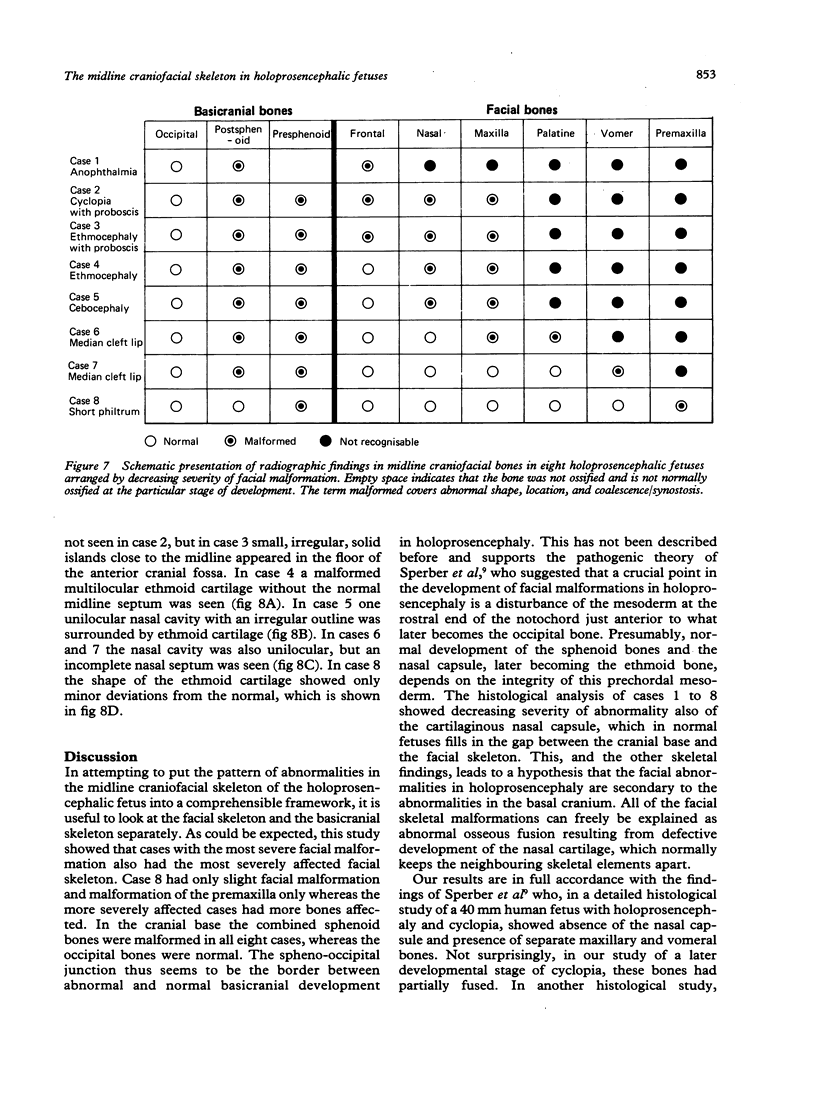
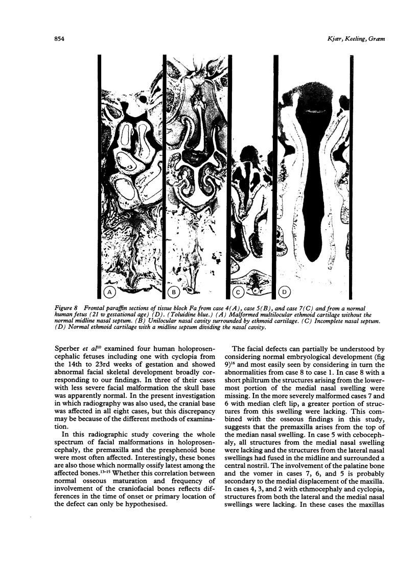
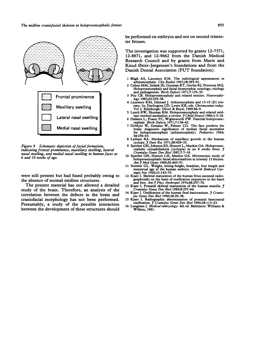
Craniofacial skeletal development in eight human holoprosencephalic fetuses from second trimester abortions were examined by radiography and histology. The whole spectrum of associated facial malformations from anophthalmia through cyclopia, ethmocephaly, cebocephaly, and median cleft lip to short philtrum was represented. Cases with the most severe facial malformations also had the most severely affected facial skeleton. In the facial skeleton, the premaxilla was most often affected; it was absent in seven cases and malformed in the one with only a short philtrum. This and other facial skeletal malformations can be explained as abnormal fusion of the facial bones because of defective development of the nasal cartilage. The occipital bones were normal, but the basicranial skeleton anterior to the spheno-occipital junction was affected in all cases. The findings support the hypothesis that the facial malformations in holoprosencephaly result from disturbance in embryonal life of the mesoderm at the rostral end of the notochord.
Full text
PDF
Images in this article
Selected References
These references are in PubMed. This may not be the complete list of references from this article.
- Bligh A. S., Laurence K. M. The radiological appearances in arhinencephaly. Clin Radiol. 1967 Oct;18(4):383–393. doi: 10.1016/s0009-9260(67)80044-8. [DOI] [PubMed] [Google Scholar]
- Cohen M. M., Jr, Jirásek J. E., Guzman R. T., Gorlin R. J., Peterson M. Q. Holoprosencephaly and facial dysmorphia: nosology, etiology and pathogenesis. Birth Defects Orig Artic Ser. 1971 Jun;7(7):125–135. [PubMed] [Google Scholar]
- DEMYER W., ZEMAN W., PALMER C. G. THE FACE PREDICTS THE BRAIN: DIAGNOSTIC SIGNIFICANCE OF MEDIAN FACIAL ANOMALIES FOR HOLOPROSENCEPHALY (ARHINENCEPHALY). Pediatrics. 1964 Aug;34:256–263. [PubMed] [Google Scholar]
- Dallaire L., Fraser F. C., Wiglesworth F. W. Familial holoprosencephaly. Birth Defects Orig Artic Ser. 1971 Jun;7(7):136–142. [PubMed] [Google Scholar]
- Fitz C. R. Holoprosencephaly and related entities. Neuroradiology. 1983;25(4):225–238. doi: 10.1007/BF00540235. [DOI] [PubMed] [Google Scholar]
- In memoriam. L. Stefan Levin. 1939-1989. J Craniofac Genet Dev Biol. 1990;10(1):1–6. [PubMed] [Google Scholar]
- Kjaer I. Ossification of the human fetal basicranium. J Craniofac Genet Dev Biol. 1990;10(1):29–38. [PubMed] [Google Scholar]
- Kjaer I. Prenatal skeletal maturation of the human maxilla. J Craniofac Genet Dev Biol. 1989;9(3):257–264. [PubMed] [Google Scholar]
- Kjar I. Skeletal maturation of the human fetus assessed radiographically on the basis of ossification sequences in the hand and foot. Am J Phys Anthropol. 1974 Mar;40(2):257–275. doi: 10.1002/ajpa.1330400211. [DOI] [PubMed] [Google Scholar]
- Latham R. A. Mechanism of maxillary growth in the human cyclops. J Dent Res. 1971 Jul-Aug;50(4):929–933. doi: 10.1177/00220345710500042401. [DOI] [PubMed] [Google Scholar]
- Leech R. W., Shuman R. M. Holoprosencephaly and related midline cerebral anomalies: a review. J Child Neurol. 1986 Jan;1(1):3–18. doi: 10.1177/088307388600100102. [DOI] [PubMed] [Google Scholar]
- Sperber G. H., Honoré L. H., Machin G. A. Microscopic study of holoprosencephalic facial anomalies in trisomy 13 fetuses. Am J Med Genet. 1989 Apr;32(4):443–451. doi: 10.1002/ajmg.1320320402. [DOI] [PubMed] [Google Scholar]
- Sperber G. H., Johnson E. S., Honoré L., Machin G. A. Holoprosencephalic synophthalmia (cyclopia) in an 8 week fetus. J Craniofac Genet Dev Biol. 1987;7(1):7–18. [PubMed] [Google Scholar]


