Abstract
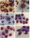
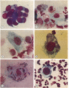
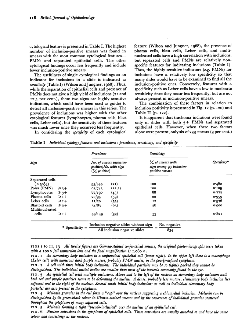
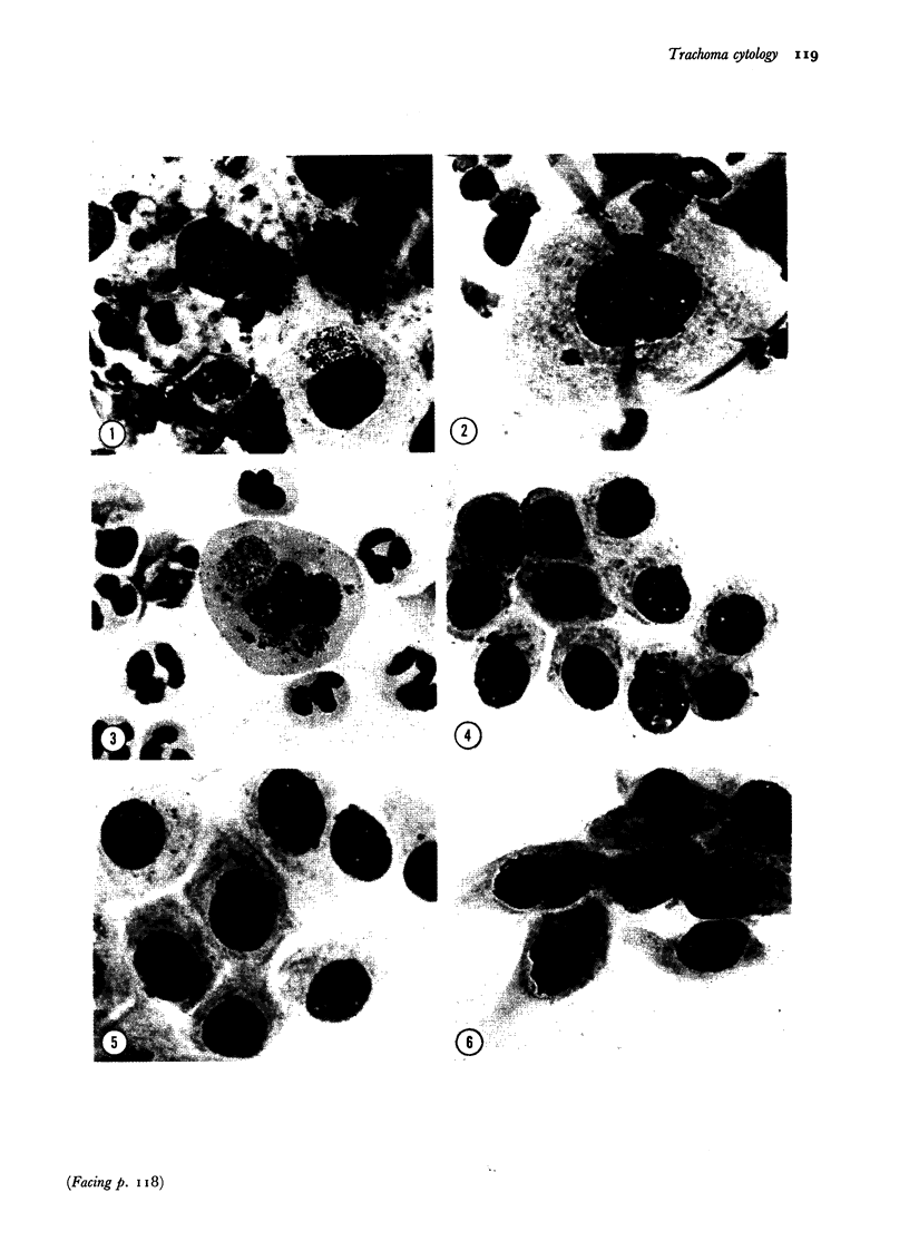
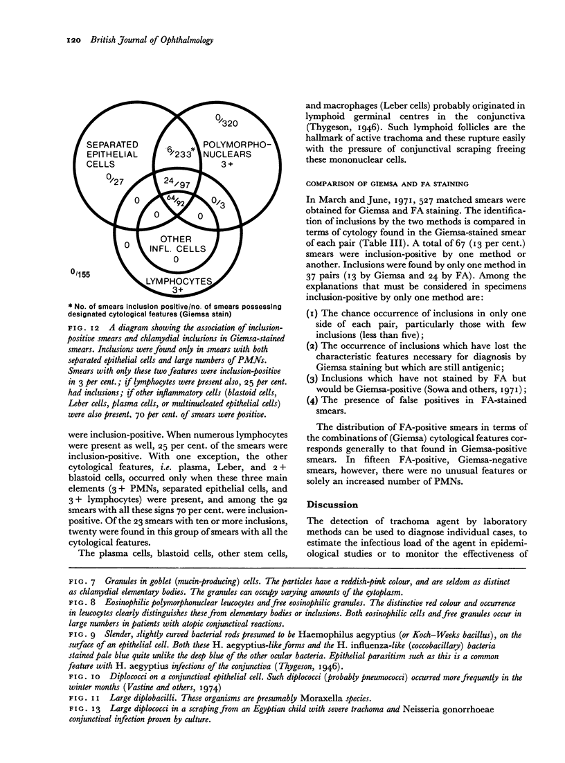
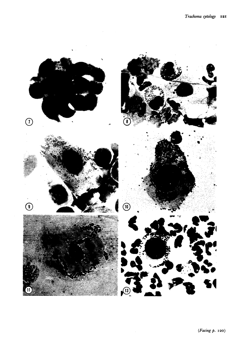
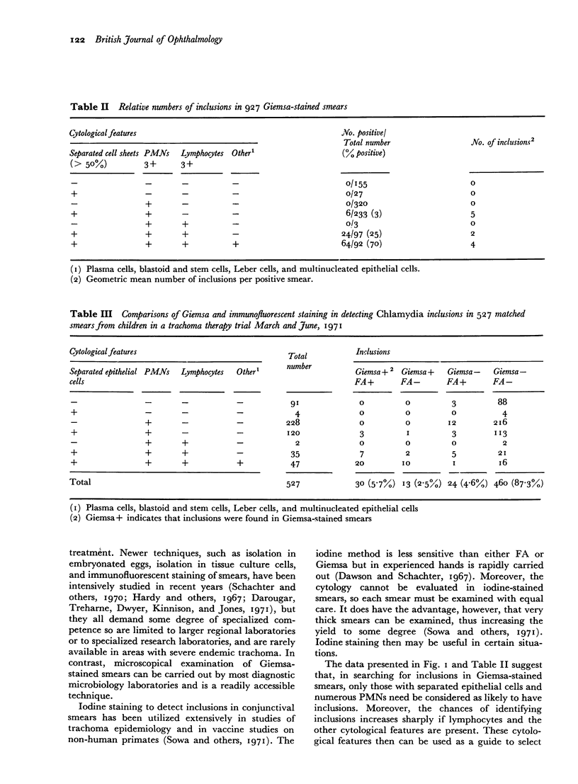
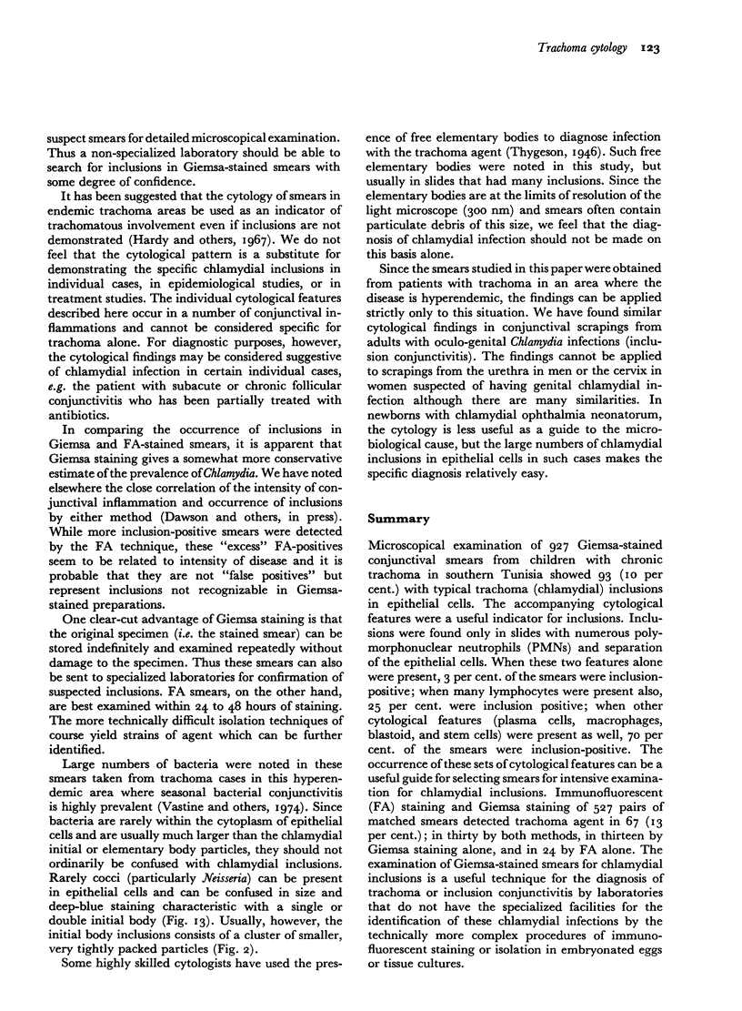
Microscopical examination of 927 Giemsa-stained conjunctival smears from children with chronic trachoma in southern Tunisia showed 93 (10 per cent.) with typical trachoma (chlamydial) inclusions in epithelial cells. The accompanying cytological features were a useful indicator for inclusions. Inclusions were found only in slides with numerous polymorphonuclear neutrophils (PMNs) and separation of the epithelial cells. When these two features alone were present, 3 per cent. of the smears were inclusion-positive; when many lymphocytes were present also, 25 per cent. were inclusion positive; when other cytological features (plasma cells, macrophages, blastoid, and stem cells) were present as well, 70 per cent. of the smears were inclusion-positive. The occurrence of these sets of cytological features can be a useful guide for selecting smears for intensive examination for chlamydial inclusions. Immunofluorescent (FA) staining and Giemsa staining of 527 pairs of matched smears detected trachoma agent in 67 (13 per cent.); in thirty by both methods, in thirteen by Giemsa staining alone, and in 24 by FA alone. The examination of Giemsa-stained smears for chlamydial inclusions is a useful technique for the diagnosis of trachoma or inclusion conjunctivitis by laboratories that do not have the specialized facilities for the identification of these chlamydial infections by the technically more complex procedures of immunofluorescent staining or isolation in embryonated eggs or tissue cultures.
Full text
PDF
Images in this article
Selected References
These references are in PubMed. This may not be the complete list of references from this article.
- Dawson C. R., Daghfous T., Messadi M., Hoshiwara I., Vastine D. W., Yoneda C., Schacter J. Severe endemic trachoma in tunisia. II. A controlled therapy trial of topically applied chlortetracycline and erythromycin. Arch Ophthalmol. 1974 Sep;92(3):198–203. doi: 10.1001/archopht.1974.01010010206003. [DOI] [PubMed] [Google Scholar]
- Gordon F. B., Harper I. A., Quan A. L., Treharne J. D., Dwyer R. S., Garland J. A. Detection of Chlamydia (Bedsonia) in certain infections of man. I. Laboratory procedures: comparison of yolk sac and cell culture for detection and isolation. J Infect Dis. 1969 Oct;120(4):451–462. doi: 10.1093/infdis/120.4.451. [DOI] [PubMed] [Google Scholar]
- Hardy D., Surman P. G., Howarth W. H. A system of representation of cytologic features of external eye infections with special reference to trachoma. Am J Ophthalmol. 1967 May;63(5 Suppl):1535–1537. doi: 10.1016/0002-9394(67)94143-8. [DOI] [PubMed] [Google Scholar]
- Schachter J., Dawson C. R., Balas S., Jones P. Evaluation of laboratory methods for detecting acute TRIC agent infection. Am J Ophthalmol. 1970 Sep;70(3):375–380. doi: 10.1016/0002-9394(70)90097-8. [DOI] [PubMed] [Google Scholar]
- Schachter J. Recommended criteria for the identification of trachoma and inclusion conjunctivitis agents. J Infect Dis. 1970 Jul-Aug;122(1):105–107. doi: 10.1093/infdis/122.1-2.105. [DOI] [PubMed] [Google Scholar]
- Sowa J., Collier L. H., Sowa S. A comparison of the iodine and fluorescent antibody methods for staining trachoma inclusions in the conjunctiva. J Hyg (Lond) 1971 Dec;69(4):693–708. doi: 10.1017/s0022172400021963. [DOI] [PMC free article] [PubMed] [Google Scholar]
- Vastine D. W., Dawson C. R., Daghfous T., Messadi M., Hoshiwara I., Yoneda C., Nataf R. Severe endemic trachoma in Tunisia. I. Effect of topical chemotherapy on conjunctivitis and ocular bacteria. Br J Ophthalmol. 1974 Oct;58(10):833–842. doi: 10.1136/bjo.58.10.833. [DOI] [PMC free article] [PubMed] [Google Scholar]


