

Abstract
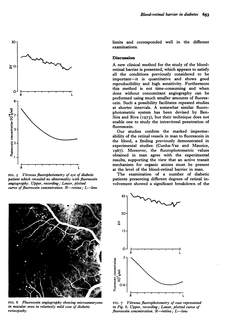
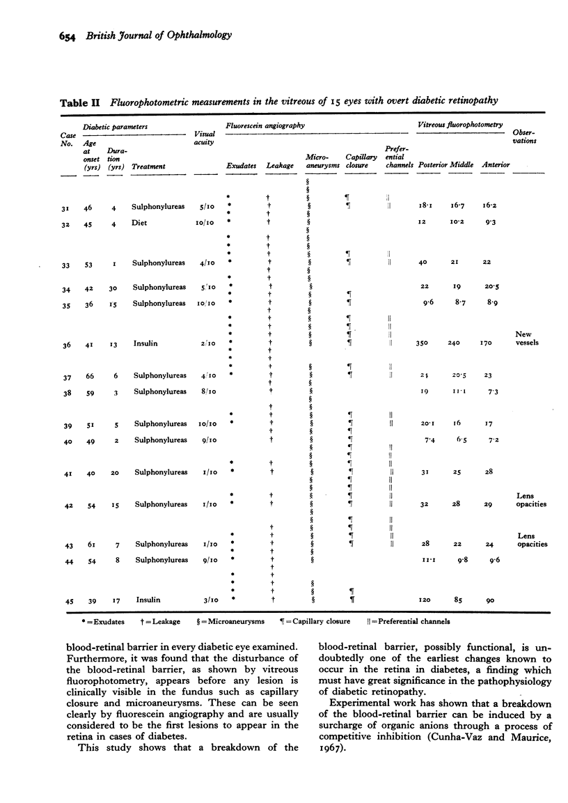
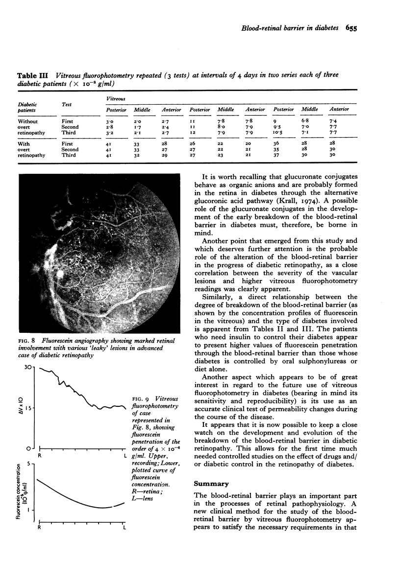
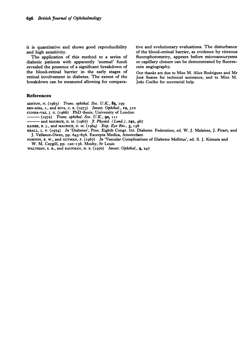
The blood-retinal barrier plays an important part in the processes of retinal pathophysiology. A new clinical method for the study of the blood-retinal barrier by vitreous fluorophotometry appears to satisfy the necessary requirements in that it is quantitative and shows good reproducibility and high sensitivity. The application of this method to a series of diabetic patients with apparently "normal" fundi revealed the presence of a significant breakdown of the blood-retinal barrier in the early stages of retinal involvement in diabetes. The extent of the breakdown can be measured allowing for comparative and evolutionary evaluations. The disturbance of the blood-retinal barrier, as evidence by vitreous fluorophotometry, appears before microaneurysms or capillary closure can be demonstrated by fluorescein angiography.
Full text
PDF
Images in this article
Selected References
These references are in PubMed. This may not be the complete list of references from this article.
- Ashton N. Bowman lecture. The blood-retinal barrier and vaso-glial relationships in retinal disease. Trans Ophthalmol Soc U K. 1965;85:199–230. [PubMed] [Google Scholar]
- Ben-Sira I., Riva C. E. Fluorophotometric recording of fluorescein dilution curves in human retinal vessels. Invest Ophthalmol. 1973 Apr;12(4):310–312. [PubMed] [Google Scholar]
- Cunha-Vaz J. G., Maurice D. M. The active transport of fluorescein by the retinal vessels and the retina. J Physiol. 1967 Aug;191(3):467–486. doi: 10.1113/jphysiol.1967.sp008262. [DOI] [PMC free article] [PubMed] [Google Scholar]
- Waltman S. R., Kaufman H. E. A new objective slit lamp fluorophotometer. Invest Ophthalmol. 1970 Apr;9(4):247–249. [PubMed] [Google Scholar]