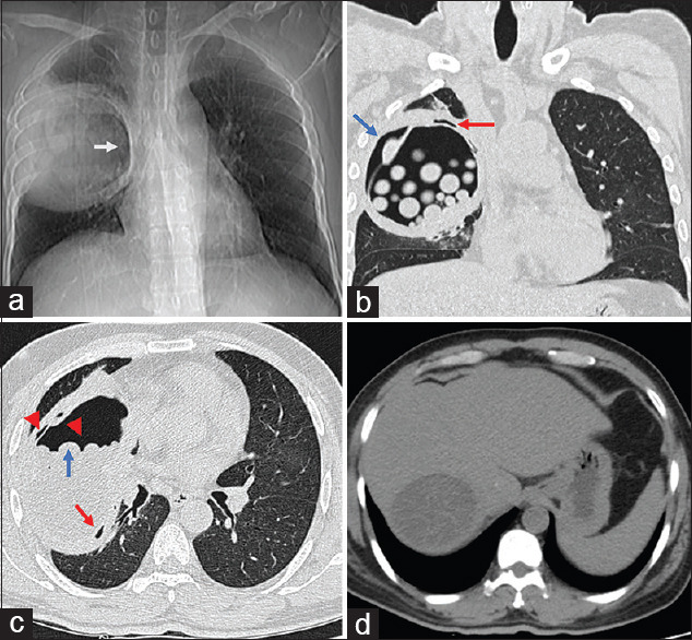
A 48-year-old male patient presented with productive cough, dyspnea and abdominal pain of 2 months duration. He was diagnosed with diabetes mellitus 6 months ago and had not been regular with his medications. Clinical examination revealed a respiratory rate of 18 per min with room air saturation of 95%, and a paucity of movements with bronchial breath sounds identified on inspection and auscultation, respectively, over the right hemithorax. Chest skiagram and computed tomography images of his thorax are shown below [Figure 1].
Figure 1.

(a) shows chest skiagram and (b–d) shows high-resolution computed tomography images of the chest. A description of the images is in the accompanying text
Question: What is the diagnosis?
Answer: Pulmonary and hepatic hydatid disease.
DISCUSSION
The chest skiagram [Figure 1a] revealed a well-defined round radio-opacity in the right mid zone with a crescentic hyperlucent medial margin, called the crescent sign (white arrow). Figure 1b is a coronal view of high-resolution computed tomography (HRCT) scan of the chest lung window, which shows a large cyst in the right lung, with multiple small rounded opacities with fluid attenuation, some of which appear to be floating within it, appearing like a sac of marbles. This is consistent with a diagnosis of a hydatid cyst with multiple daughter cysts within it. There are pockets of air seen between the outer pericyst and inner endocyst, aptly named ‘air-crescent sign’ (blue arrow) and ‘signet ring sign’ (red arrow), which suggest a ruptured or complicated hydatid cyst. Figure 1c, the sagittal section of the HRCT scan of the chest lung window at the level of the aortic outflow tract, shows the ruptured hydatid cyst in the right lung. The daughter cysts have collected in the dependent area of the larger cyst, with some appearing to ‘rise’ over the rest, or the ‘rising-sun sign’ (blue arrow). Also seen is a small air pocket inferiorly (red arrow) between the pericyst and endocyst, called the ‘air-bubble sign’. The presence of air on either side of the endocyst (red arrowheads) gives the appearance of an ‘onion-peel sign’, or the ‘cumbo sign’. Figure 1d shows a large well-defined homogenous cyst in the right lobe of the liver.[1]
Blood investigations revealed uncontrolled blood sugar levels. Armed with the characteristic radiology above, and supplemented by a positive serum latex agglutination test for Echinococcus antigen, he was diagnosed with pulmonary and hepatic hydatid disease. He was started on oral albendazole and subsequently underwent pericystectomy of lung cyst along with partial pericystectomy of cyst in the liver. Post-surgery, he is at present asymptomatic and is on regular follow-up.
Hydatid disease is a zoonosis, commonly caused by the accidental ingestion of ova of the tapeworm Echinococcus granulosus, which subsequently develop into larvae in the host intestine and migrate to other parts of the body where they develop into embryos, with the liver and lung being the most commonly involved organs.[1] Characteristic thoracic radiology should lead clinicians to suspect pulmonary hydatid disease, especially when dealing with patients with non-resolving pneumonia or lung mass.
Financial support and sponsorship
Nil.
Conflicts of interest
There are no conflicts of interest.
REFERENCE
- 1.Garg MK, Sharma M, Gulati A, Gorsi U, Aggarwal AN, Agarwal R, et al. Imaging in pulmonary hydatid cysts. World J Radiol. 2016;8:581–7. doi: 10.4329/wjr.v8.i6.581. [DOI] [PMC free article] [PubMed] [Google Scholar]


