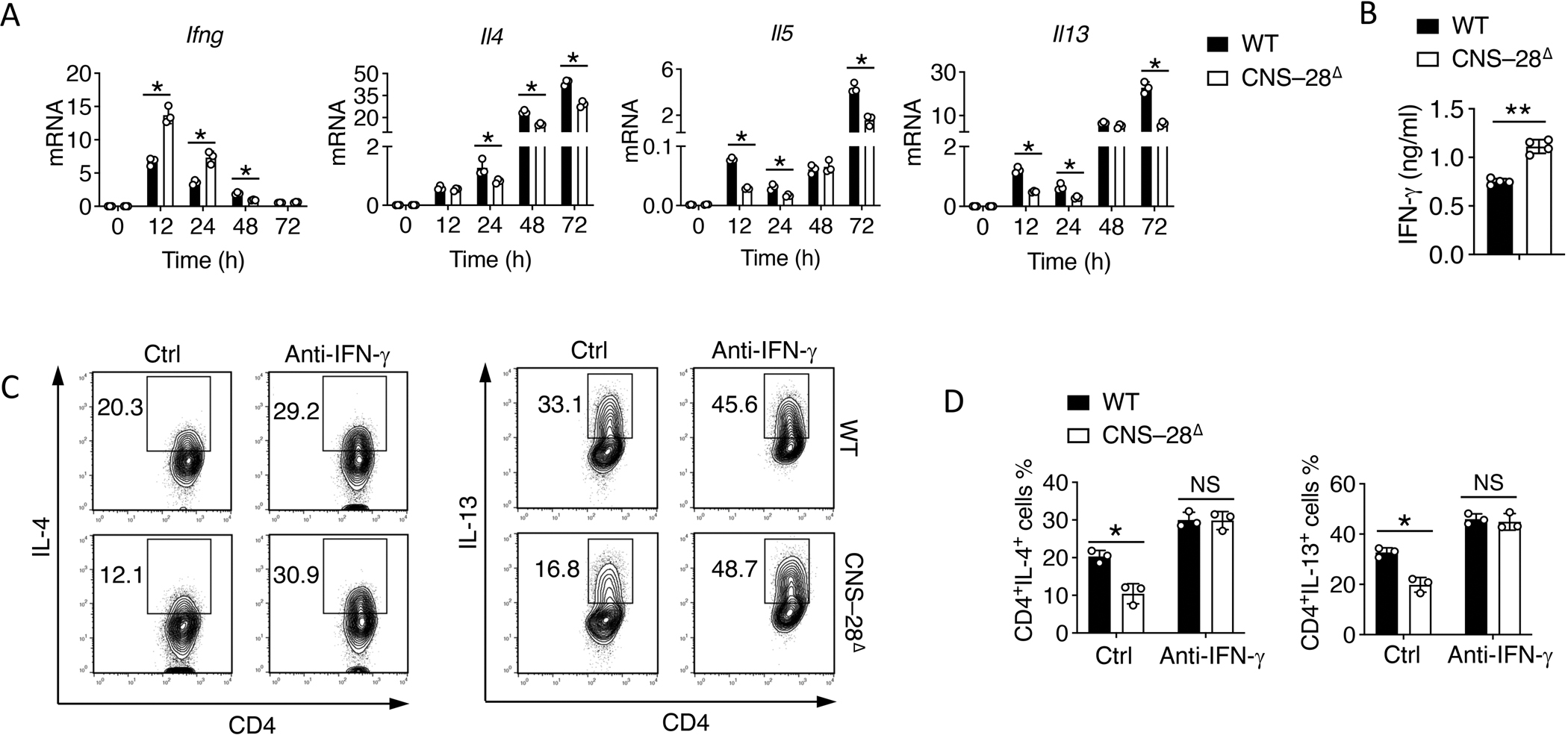
Fig 7. CNS–28 represses type 2 responses due to enhanced IFN-γ production.

(A) qPCR analysis of mRNA during differentiation of WT and CNS–28Δ naïve CD4+ T cells under Th2 conditions for 12, 24, 48 or 72 h (horizontal axis); results are presented relative to those of Gapdh.
(B) ELISA assessment of IFN-γ in the culture medium in WT and CNS–28Δ differentiated Th2 cells.
(C-D) Activated WT and CNS–28Δ naive CD4+ T cells were stimulated with combinations of IL-4 with anti-IFN-γ antibody for 3 days. Intracellular staining of type 2 cytokines was measured by (C) flow cytometry and (D) Quantification.
Data are representative of at least two independent experiments (A-D). *p < 0.05, ***p < 0.01, NS: not statistically significant. (A, D, Two-way ANOVA with Tukey’s multiple comparison test; B, Student’s t test, error bars represent SD).
