Abstract
Objectives:
The purpose of this study was to investigate the effect of Olea europaea L. leaf extract (OLE) on senescence and senescence-associated secretory phenotype (SASP) caused by temozolomide (TMZ) in glioblastoma (GB).
Materials and Methods:
A senescence β-galactosidase assay and a colony formation assay were used to determine the effects of OLE, TMZ, and OLE + TMZ on the cellular senescence and aggressiveness of GB cell lines T98G and U87MG. mRNA expression levels of p53, a senescence factor, interleukin (IL)-6, matrix metalloproteinases (MMP)-9, and nuclear factor kappa B1 (NF-κB1) as SASP factors and Bcl-2 and Bax as senolytic markers were assessed using quantitative reverse transcription-real-time polymerase chain reaction. Cells were double-stained with acridine orange and propidium iodide to observe the cell morphology.
Results:
TMZ increased the senescence rate of GB cells (p<0.001). Besides, OLE + TMZ reduced the proportion of senescent cells (p<0.001) and their capability to form colonies compared to TMZ-only-treated cells. Additionally, OLE + TMZ co-treatment elevated the mRNA expression levels of MMP-9, IL-6, NF-κB1, p53, and the Bax/Bcl-2 ratio compared to TMZ-only treatment. Especially in U87MG cells, involvement of OLE in TMZ treatments increased more than six times in the Bax/Bcl-2 ratio compared to TMZ-only, which induced the apoptosis-like morphological features (p<0.0001).
Conclusion:
Collectively, our findings presented the inhibitory effect of OLE on TMZ-mediated SASP-factor production in GB and, accordingly, its potential contribution to elongate the time of recurrence.
Keywords: Glioblastoma, Olea europaea leaf extract, temozolomide, senescence, SASP
INTRODUCTION
Cellular senescence has been recognized as an essential tumor suppressor mechanism with its ability to the cessation of cell division.1,2 On the other hand, recent studies have evidenced a contrasting effect of cellular senescence by promoting tumor growth with stimulating growth factors, matrix proteases, and pro-inflammatory proteins, which are described as senescence-associated secretory phenotype (SASP).2,3,4 Some chemotherapeutic agents, such as 5-fluorouracil, gemcitabine, doxorubicin, irinotecan, and methotrexate, induce cellular senescence.5,6,7,8 It appeared that chemotherapeutic drug-induced aging could be beneficial with its cell proliferation inhibitory effect. However, therapy-induced senescent (TIS) cells give the tumor ability to a future relapse by producing pro-inflammatory and matrix-destroying molecules known as SASP, which alters the tumor cells’ metabolism in a way that reveals an aggressive cell phenotype.9 Therefore, the effect of chemotherapy-induced cellular senescence on tumor progression could be two-sided and might be a reason for acquired therapy resistance and tumor recurrence.
Glioblastoma (GB) is one of the deadliest cancer types and the DNA-methylating drug temozolomide (TMZ) is the most widely used chemotherapy agent for treating GB. TMZ was shown to induce senescence by the specific DNA lesion O6-methylguanine, which fails recognition of DNA damage, activation of the DNA damage response (DDR) pathway, and arrest of cells in the G2-M phase.10,11 Considering the potential of GB cells to escape and become drug-resistant after TMZ administration due to being arrested in senescence, involvement of an additive agent with anti-senescence features in TMZ therapy is expected to reduce the risk of senescence-related symptoms and tumor relapse.12 The therapeutic effect of various natural compounds, such as quercetin, fisetin, and curcumin, and their analogs in age-related diseases was explained with their senescence cell killing and senolytic effects.13 Similarly, oleuropein aglycone was reported to modulate angiogenesis on senescent fibroblasts.14 An individual study showed that oleuropein exhibits senescence inhibitory features by retaining proteasome function during replicative senescence in human embryonic fibroblasts.15 Our previous studies indicated that Olea europaea L. (Oleaceae) leaf extract (OLE), which contains a high amount of oleuropein, increases the therapeutic features of TMZ in GB.16,17,18,19 Although the effect of oleuropein on cellular senescence was described in fibroblast, its effect was not described in GB. Additionally, one of our previous studies indicated that OLE consists of several additional bioactive components, such as secoiridoids, triterpenes, and flavonoids, in trace amounts, and these minor compounds in OLE could play critical anticancer roles.17 However, the effect of OLE on TMZ-induced SASP expression and its effect on tumor progression in GB remains unknown. Therefore, in this study, we investigated the effect of OLE on senescent cells induced by TMZ in GB cell lines with different TMZ sensitivity using in vitro functional analyses. Our findings identified that OLE attenuates TIS cell-promoted expression of proinflammatory SASP factors and attenuated aggressive characteristics in GB cells, independent of their TMZ sensitivity.
MATERIALS AND METHODS
Cell culture and reagents
Human GB TMZ-resistant T98G cells and TMZ-sensitive U87MG cells, and control HUVEC cells, which was used to determine the drug cytotoxicity, were obtained from Medical Biology Department, Faculty of Medicine, Bursa Uludağ University (Bursa, Türkiye). All cell lines were maintained in a Dulbecco’s Modified Eagle’s Medium-F12 (DMEMF12; HyClone, Utah, USA) containing L-glutamine supplemented with 10% fetal bovine serum (FBS, BIOCHROME, Berlin, Germany), 1 mM sodium pyruvate, 100 mg/mL streptomycin, and 100 U/mL penicillin. All cells were incubated in a humidified atmosphere at 37°C and 5% CO2.
The standardized OLE (05.06.2007, 10-00014-00- 015-0) was kindly provided by Kale Naturel (Edremit, Balıkesir, Türkiye) and prepared as described in our previous study.19 An Agilent 1200 High Performance Liquid Chromatography system (Waldbronn, Germany) identified 19.419 mg/mL of oleuropein among the phenolic compounds of the OLE fractions at 280 nm wavelength (Figure 1A).19,20 TMZ was provided by Sigma, USA. Oleuropein was detected as 19.419 mg/mL in the standardized OLE used in this study.
Figure 1.
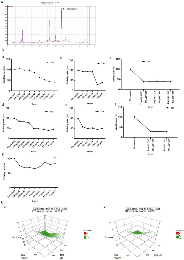
(A) The oleuropein content of OLE. The oleuropein concentration of OLE was detected using HPLC/DAD analyses. (B) T98G and U87MG cells viability rates of determined after treatment with OLE, TMZ, and OLE + TMZ and HUVEC cell viability rates of determined after treatment with OLE for 24 h. OLE (a), TMZ (b), OLE + TMZ (c) treatment in T98G. OLE (d), TMZ (e), OLE + TMZ (f) treatment in U87MG. OLE (g) treatment in HUVEC (p<0.001, one-sample t-test). (C) The combined effect of OLE and TMZ in T98G and U87MG cell lines. The additive effects of OLE and TMZ were detected according to the zero interaction strength synergy scoring system. The effect of OLE + TMZ in T98G cells is shown in “a”, and in U87MG cells was shown in “b”
OLE: Olea europaea leaf extract, HPLC/DAD: High performance liquid chromatography/Diode-array detection, TMZ: Temozolomide
Investigation of cell cytotoxicity and proliferation
The cytotoxicity of OLE and TMZ and their effect on cell proliferation was assessed using a cell proliferation kit (WST-1, Roche Applied Sciences, Mannheim, Germany) as described previously.18 The inhibitory concentrations at which 50% of the cells die (IC50) were selected to treat cells for the experiment set up. The possible additive, antagonist, and synergistic effects of OLE on TMZ treatment were calculated using a web-based tool, i.e. SynergyFinder (version 2.0).21
Apoptosis induction assay [fluorescent microscopy; acridine orange (AO)/propidium iodide (PI) double staining]
The effect of OLE on morphological features of T98G and U87MG cells was assessed by AO/PI, a double-fluorescent dye staining method.22,23 The morphology of the cells was analyzed with an inverted fluorescent microscope (Olympus, Tokyo, Japan). Cells with intact green nuclei were considered viable; cells with dense green chromatin condensation areas in the nucleus were considered early apoptotic, cells with dense orange chromatin condensation areas were considered late apoptotic, and cells with intact orange nuclei were considered secondary necrotic.22,23
Evaluation of tumor aggressiveness
The CellMATM Clonogenic assay kit (BioPioneer, USA) was used to determine the effect of OLE on colony formation in T98G and U87MG cells. The blue-colored colonies were counted under an inverted microscope under 20X magnification.
Detection of senescence-associated β-galactosidase
The rate of senescence in GB cell lines was detected using a senescence β-galactosidase staining kit (9860, Cell Signaling Technology, Danvers, MA, USA), according to the manufacturer’s instructions. The blue-colored cells were visualized and counted under light microscopy using a 10X magnification.
Examination of the expression levels of senescence factors
Total RNA was isolated using a Zymo Research RNA isolation kit (Thermo Fisher Scientific, Glasgow, UK). RNA quantity and quality were assessed using a NanoDrop 2000 Spectrophotometer (Beckman Coulter, California, USA). RNA samples with a total concentration between 200 and 400 ng/µL were selected for cDNA synthesis (High-Capacity cDNA Reverse Transcription Kit; Thermo Fisher Scientific, Glasgow, UK). Quantitative reverse transcription-real-time polymerase chain reaction (RT-qPCR) analysis was performed using TaqMan gene expression assays specific to the genes related to senescence factors [p53(DM02154335-g1)], SASP factors [interleukin (IL)-6 (Hs00174131_m1), matrix metalloproteinases (MMP)-9 (Hs00957562_m1) and nuclear factor kappa B1 (NF-κB1) (Hs00428211-m1)] and senolytic effect [Bcl-2 (Hs00608023_m1) and Bax (Hs00180269 _ m1)]. Expression results were normalized to the expression of a housekeeping gene GAPDH (Hs00957562_m1). The threshold cycle for each RNA expression was determined using a StepOne RT-PCR system (Applied Biosystems, Warrington, UK). 2-ΔCt Method calculated the fold change in RNA expression.
Statistical analysis
One-Way ANOVA was used to evaluate the findings of the WST-1 assay, and independent t-test analyses evaluated the difference in the number of cell colonies. The independent t-test analyzed the findings of the senescence-associated β-galactosidase assay. The independent t-test evaluated the findings of RT-qPCR. Data are presented as mean ± standard error of mean. Significance was established at a value of p<0.05. All statistical analyzes were performed using the IBM SPSS Statistics for Windows, version 23.0 (IBM Corp., Armonk, NY, USA). The findings were interpreted as graphs using the GraphPad Prism 6 (GraphPad Software Inc, San Diego, California, USA).
RESULTS
OLE inhibits the proliferation of GB cells
Previously determined effective concentrations of OLE and TMZ were confirmed using current OLE extract in laboratory conditions. OLE, TMZ, and their co-treatment reduced tumor cell proliferation in GB cell lines, T98G, and U87MG, similar to our previous findings, the effective doses of OLE + TMZ treatments were determined as 2 mg/mL OLE and 400 µM TMZ for T98G cells, while they were 1 mg/mL OLE and 300 µM TMZ for U87MG during 24 h of incubation time (Figure 1B, a-f).24 In addition, non of the applied concentrations of OLE caused a notable cytotoxic effect on HUVEC, a non-tumor endothelial cell line (Figure 1B, g).24 Therefore, to investigate the effect of OLE and TMZ on senescence and aggressiveness of GB cell lines, 2 mg/mL OLE and 400 µM TMZ were used for T98G, while 1 mg/mL OLE and 300 µM TMZ were used for U87MG cells for the remaining in vitro analysis. According to the findings, OLE + TMZ treatment showed an additive effect in T98G (SC: -8.253) and U87MG (SC: -2.078) (Figure 1C) and increased the number of cells with apoptotic morphology compared to TMZ-only treatment, suggesting that OLE provokes cell death independent from TMZ sensitivity of GB cells (Figure 2).
Figure 2.
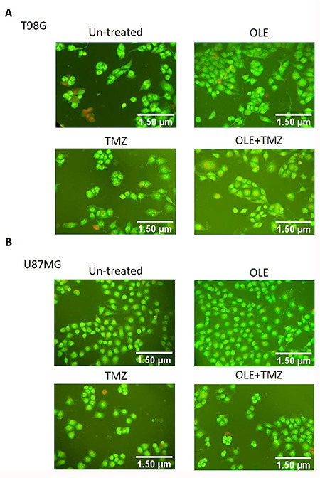
The effect of OLE and TMZ on GB cell morphology. Findings of AO/PI staining showed that OLE and OLE + TMZ increased the apoptosis rate in T98G (A) and U87MG cells (B). Living cells: cells with intact green nuclei; early apoptotic cells: cells with dense green chromatin condensation areas in the nucleus; late apoptotic cells: cells with dense orange chromatin condensation areas; secondary necrotic cells: cells with intact orange nuclei
OLE: Olea europaea leaf extract, TMZ: Temozolomide, AO: Acridine orange, PI: Propidium iodide
OLE inhibits GB tumor aggressiveness
OLE, TMZ, and their co-treatment led to a significant decrease in the number of tumor colonies of both T98G and U87MG cells compared with untreated cells (Figure 3). The number of cell colonies was 216 in untreated T98G cells, while it decreased to 38 after OLE-only, 98 after TMZ-only, and 36 after OLE + TMZ treatments (p<0001, Figure 3A). Additionally, the number of colonies was 235 in untreated U87MG cells, and OLE-only, TMZ-only, and OLE + TMZ treatments decreased it to 49, 64, and 46, respectively (p<0.001, Figure 3B), suggesting that OLE-only treatment tented to reduce the number of colonies than that of TMZ-only treatment in both T98G and U87MG cells. Additionally, the co-treatment of OLE + TMZ reduced the colony formation capacity of cells compared to TMZ-only-treated T98G cells. OLE + TMZ treatment 2.84 fold reduced the number of colonies formed by T98G cells compared to the number of colonies after TMZ-only treatment (p<0.001). Moreover, after alone and complementary usage of OLE, the colonies’ size was smaller than those of untreated and TMZ-only-treated T98G cells. Interestingly, in U87MG, while alone usage of OLE reduced the size of colonies compared to untreated cells, it did not affect the size of TMZ-treated cells (Figure 3).
Figure 3.
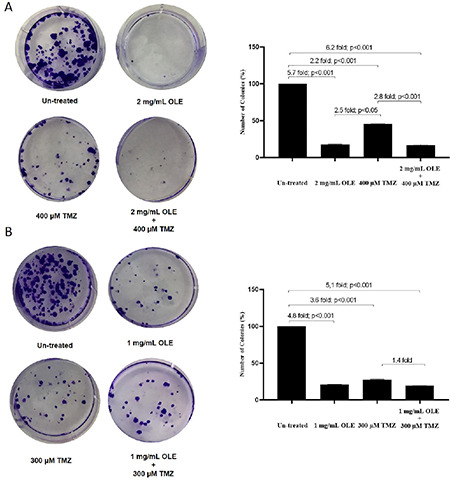
Effects of OLE, TMZ, and OLE + TMZ combination on colony formation of GB cells. in T98G cells OLE leaded 6.25-fold (p<0.001); TMZ: 2.20-fold (p<0.001) and OLE + TMZ 5.71-fold (p<0.001) (A) in U87MG cells OLE leaded 4.8-fold (p<0.001); TMZ: 3.60-fold (p<0.001) and OLE + TMZ 5.1 (p<0.001) (B) fold decrease compared to those of untreated cells. P values were calculated using an independent t-test (n: 3)
OLE: Olea europaea leaf extract, TMZ: Temozolomide, GB: Glioblastoma
OLE reduces the senescence caused by TMZ
Untreated senescent cells were 0.8% in T98G and 3.1% in U87MG cells, respectively (Table 1). TMZ-only treatment caused a significant increase in the number of senescent cells in both T98G and U87MG cells compared with those of untreated cells (the rate of senescent cells after TMZ treatment in T98G cells: 47%, p<0.001; in U87MG cells: 13.4%, p<0.001). Although OLE-only treatment resulted in more senescent cells in both T98G and U87MG cells, this increase was lower than that after TMZ-only treatment (OLE-only treatment, 11% induced senescent cells in T98G cells and 5.1% in U87MG cells). Likewise, while OLE + TMZ treatment increased the number of senescent cells compared to untreated cells (in T98G cells: 30%, p<0.001; in U87MG: 4.5%, p<0.001, compared to untreated cells), the rate of this increase was lower than that of caused by TMZ-only treatment (p<0.0001; Figure 4). These findings showed that the involvement of OLE in the TMZ treatment reduced the senescence-provoking capacity of TMZ (Figure 4).
Table 1. The effect of combination of OLE, TMZ, and OLE + TMZ on cell aging in T98G and U87MG cells.
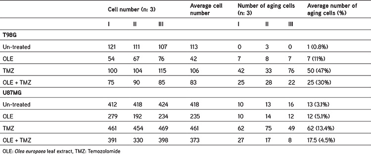
Figure 4.
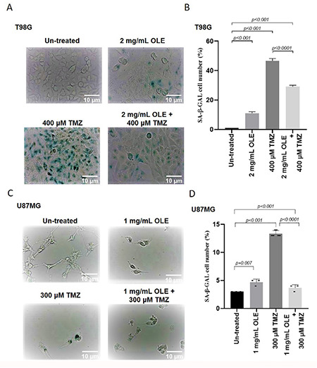
Effects of OLE and TMZ on cellular senescence in T98G (A) and U87MG (B) (a light microscope was analyzed using 20X objectives for T98G cells and 10X objectives for U87MG cells). A comparison of the effects of OLE and TMZ treatments on senescence in T98G (A) and U87MG cells (B)
OLE: Olea europaea leaf extract, TMZ: Temozolomide
OLE suppresses the expression of SASP and senescence factors in GB cells
TMZ-only treatment increased the mRNA expression levels of SASP-related genes, IL-6, NF-κB1, and MMP-9, compared with untreated cells (TMZ induced IL-6: 2.03-fold; p=0.003, NF-κB1: 3.26-fold; p<0.001 and MMP-9: 6.14-fold; p<0.001; Figure 5). In contrast, OLE-only treatment slightly increased MMP-9 (1.54-fold, p<0.001) and did not affect NF-κB1 and IL-6. Additionally, a co-treatment with OLE + TMZ significantly attenuated the mRNA expression of these genes (OLE + TMZ reduced IL-6: 1.81-fold; p=0.017, NF-κB1: 2.35-fold; p<0.001 and MMP-9: 3.07-fold; p<0.0001 compared to TMZ-only treatment; Figure 5). Similarly, TMZ-only treatment significantly induced the mRNA expression of IL-6 (5.73-fold; p<0.0001), NF-κB1 (8.83-fold; p<0.0001), and MMP-9 (6.00-fold; p<0.001) in U87MG cells, compared with untreated cells. Additionally, OLE-only treatment decreased the expression of IL-6 (2.22-fold; p<0.003) and NF-κB1 (3.1-fold; p=0.001) and did not affect MMP-9 compared with untreated cells. A co-treatment with OLE + TMZ significantly reduced the expression of these genes compared with TMZ-only treatment (OLE + TMZ reduced IL-6: 14.3-fold; p<0.0001, NF-κB1: 13.23-fold; p<0.0001 and MMP-9: 2.21-fold; p<0.001; Figure 5). These findings showed that a mechanism of OLE to reduce GB tumor aggressive phenotype is by reducing the expression of TMZ-induced SASP factors.
Figure 5.
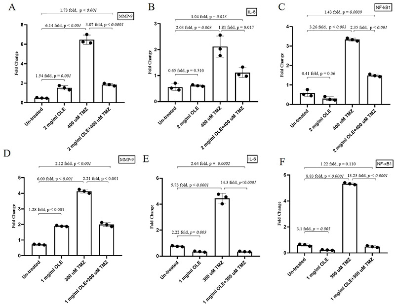
The effect of T98G and U87MG cells treated with OLE, TMZ, and OLE + TMZ on the expression levels of SASP factors. MMP-9 (A), IL-6 (B), and NF-κB1 (C) expression levels in T98G. MMP-9 (D), IL-6 (E), and NF-κB1 (F) expression levels in U87MG
OLE: Olea europaea leaf extract, TMZ: Temozolomide, SASP: Senescence-associated secretory phenotype, MMP: Matrix metalloproteinase, IL: Interleukin, NF-κB1: Nuclear factor kappa B1
Beside the reducing effect on SASP factors, OLE + TMZ significantly reduced the expression level of p53, a senescence factor, whereas it induced the ratio of Bax/Bcl-2, a senolytic effective factor compared to TMZ-only in both T98G and U87MG cells (OLE + TMZ reduced p53: 1.27-fold; p=0.002, and Bcl-2: 1.45-fold; p=0.002, while it induced Bax: 2.02-fold; p=0.0007 and Bax/Bcl-2: 1.3-fold p>0.05 in T98G and it reduced p53: 4.66-fold; p<0.0001, and Bcl-2: 1.47-fold; p=0.0001 and induced Bax: 7.75-fold; p<0.0001 and Bax/Bcl-2: 6-fold; p<0.0001 in U87MG compared to TMZ) (Figure 6). These data provide evidence of the diverting effect of OLE on apoptosis on senescent GB cells due to TMZ treatment.
Figure 6.
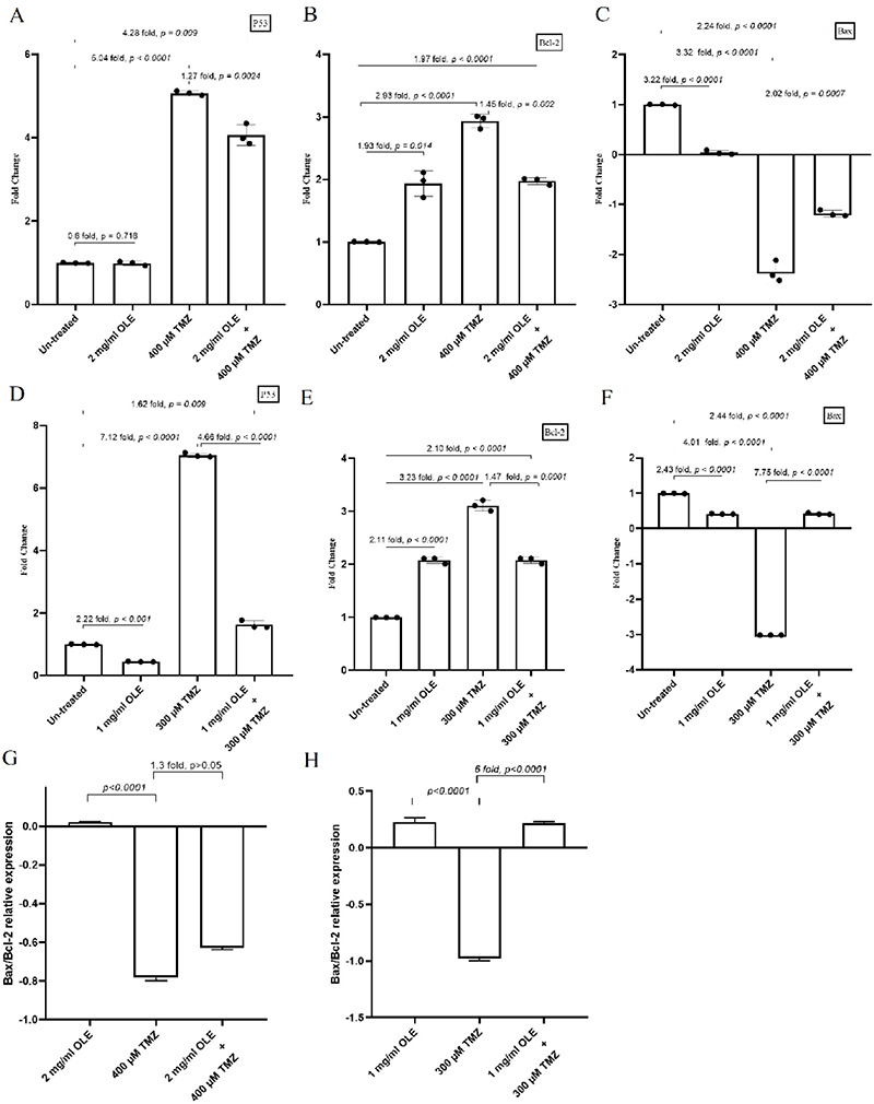
The effect of T98G and U87MG cells treated with OLE, TMZ, and OLE + TMZ on the expression levels of senescence factors and senolytic effect. p53 (A), Bcl-2 (B), Bax (C) expression levels in T98G. p53 (D), Bcl-2 (E), and Bax (F) expression levels in U87MG. Bax/Bcl-2 relative expression of T98G (G) and U87MG (H) cells treated with OLE, TMZ, and OLE + TMZ
OLE: Olea europaea leaf extract, TMZ: Temozolomide
DISCUSSION
Cellular senescence arrests the cell cycle by phosphorylating p53 and the expression of p21.2,3,4 One of our previous studies showed that TMZ-only treatment induces mRNA expression of p53 in GB tumors.19 Confirming our previous findings, TMZ-only treatment induced P53 expression in T98G and U87MG cell lines in this study. Although the well-described function of p53 is activating apoptosis,25 recent studies highlighted its regulatory role in cellular senescence via DDR activation.26,27,28 Studies evidenced that p53 could stimulate apoptosis as a response to overwhelming stress, while it could stimulate senescence as a response to less severe damage by failing to induce pro-apoptotic factors and leading to over-expression of the pro-survival gene Bcl-2.29,30,31 A very recent study of Aasland et al.11 linked TMZ-induced p53 expression to initiation of senescence in O6-methylguanine DNA-methyltransferase (MGMT) expressing GB cell lines. Although MGMT is expressed in T98G cells, it is absent in U87MG.32 In this study, after TMZ-only treatment, P53 and Bcl-2 expression were induced in these cell lines. Unlike apoptosis, p53-initiated cellular senescence produces diverse bioactive factors SASP.33 Activation of SASP requires a NF-κB and C/EBPβ pathways-mediated sustained DDR. NF-κB regulates the production of IL-6, which plays a role in the maintenance and propagation of the SASP response in the tumor microenvironment.34,35 Besides, NF-κB leads to transcriptional activation of pro-inflammatory cytokines and MMPs, such as MMP-9, a key modulator of tumor aggressiveness, in GB.36,37 The findings of this study displayed that TMZ-only treatment induced the expression levels of NF-κB, IL-6, and MMP9. Additionally, SA-β-gal activity, which was mostly detected in senescent cells,38 was induced after TMZ-only treatment in both cells, suggesting that TMZ-induced p53 dependent cellular senescence may be independent of MGMT in GB.
Conversely, while OLE-only did not affect p53 and slightly induced BCL2 in MGMT expressing T98G cells, it reduced p53 and had a higher capacity to induce Bcl-2 in MGMT-methylated U87MG cells. These findings indicate that the level of MGMT expression affects the expression of p53 in GB. According to our previous studies, OLE induces a methylation level of MGMT, while reducing p53 and promoting apoptosis.39 Similarly, in this study, the complementary usage of OLE with TMZ increased the Bax/Bcl-2 ratio, which is widely used to predict the cells undergoing apoptosis.
Evidence indicated that cellular senescence promotes tumor aggressiveness and drug resistance.40 However, in this study, OLE reduces the colony-forming ability of GB cells compared to TMZ-only-treated cells, suggesting that OLE promotes apoptosis rather than senescence. Supporting this, based on the current findings, OLE did not affect NF-κB and IL-6 in T98G cells and reduced the expression of these genes in U87MG cells. Additionally, upon senescence induced by TMZ, OLE reduced the senescence markers, including NF-κB, IL-6, and MMP-9, and β-galactosidase staining in both GB cell lines independent of MGMT expression level. Therefore, these findings provide evidence of the effect of OLE on directing TIS-GB cells to apoptosis.
CONCLUSION
This study uniquely exhibited that the enhancing effect of OLE on TMZ sensitivity in GB tumors might be associated with its attenuating capacity to TMZ-induced senescent. Advanced studies are recommended for a better understanding of the effecting mechanism of OLE on TIS and to exhibit the potential of OLE to be a complementary therapy as a senolytic agent for patients with GB.
Acknowledgments
We thank Prof. Ferah Budak from Bursa Uludağ University, Faculty of Medicine, Immunology Department and Assoc. Prof. Senem Kamiloğlu Beştepe from Bursa Uludağ University, Faculty of Agriculture Department of Food Sciences for their assistance in the experimental process. This work was supported by the Scientific and Technical Research Council of Türkiye (TÜBİTAK) under grant no: 118S799. One of the authors (M.E.) is a 100/2000 The Council of Higher Education Ph. D. Scholars in Molecular Biology and Genetics (Gene Therapy and Genome Studies).
Footnotes
Ethics
Ethics Committee Approval: Not applicable.
Informed Consent: Not applicable.
Peer-review: Externally peer-reviewed.
Authorship Contributions
Concept: M.E., B.T., Design: M.E., B.T., S.A.A., Data Collection or Processing: M.E., B.T., Ç.T., Analysis or Interpretation: M.E., Ç.T., Literature Search: M.E., B.T., S.A.A., Ç.T., G.T., Writing: M.E., B.T.
Conflict of Interest: No conflict of interest was declared by the authors.
Financial Disclosure: The authors declared that this study received no financial support.
References
- 1.Schmitt R. Senotherapy: growing old and staying young? Pflugers Arch. 2017;469:1051–1059. doi: 10.1007/s00424-017-1972-4. [DOI] [PubMed] [Google Scholar]
- 2.Foster SA, Wong DJ, Barrett MT, Galloway DA. Inactivation of p16 in human mammary epithelial cells by CpG island methylation. Mol Cell Biol. 1998;18:1793–1801. doi: 10.1128/mcb.18.4.1793. [DOI] [PMC free article] [PubMed] [Google Scholar]
- 3.Noda A, Ning Y, Venable SF, Pereira-Smith OM, Smith JR. Cloning of senescent cell-derived inhibitors of DNA synthesis using an expression screen. Exp Cell Res. 1994;211:90–98. doi: 10.1006/excr.1994.1063. [DOI] [PubMed] [Google Scholar]
- 4.Coppé JP, Desprez PY, Krtolica A, Campisi J. The senescence-associated secretory phenotype: the dark side of tumor suppression. Annu Rev Pathol. 2010;5:99–118. doi: 10.1146/annurev-pathol-121808-102144. [DOI] [PMC free article] [PubMed] [Google Scholar]
- 5.Chakrabarty A, Chakraborty S, Bhattacharya R, Chowdhury G. Senescence-induced chemoresistance in triple negative breast cancer and evolution-based treatment strategies. Front Oncol. 2021;11:674354. doi: 10.3389/fonc.2021.674354. [DOI] [PMC free article] [PubMed] [Google Scholar]
- 6.Altieri P, Murialdo R, Barisione C, Lazzarini E, Garibaldi S, Fabbi P, Ruggeri C, Borile S, Carbone F, Armirotti A, Canepa M, Ballestrero A, Brunelli C, Montecucco F, Ameri P, Spallarossa P. 5-fluorouracil causes endothelial cell senescence: potential protective role of glucagon-like peptide 1. Br J Pharmacol. 2017;174:3713–3726. doi: 10.1111/bph.13725. [DOI] [PMC free article] [PubMed] [Google Scholar]
- 7.Song Y, Baba T, Mukaida N. Gemcitabine induces cell senescence in human pancreatic cancer cell lines. Biochem Biophys Res Commun. 2016;477:515–519. doi: 10.1016/j.bbrc.2016.06.063. [DOI] [PubMed] [Google Scholar]
- 8.Bojko A, Czarnecka-Herok J, Charzynska A, Dabrowski M, Sikora E. Diversity of the senescence phenotype of cancer cells treated with chemotherapeutic agents. Cells. 2019;8:1501. doi: 10.3390/cells8121501. [DOI] [PMC free article] [PubMed] [Google Scholar]
- 9.Faheem MM, Seligson ND, Ahmad SM, Rasool RU, Gandhi SG, Bhagat M, Goswami A. Convergence of therapy-induced senescence (TIS) and EMT in multistep carcinogenesis: current opinions and emerging perspectives. Cell Death Discov. 2020;6:51. doi: 10.1038/s41420-020-0286-z. [DOI] [PMC free article] [PubMed] [Google Scholar]
- 10.Villalva C, Cortes U, Wager M, Tourani JM, Rivet P, Marquant C, Martin S, Turhan AG, Karayan-Tapon L. O6-Methylguanine-methyltransferase (MGMT) promoter methylation status in glioma stem-like cells is correlated to temozolomide sensitivity under differentiation-promoting conditions. Int J Mol Sci. 2012;13:6983–6994. doi: 10.3390/ijms13066983. [DOI] [PMC free article] [PubMed] [Google Scholar]
- 11.Aasland D, Götzinger L, Hauck L, Berte N, Meyer J, Effenberger M, Schneider S, Reuber EE, Roos WP, Tomicic MT, Kaina B, Christmann M. Temozolomide induces senescence and repression of DNA repair pathways in glioblastoma cells via activation of ATR-CHK1, p21, and NF-κB. Cancer Res. 2019;79:99–113. doi: 10.1158/0008-5472.CAN-18-1733. [DOI] [PubMed] [Google Scholar]
- 12.Demaria M, O’Leary MN, Chang J, Shao L, Liu S, Alimirah F, Koenig K, Le C, Mitin N, Deal AM, Alston S, Academia EC, Kilmarx S, Valdovinos A, Wang B, de Bruin A, Kennedy BK, Melov S, Zhou D, Sharpless NE, Muss H, Campisi J. Cellular senescence promotes adverse effects of chemotherapy and cancer relapse. Cancer Discov. 2017;7:165–176. doi: 10.1158/2159-8290.CD-16-0241. [DOI] [PMC free article] [PubMed] [Google Scholar]
- 13.Yousefzadeh MJ, Zhu Y, McGowan SJ, Angelini L, Fuhrmann-Stroissnigg H, Xu M, Ling YY, Melos KI, Pirtskhalava T, Inman CL, McGuckian C, Wade EA, Kato JI, Grassi D, Wentworth M, Burd CE, Arriaga EA, Ladiges WL, Tchkonia T, Kirkland JL, Robbins PD, Niedernhofer LJ. Fisetin is a senotherapeutic that extends health and lifespan. EBioMedicine. 2018;36:18–28. doi: 10.1016/j.ebiom.2018.09.015. [DOI] [PMC free article] [PubMed] [Google Scholar]
- 14.Margheri F, Menicacci B, Laurenzana A, Del Rosso M, Fibbi G, Cipolleschi MG, Ruzzolini J, Nediani C, Mocali A, Giovannelli L. Oleuropein aglycone attenuates the pro-angiogenic phenotype of senescent fibroblasts: a functional study in endothelial cells. J Funct Foods. 2019;53:219–226. [Google Scholar]
- 15.Katsiki M, Chondrogianni N, Chinou I, Rivett AJ, Gonos ES. The olive constituent oleuropein exhibits proteasome stimulatory properties in vitro and confers life span extension of human embryonic fibroblasts. Rejuvenation Res. 2007;10:157–172. doi: 10.1089/rej.2006.0513. [DOI] [PubMed] [Google Scholar]
- 16.Tunca B, Tezcan G, Cecener G, Egeli U, Ak S, Malyer H, Tumen G, Bilir A. Olea europaea leaf extract alters microRNA expression in human glioblastoma cells. J Cancer Res Clin Oncol. 2012;138:1831–1844. doi: 10.1007/s00432-012-1261-8. [DOI] [PubMed] [Google Scholar]
- 17.Tezcan G, Aksoy SA, Tunca B, Bekar A, Mutlu M, Cecener G, Egeli U, Kocaeli H, Demirci H, Taskapilioglu MO. Oleuropein modulates glioblastoma miRNA pattern different from Olea europaea leaf extract. Hum Exp Toxicol. 2019;38:1102–1110. doi: 10.1177/0960327119855123. [DOI] [PubMed] [Google Scholar]
- 18.Mutlu M, Tunca B, Ak Aksoy S, Tekin C, Egeli U, Cecener G. Inhibitory effects of Olea europaea leaf extract on mesenchymal transition mechanism in glioblastoma cells. Nutr Cancer. 2021;73:713–720. doi: 10.1080/01635581.2020.1765260. [DOI] [PubMed] [Google Scholar]
- 19.Tezcan G, Tunca B, Bekar A, Budak F, Sahin S, Cecener G, Egeli U, Taskapılıoglu MO, Kocaeli H, Tolunay S, Malyer H, Demir C, Tumen G. Olea europaea leaf extract improves the treatment response of GBM stem cells by modulating miRNA expression. Am J Cancer Res. 2014;4:572–590. [PMC free article] [PubMed] [Google Scholar]
- 20.Kamiloglu S. Effect of different freezing methods on the bioaccessibility of strawberry polyphenols. Int J Food Sci Technol. 2019;54:2652–2660. [Google Scholar]
- 21.Ianevski A, Giri AK, Aittokallio T. SynergyFinder 2 0: visual analytics of multi-drug combination synergies. Nucleic Acids Res. 2020;48:W488–W493. Erratum in: Nucleic Acids Res. 2022;50:7198.. doi: 10.1093/nar/gkaa216. [DOI] [PMC free article] [PubMed] [Google Scholar]
- 22.Ciapetti G, Granchi D, Savarino L, Cenni E, Magrini E, Baldini N, Giunti A. In vitro testing of the potential for orthopedic bone cements to cause apoptosis of osteoblast-like cells. Biomaterials. 2002;23:617–627. doi: 10.1016/s0142-9612(01)00149-1. [DOI] [PubMed] [Google Scholar]
- 23.Anasamy T, Abdul AB, Sukari MA. A phenylbutenoid dimer, cis-3-(3’,4’-dimethoxyphenyl)-4-[(e)-3’’’,4’’’-dimethoxystyryl] cyclohex-1-ene, exhibits apoptogenic properties in T-acute lymphoblastic leukemia cells via induction of p53-independent mitochondrial signalling pathway. Evid-Based Complement Alternat Med. 2013;93:9810. doi: 10.1155/2013/939810. [DOI] [PMC free article] [PubMed] [Google Scholar]
- 24.Mutlu M, Tunca B, Aksoy SA, Tekin C, Cecener G, Egeli U. Olea europaea leaf extract decreases tumour size by affecting the LncRNA expression status in glioblastoma 3D cell cultures. Eur J Integr Med. 2021;45:101345. [Google Scholar]
- 25.Fridman JS, Lowe SW. Control of apoptosis by p53. Oncogene. 2003;22:9030–9040. doi: 10.1038/sj.onc.1207116. [DOI] [PubMed] [Google Scholar]
- 26.Giordano A, Macaluso M. Fenofibrate triggers apoptosis of glioblastoma cells in vitro: new insights for therapy. Cell Cycle. 2012;11:3154. doi: 10.4161/cc.21719. [DOI] [PMC free article] [PubMed] [Google Scholar]
- 27.Surova O, Zhivotovsky B. Various modes of cell death induced by DNA damage. Oncogene. 2013;32:3789–3797. doi: 10.1038/onc.2012.556. [DOI] [PubMed] [Google Scholar]
- 28.Mijit M, Caracciolo V, Melillo A, Amicarelli F, Giordano A. Role of p53 in the regulation of cellular senescence. Biomolecules. 2020;10:420. doi: 10.3390/biom10030420. [DOI] [PMC free article] [PubMed] [Google Scholar]
- 29.Vousden KH, Lane DP. p53 in health and disease. Nat Rev Mol Cell Biol. 2007;8:275–283. doi: 10.1038/nrm2147. [DOI] [PubMed] [Google Scholar]
- 30.Childs BG, Baker DJ, Kirkland JL, Campisi J, van Deursen JM. Senescence and apoptosis: dueling or complementary cell fates? EMBO Rep. 2014;15:1139–1153. doi: 10.15252/embr.201439245. [DOI] [PMC free article] [PubMed] [Google Scholar]
- 31.Tavana O, Benjamin CL, Puebla-Osorio N, Sang M, Ullrich SE, Ananthaswamy HN, Zhu C. Absence of p53-dependent apoptosis leads to UV radiation hypersensitivity, enhanced immunosuppression and cellular senescence. Cell Cycle. 2010;9:3328–3336. doi: 10.4161/cc.9.16.12688. [DOI] [PMC free article] [PubMed] [Google Scholar]
- 32.Lee SY. Temozolomide resistance in glioblastoma multiforme. Genes Dis. 2016;3:198–210. doi: 10.1016/j.gendis.2016.04.007. [DOI] [PMC free article] [PubMed] [Google Scholar]
- 33.Sheekey E, Narita M. p53 in senescence - It’s a marathon, not a sprint. FEBS J. 2023;290:1212–1220. doi: 10.1111/febs.16325. [DOI] [PubMed] [Google Scholar]
- 34.Nakajima K, Yamanaka Y, Nakae K, Kojima H, Ichiba M, Kiuchi N, Kitaoka T, Fukada T, Hibi M, Hirano T. A central role for Stat3 in IL-6-induced regulation of growth and differentiation in M1 leukemia cells. EMBO J. 1996;15:3651–3658. [PMC free article] [PubMed] [Google Scholar]
- 35.Ershler WB, Keller ET. Age-associated increased interleukin-6 gene expression, late-life diseases, and frailty. Annu Rev Med. 2000;51:245–270. doi: 10.1146/annurev.med.51.1.245. [DOI] [PubMed] [Google Scholar]
- 36.Soubannier V, Stifani S. NF-κB signalling in glioblastoma. Biomedicines. 2017;5:29. doi: 10.3390/biomedicines5020029. [DOI] [PMC free article] [PubMed] [Google Scholar]
- 37.Curran S, Murray GI. Matrix metalloproteinases: molecular aspects of their roles in tumour invasion and metastasis. Eur J Cancer. 2000;36:1621–1630. doi: 10.1016/s0959-8049(00)00156-8. [DOI] [PubMed] [Google Scholar]
- 38.Kurz DJ, Decary S, Hong Y, Erusalimsky JD. Senescence-associated (beta)-galactosidase reflects an increase in lysosomal mass during replicative ageing of human endothelial cells. J Cell Sci. 2000;113:3613–3622. doi: 10.1242/jcs.113.20.3613. [DOI] [PubMed] [Google Scholar]
- 39.Tezcan G, Tunca B, Demirci H, Bekar A, Taskapilioglu MO, Kocaeli H, Egeli U, Cecener G, Tolunay S, Vatan O. Olea europaea leaf extract improves the efficacy of temozolomide therapy by inducing MGMT methylation and reducing P53 expression in glioblastoma. Nutr Cancer. 2017;69:873–880. doi: 10.1080/01635581.2017.1339810. [DOI] [PubMed] [Google Scholar]
- 40.Yang L, Fang J, Chen J. Tumor cell senescence response produces aggressive variants. Cell Death Discov. 2017;3:17049. doi: 10.1038/cddiscovery.2017.49. [DOI] [PMC free article] [PubMed] [Google Scholar]


