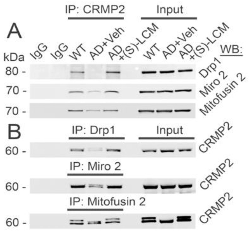
Figure 5.
CRMP2 interacts with Drp1, Miro 2, and Mitofusin 2 in cultured cortical neurons (14 DIV) from APP/PS1 (AD) and WT mice. In AD neurons, CRMP2 interaction with Drp1, Miro 2, and Mitofusin 2 was disrupted. (S)-LCM) precluded CRMP2 dissociation from these proteins. In (A), immunoprecipitation (IP) of CRMP2 with Miro 2, Drp1, and Mitofusin 2 using pull-down procedure with anti-CRMP2 antibody followed by immunoblotting with anti-Miro 2, anti-Drp1, and anti-Mitofusin 2 antibodies. In (B), IP of Miro 2, Drp1, and Mitofusin 2 with CRMP2 using pull-down procedure with anti-Miro 2, anti-Drp1, and anti-Mitofusin 2 antibodies followed by immunoblotting with anti-CRMP2 antibody. Cells were exposed to either a vehicle (Veh, 0.01% DMSO) or 10 µM (S)-LCM for 7 days prior to analysis. Cortical neurons were isolated from P1 APP/PS1 (AD) and WT mice of both sexes and cultured for 12–14 days in vitro (12–14 DIV). For the input, 5% of total protein was used in the immunoprecipitation procedure. Representative data from N = 5 biological repeats is shown.

