Abstract
Mucin 5AC (MUC5AC) glycoprotein plays a crucial role in carcinogenesis and drug sensitivity in pancreatic ductal adenocarcinoma (PDAC), both individually and in combination with other mucins. Its function and localization are glycoform-specific. The immature isoform (detected by the CLH2 monoclonal antibody, or mab) is usually in the perinuclear (cytoplasmic) region, while the mature (45 M1, 2-11, Nd2) variants are in apical and extracellular regions. There is preclinical evidence suggesting that mature MUC5AC has prognostic and predictive (response to treatment) value. However, these findings were not validated in clinical studies. We propose a MUC5AC signature with three components of MUC5AC—localization, variant composition, and intensity—suggesting a reliable marker in combination of variants than with individual MUC5AC variants alone. We also postulate a theory to explain the occurrence of different MUC5AC variants in abnormal pancreatic lesions (benign, precancerous, and cancerous). We also analyzed the effect of mature MUC5AC on sensitivity to drugs often used in PDAC management, such as gemcitabine, 5-fluorouracil, oxaliplatin, irinotecan, cisplatin, and paclitaxel. We found preliminary evidence of its predictive value, but there is a need for large-scale studies to validate them.
Keywords: pancreatic ductal adenocarcinoma, MUC5AC, biomarker, predictive, gemcitabine, chemoresistance
1. Introduction
Pancreatic ductal adenocarcinoma (PDAC) is one of the aggressive cancers without significant progress on the therapeutic front for a long time [1]. Recently presented NAPOLI-3 results showed the survival advantage of nano-liposomal irinotecan-based therapy (NALIRIFOX) over gemcitabine/nab-paclitaxel (Gem-NP) [2]. It could be the standard of care in selected patients, but it may not change overall PDAC outcomes, as the median overall survival is similar to that from the PRODIGE trial (11.1 months) with FOLFIRINOX. Unfortunately, immune checkpoint inhibitors and targeted therapies have not yet revolutionized PDAC management, as they have in other cancer types, such as breast and lung cancer.
One of the inherent challenges in the management of PDAC is the lack of effective predictive biomarkers to help guide the selection of chemotherapy regimens, such as 5FU-based regimens, e.g., FOLFIRINOX or NALIRIFOX, versus gemcitabine-based regimens, such as Gem-NP. BRCA mutations can help identify patients who may benefit from platinum-based treatments, but these mutations are rare in PDAC (<5%) [3]. Currently, the decision of treatment is based on functional status and comorbidities that may not reflect the potential response to a particular chemotherapeutic regimen. Having an effective biomarker in this regard is helpful to gear treatment in a personalized approach. In this review, we focus on a novel biomarker, MUC5AC, that has the potential to improve PDAC management.
Mucin 5AC (MUC5AC) is a gel-forming, glycosylated, high-molecular-weight protein expressed in abnormal pancreatic tissues, including PDAC [4,5]. In our previous publication, we discussed different MUC5AC variants and reviewed evidence on the prognostic value of immature MUC5AC (detected by anti-mucin 5AC CLH2 monoclonal antibody, or mab), which was inconclusive [6]. Post-transcriptional changes of MUC5AC in pancreatic cells, specifically N-glycosylation, promote carcinogenesis, as demonstrated in a study by Pan et al. [7]. Such changes include following a sequence of steps, starting from the perinuclear region, to the apical region, dimerization of the unglycosylated MUC5AC monomer, the addition of N-acetyl galactosamine residues (maturation by glycosylation), multimerization, and finally secretion of mature MUC5AC into the duct or inter-cellular regions [8].
MUC5AC isoforms can be broadly divided into immature and mature MUC5AC variants for practical purposes [9,10,11,12,13,14,15,16,17]. Immature MUC5AC is the initial un- or less glycosylated variant in the perinuclear/cytoplasmic region. It can be detected by the CLH2 mab. Mature MUC5AC is a heavily glycosylated MUC5AC variant detected by 45M1 or 2-11M1 or Nd2 mabs, and is localized primarily in apical, extracellular (secreted or inter-cellular) regions. When subjected to growth factors, pancreatic cancer cell lines (PCLs) produced more mature than immature MUC5AC isoforms, indicating the difference in their function and malignant potential [17]. Immunogenic MUC5AC refers to a subtype of mature MUC5AC variant with an epitope capable of eliciting an immune reaction detected by NPC-1C and PAM4 mabs [9,10]. Some studies also identified other immunogenic peptides, such as MUC5AC-A02-1398 (FLNDAGACV) and MUC5AC-A24-716 (TCQPTCRSL) on MUC5AC that can provoke cell-mediated toxicity [18]. The CLH2 mab targets the sequence TTSTTSAP within the tandem repeat (backbone) of MUC5AC. It can recognize glycosylated and unglycosylated MUC5AC variants, except when galactosamine residues cover this region. The epitopes of 45M1 (C-terminal), 21-M1 (N-terminal), and Nd2 are on glycosylated MUC5AC, but can partially react to immature forms [19,20,21]. Therefore, it is safe to conclude that glycosylation, localization, immunoreactivity to specific mabs, and the function of MUC5AC are interlinked.
In this paper, we introduce the concept of a ’MUC5AC signature for PDAC’, discuss MUC5AC’s role in carcinogenesis and promoting distant metastases, and discuss its regulation. We examined the available indirect evidence of MUC5AC’s predictive value in the PCLs.
2. MUC5AC Signature
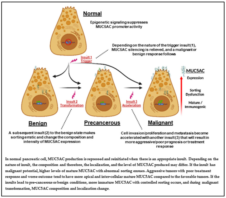
Inaguma et al. proposed that MUC5AC ‘sorting’ (distribution to apical vs. extracellular vs. perinuclear or cytoplasmic) is well controlled in gastric/respiratory cells, where it is normally produced [22]. The control of such a process is lost in lung cancers, cholangiocarcinoma, and PDAC. This does not explain what triggers MUC5AC expression and why it does not transform all pancreatic cells into cancerous ones (MUC5AC is detected in some benign diseases, such as intraductal papillary mucinous neoplasms or IPMN). Spinning off from this hypothesis, we propose the following sequence of events that can happen in the malignant transformation of pancreatic cells and the acceleration of malignant disease, ultimately shaping the MUC5AC signature.
The series of events can be broadly divided into three interlinked stages—trigger response, malignant transformation, and malignant acceleration—with three components in each stage—sorting, MUC5AC variant composition, and level or intensity of expressed MUC5AC. We did not consider MU5AC mRNA in this theory, as the maturation, or acquired immunogenicity, is a post-translational change, and mRNA expression level may not make a significant difference. However, sorting and MUC5AC variants (through glycosylation) may be related. MUC5AC is believed to support/promote malignant transformation and acceleration of metastasis [22]. Figure 1 illustrates the proposed theory based on our hypothesis. We acknowledge that multiple factors, including mutations, initiate and promote carcinogenesis. In this model, we referred to them as triggers.
Figure 1.
MUC5AC signature modeling in pancreatic adenocarcinoma based on the proposed hypothesis.
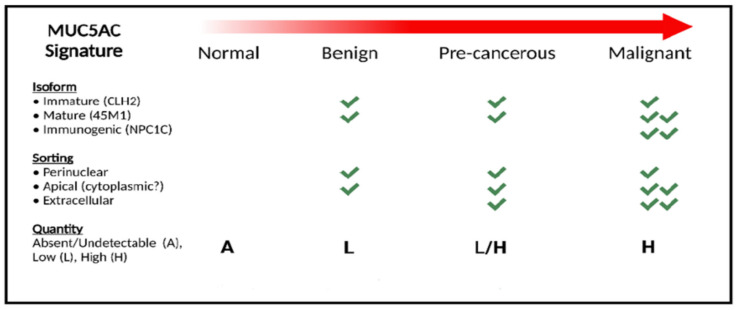
The proposed sequence of events can explain the detection of MUC5AC in benign, precancerous, and cancerous lesions; a differential pattern of expression and variants of MUC5AC in cancerous vs. precancerous forms; the malignant transformation of the latter to the former; and may explain MUC5AC’s prognostic and predictive value. Based on this hypothesis, we propose the concept of classifying PDAC by the MUC5AC signature, which incorporates three key elements of MUC5AC: composition of MUC5AC variant identified; localization of the MUC5AC variant (sorting); and intensity of the expression (Figure 2).
Figure 2.
Classification of MUC5AC in pancreatic ductal adenocarcinoma.
Strengthening Our Hypothesis Using MUC5AC Signature
To prove our model with available evidence in the literature, we start with two interlinked components: sorting and MUC5AC variants. There are limited studies that compare immunohistochemical staining of mature and immature MUC5AC among the spectrum of pancreatic tissues, ranging from normal, to pancreatitis, PanIN, IPMN, and tumors. CLH2 mab was used in most of the studies. Based on localization, we drew conclusions on the MUC5AC isoform. To date, there are no studies that examined the staining of NPC1C and PAM4 in benign/precancerous pancreatic pathologies.
Kim et al. published a study in 2002 that supports sorting and the MUC5AC variant components discussed here [16]. Staining was predominantly apical and extracellular with mature (21 M1/Nd2 mab) and perinuclear/cytoplasmic with immature (CLH2 mab) MUC5AC across PanINs and cancers, proving that they exist in precancerous (PanIN and IPMN) and PDAC tissues. In another study, immature MUC5AC (by CLH2 mab) expression pattern and frequency were compared in PanIN lesions derived from surgical specimens from patients with adjacent normal (N-PanIN) and PDAC (C-PanIN) tissues. CLH2 staining was cytoplasmic (perinuclear), as well as apical/extracellular in both N-PanIN and C-PanIN, indicating possible cross-reactivity of CLH2 mab with mature MUC5AC [23]. The frequency of immature MUC5AC was significantly different in PanIN-1A (p < 0.0001) and PanIN 2 (p < 0.005) lesions in N-PanIN and C-PanIN patients. Interestingly, there were no morphological differences among the PanIN groups, suggesting a minimal effect of MUC5AC production on the morphology (discussed in Section 4.3). The staining pattern for immunogenic MUC5AC is similar to that of mature MUC5AC (and different from immature) [9,10,11,12]. Finally, MUC5AC produced when stimulated by growth factors such as vasoactive peptide (VIP), or transcription factors (TFs) such as GLI1, is primarily apical or extracellular, further supporting the sorting process (discussed in Section 3 below) [17,22].
The intensity of staining was compared between PanIN and PDAC in a study by Ochinuda et al. [24]. Strong MUC5AC expression was seen in 48% of PDACs, vs. 0% of PanINs. Moderate expression was reported in 35% of PDACs vs. 65% of PanINs, and weak in 17% of PDACs vs. 36% of PanINs. None of the samples were negative. Overall, strong/moderate expression was 83% in PDAC vs. 64% in PanINs. Compared to non-tumoral tissues, malignant tissues had a 2.8-fold greater expression of mRNA, but this differential expression was not statistically significant. Immature MUC5AC staining was focal and cytoplasmic in PanIN, but it was strongly positive in PDAC in another study [25]. PanIN and atypical duct areas in non-neoplastic tissues (with negative staining) showed strong MUC5AC expression, hinting at the initiation of pre-malignant changes with MUC5AC expression.
The MUC5AC signature for the PDAC model can explain the wide range of outcomes reported by studies that used CLH2 mab, which we summarized in our previous publication [6]. Most studies reported cytoplasmic staining, and patients were classified positive or negative based on the extent of staining, making it difficult to assess the prognostic value of the MUC5AC signature. When the thresholds to classify were low (5 or 10%) immature MUC5AC expression was a good prognostic marker [26,27]. Alternatively, when the thresholds were high (25% or high-H score), the outcomes were poor with its detection [28,29]. This underscores the importance of using all three components (isoform, localization, and intensity) in the proposed MUC5AC signature.
3. Regulation of MUC5AC Expression
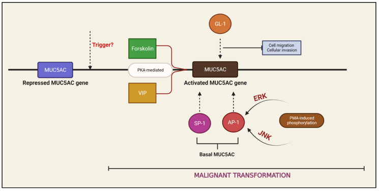
In PDAC, the influence of MUC5AC can be simplified, as shown in Supplementary Figure S1. It stimulates tumor cell proliferation and distant metastasis, protects the tumor from host defenses, and reduces sensitivity to chemotherapeutic agents, such as gemcitabine (gem) [22,30,31,32,33]. The regulation of MUC5AC production is not well understood, but two important regulators identified are the growth factors (GFs) and TFs (Figure 3).
Figure 3.
Regulation of MUC5AC production. VIP—vasoactive intestinal peptide; Sp-1—specificity protein 1; AP-1—activator protein 1; PMA—Phorbol 12-myrisate 13-acetate; PKC—protein kinase C; JNK—c-Jun N-terminal kinases; ERK—extracellular signal-regulated kinase.
MUC5AC gene expression is constitutively repressed in normal pancreatic cells and, therefore, the respective protein is not detected in them [34]. Epigenetic silencing, by CpG methylation and H3 Lysine 9 modification of the promoter, is believed to be one of the principal processes of such suppression [35,36]. Supporting this theory, Yamada et al. reported higher methylation levels in MUC5AC-deficient compared to MUC5AC-positive lines [36]. Interestingly, demethylation of the CpG sites immediately proximal (5′) to the MUC5AC gene transcription site by 5-aza-2’-deoxycytidine (5AZA) could not increase MUC5AC production in a preclinical study published in 2003 [37]. This was confirmed by other studies, and could mean that MUC5AC gene expression is controlled by other sites/mechanisms, and epigenetics is not the primary mechanism [35].
Kato et al. later identified two such regions corresponding to binding sites for TFs, specificity protein 1 (Sp-1) and activator protein 1 (AP-1), as regulatory sites for MUC5AC gene transcription [38]. Both of the TFs participate in basal MUC5AC production in a malignant pancreatic cell, while AP-1 also takes part in Phorbol 12-myrisate 13-acetate (PMA)-induced MUC5AC production. PMA stimulation phosphorylates sub-units of AP-1 (cFos/cJun) via protein kinase C (PKC)/ERK/AP-1 and PKC/JNK/AP-1 pathways that, in turn, upregulate MUC5AC promoter activity (to produce mRNA). ERK and JNK inhibitors downregulate this process, suggesting their role in MUC5AC regulation.
Likewise, Krüppel-like zinc-finger protein GLI-1 also promotes MUC5AC production by activating its gene promoter [22]. GLI-1-induced MUC5AC’s function seems to be different from that of Sp-1- or AP-1-induced MUC5AC in the following ways: (1) it reduces the amount of E-cadherin at the intercellular membranes, thus promoting cell migration; and (2) it increases the nuclear accumulation of beta-catenin and excess target gene expression, thus promoting cellular invasion. GLI-1 expression correlates with MUC5AC expression in PanIN and PDAC tissues (low to undetectable in PanIn-1A/1B and high in PanIn-2/3 and PDAC). GLI-1′s expression statistically correlated with altered E-catherin (loss of membrane localization) and beta-catenin (increased nuclear localization) in PanIN-3 and PDAC tissues. GLI-1 does not appear to be responsible for malignant transformation, but some critical aberrations frequently associated with PDAC carcinogenesis, such as KRAS mutations and irregularities in the hedgehog signaling pathway, promote its production as well [39,40,41].
GFs are known to promote pancreatic cell growth, migration, and invasion. GFs such as forskolin and VIP also promote MUC5AC production and release in malignant pancreatic tissues [17,42,43,44,45]. They exert their effect through cyclic adenosine monophosphate (cAMP)-dependent kinases, also known as protein kinase A (PKA), just as epidermal growth factor receptors (EGF) are overexpressed in PDAC [46,47]. In vitro studies have even demonstrated enhanced VIP-induced MUC5AC production in the presence of EGF [43]. A study published in 2006 illustrated that GF-induced MUC5AC is mature, with the following observations: (1) In an unstimulated PCL, the CLH2 mab stained immature variants in the perinuclear region intensely, while the mature (Nd2) mab stained both perinuclear and cytoplasmic (peripheral) regions. When stimulated by forskolin, CLH2 continued to stain the perinuclear region, but mature mabs (45 M1, Nd2, and 21 M1) stained the mucins that accumulate in apical and extracellular regions (even between cells). (2) The VIP-induced release of mature MUC5AC is more on the cells’ luminal (apical) side than on the basolateral side. Interestingly, basolateral VIP receptors appear to significantly affect VIP-induced MUC5AC when stimulated or inhibited (by PKA inhibitors). (3) In PDAC tumor tissue, CLH2 could stain only in the cytoplasm, while 45M1, Nd2, and 2-11M could stain both luminal and cytoplasmic regions. CLH2 noticeably did not stain the mucin in the lumen (secreted). Therefore, it can be deduced that GF-induced MUC5AC is predominantly heavily glycosylated, or promotes glycosylation. Interestingly, after forskolin-induced MUC5AC production, inhibition of O-glycosylation increased the immunoreactivity of the CHL2 mab in PCLs, but there was no change for other mabs, further strengthening the concept that N-glycosylation is important in carcinogenesis [7].
In summary, MUC5AC is not expressed in a normal pancreatic cell, likely due to epigenetic silencing. The expression is mediated via PKA/PKC pathways, with GF and TF as key regulators; however, the etiology of the triggers that start and propagate its production in pancreatic cells is unknown. Detection of GLI-1 and GNAS mutations that alter mucin gene expression profiles in pancreatic ductal cells, including the overexpression of MUC5AC through the MAPK/PIK3 pathway in precancerous lesions such as pancreatic intraepithelial neoplasia (PanIN) and IPMNs, may be an early indicator of malignant transformation [22,48]. If we extrapolate the evidence from other organs, such as the lung, nasal epithelium, and gastrointestinal tract, they can be infectious (bacterial or viral), inflammatory (through cytokines), environmental (smoking), or growth factors (epidermal growth factor) [49,50,51,52,53,54,55,56,57]. This warrants close examination of the pancreatic tumor microenvironment that plays a role in MUC5AC production or upregulation.
4. Predictive Value of MUC5AC
To understand the impact of MUC5AC on treatment response, we used the results of the preclinical studies reported by Krishn et al. as a reference [58]. This study proved MUC5AC’s role in the pancreatic cancer cell, including viability, anchorage-independent growth, motility, adhesion to the extracellular matrix, angiogenesis, apoptosis, and sensitivity to gemcitabine [58]. Through in vivo studies in nude mice, the authors suggested its role in promoting tumor growth, metastasis, and disease progression (Table 1). For immunohistochemistry (IHC) and western blot (WB), mature MUC5AC, 45M1 mab was used.
Table 1.
Summary of MUC5AC’s role in pancreatic cell lines and in vivo studies.
| Study | Results |
|---|---|
| MUC5AC expression by IHC intumor-tissues | 80% of pancreatic cancer surgical specimens (positive for histology score > 0.01) Not detected in the normal tissue Localization was not reported |
| In pancreatic cell lines (PCL) | |
| MUC5AC-mRNA expression(by quantitative-polymerase chain reaction) | Higher in PCLs compared to normal pancreatic cell lines No expression in some PCLs (MIA PaCa, PANC10.05, QGP1) |
| MUC5AC protein expression(by western blot) | Positive in some PCL (ASPC1, BXPC3, COLO357, SW1990, and T3M4) Negative in some PCL (CD18/HPAF, CAPAN1, MIA PaCa, Panc-1). |
| Localization by confocalmicroscopy) | Stain the cytoplasm and intercellular junction (typical for mature MUC5AC) |
| In MUC5AC-knockdown PCL | |
| Decreased | Pancreatic cancer cell viability, anchorage-independent growth, cell motility, adhesion to the extracellular matrix, and angiogenesis Sensitivity to Gemcitabine (β-catenin mediated resistance) |
| Increased | Apoptosis |
| Nude mice with orthotopically transplanted MUC5AC knockdown PCL in pancreas * | Lesser pancreatic tumor weight Fewer metastatic sites |
| KrasG12D; Pdx1-Cre; Muc5ac-/- mouse models ** | Delay in onset and progression of pancreatic cancer cells |
* When compared, nude mice transplanted with PCL (MUC5AC positive); ** When compared to KrasG12D; Pdx1-Cre; Muc5ac+/+ mouse.
Important takeaways from this study are as follows: (i) Mature MUC5AC is detected in most PDACs, and not detected in normal pancreatic tissues. (ii) All PCLs did not express mature MUC5AC. (iii) There is discordance between MUC5AC mRNA and protein expression in some PCLs. (iv) Inhibiting MUC5AC could improve outcomes, including sensitivity to gemcitabine. To further verify the observations of this study, we compared cell growth, migration, invasion, and drug sensitivity of PCLs with wide-ranging levels of MUC5AC. The goal of this analysis was to assess the influence of mature MUC5AC on PDAC.
4.1. Comparing PCLs Based on MUC5AC Expression
PCLs were classified based on their native MUC5AC protein and/or mRNA expression (Table 2). Among the studied PCLs, there were PCLs with concordance between mRNA and protein expression, as in COLO 357 (CO), SW 1990 (SW), BxPc3 (B), and MIA PaCa (MPC); discordant PCLs were the medium mRNA expressors with low or no protein expression, as in PANC-1 (P) and AsPc3 (A). Alternatively, in T3M4 PCL, there is high mRNA expression, but low protein expression (protein expression of SU 8686 was not reported). This matches observations in clinical samples with PanINs and PDAC, and CLH2-reactive MUC5AC; the discordance was high in PanIN1b (45% for in-situ hybridization vs. 82% IHC), PanIN2 (81% vs. 94%), and PDAC (61% vs. 100%) [16]. The concordance was 100% in PanIN3 lesions, while the discordance was minimal in PanIN1A (62% vs. 59%). To assess the influence of mature MUC5AC on various aspects of PDAC, aspects of malignant cells, including cell growth, migration, invasion, and drug sensitivity, were compared based on their expression levels (Table 3).
Table 2.
Classification of select pancreatic cell lines based on MUC5AC mRNA and protein expression.
| High | Medium | Low | No | |
|---|---|---|---|---|
| mRNA | COLO357 SW 1990 SU 8686 T3M4 |
BXPC3 PANC1 AsPc3 |
CAPAN | MIA PaCa |
| Protein | COLO357 SW 1990 |
BXPC3 |
AsPc3 T3M4 |
PANC-1 CAPAN MIA PaCa |
Table 3.
COLO357 (CO) vs. MIA PaCa (M).
| Features in CO Compared to M | Associated Features | |
|---|---|---|
| Prognostic factors [59,60,61,62] | Higher osteopontin | Invasiveness |
| Higher BMP2 | Poor survival | |
| Higher CXCR 4 (receptor and protein) | Cell survival, proliferation, migration, invasion, and metastasis. | |
| Predictive factors [63,64] | More sensitive to 5FU, Irinotecan, Cisplatin | |
| Less sensitive to Gem and Oxaliplatin |
4.2. Comparing Basic Pathological Characteristics
MUC5AC protein expression and basic characteristics are illustrated in Supplementary Table S1. There is no significant correlation between MUC5AC expression levels and basic characteristics of PCLs, such as source (primary tumor vs. metastatic), doubling time, and differentiation. PDACs stained by mature (21M1 and Nd2 mab) and immature (CHL2 mab) mabs did not differ morphologically in a retrospective study published two decades ago, supporting this observation [16]. In that study, 21M1 mab stained 100% of well-differentiated tumors, while CLH2 and Nd-2 stained 90% and 85%, respectively. The frequency of 21M1- and CLH2-positive tumors in moderately (96% vs. 92%) and poorly (59% vs. 64%) differentiated tumors was marginal. Remarkably, there were differences in staining frequencies between two mabs that identify mature MUC5AC (21M1 and Nd2), further highlighting the influence of the other two components in the MUC5AC signature (location and intensity) in PDAC.
4.3. Comparing Malignant Potential
To understand the impact of MUC5AC on the malignant potential of PCLs, COLO357 (CO) and MIA PaCa (M), representing high and no MUC5AC (mRNA and protein) expression, respectively, were selected (Table 3). The literature search was limited, as there were no head-to-head studies. We divided the search into two parts: (1) poor pathophysiological characteristics or prognostic factors, and (2) chemosensitivity or predictive factors. The results are summarized in Table 3.
We expanded the search comparing the PCLs based on Table 2 (Table 4) [59,65,66,67,68,69,70,71,72,73,74,75,76].
Table 4.
Comparison of key characteristics in pancreatic cell lines.
| Cell Lines Compared | MUC5AC Expression | Results |
|---|---|---|
| M vs. BxPC3 | No vs. M (mRNA & protein) | BxPC3—More invasion, angiogenic potential, and tumorigenicity. More sensitive to Gem and 5FU |
| ASPC-1 vs. BxPC3 | L vs. M (mRNA & protein) | BxPC3 has -More invasion, angiogenic potential, and tumorigenicity. More resistant to 5FU and less-similar resistance to Gemcitabine |
| ASPC-1 vs. CO | L vs. H (mRNA & protein) | CO—less sensitive to 5FU and more sensitive to Gem |
| SU86.86 vs. Panc-1 | H vs. L (mRNA) | SU86.86 is less adhesive, more invasive and angiogenic potential |
| M vs. SW 1990 | L vs. H (mRNA & protein) | SW1990 is more resistant to Gem and more sensitive to 5FU (IC 50 of 9 vs. 5.68 μM) |
| CO > SU86686 > BxPC3 > M | H vs. H vs. M vs. No (P) H vs. M vs. M vs. No (mRNA) |
Osteopontin influenced invasiveness |
| BxPC-3 > AsPC-1 or M | M vs. L vs. No (P and mRNA) | Tendency to invade—MPC has a minimum tendency to invade |
N—no expression; L—low expression; M—medium expression; H—high expression.
4.4. Comparing Chemosensitivity
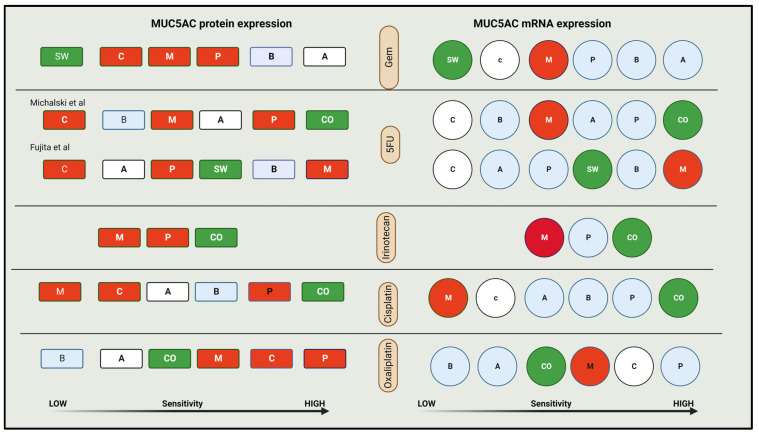
Widely used treatment regimens are 5FU (FOLFIRINOX, NALIRIFOX, FOLFOX, NALIRI) or gem-based (Gem-NP or GemOx). We attempted to establish the best treatment group based on mature MUC5AC expression levels. The chemosensitivities of PCLs to some of the standard drugs used in treating PDAC, such as gemcitabine (gem), fluorouracil (5FU), cisplatin (cis), irinotecan (iri), and oxaliplatin (Ox) were compared using three studies, and one PCL for each combination (mRNA and protein) (as in Supplementary Table S2) [63,64,75]. There were no studies that looked at sensitivity of NP or NALIRI. The difference in paclitaxel was too close to see any pattern in Michalski et al.’s study, and we did not include it in our analysis (Figure 3 and Table 5).
Table 5.
Summary of drug sensitivity and MUC5AC expression.
| Drug Tested | Sensitivity to the Drugs | |
|---|---|---|
| Protein Expression | mRNA Expression | |
| Gemcitabine | H < No < L-M | NDP |
| 5FU | NDP | NDP |
| Irinotecan | No < H | No < L-M < H |
| Oxaliplatin | L-M < H < No | H < No, NDP for L-M |
| Paclitaxel * | NDP | NDP |
| Cisplatin | No < H, no NDP for L-M | No < L-M < H |
| Nab-paclitaxel | NT | NT |
| Nanoliposomal irinotecan | NT | NT |
* Not discussed in the figure. H—high expression, No—no expression; L-M—low to moderate expression, NT—not tested, NDP—no definite pattern.
We studied the PCLs that were similar in both studies. CO was not studied by Fujita et al. and, therefore, SW was used to represent high MUC5AC protein expressors. Gem sensitivity was too close to observe a trend from Michalski et al.’s study [63]. We relied on Fujita et al.’s study (Figure 4) for our observations.
Figure 4.
MUC5AC expression and drug sensitivity in pancreatic cancer cell lines [63,75]. Color coding for expression: Red—no; Green—high; Blue—Moderate; White—low; Letters—protein expression. CO—COLO357; SW—SW1990; B—BXCP3; A—ASPC-1; M—MIA PaCa; P—PANC-1; C—CAPAN-1, Gem—gemcitabine, 5FU—5 fluorouracil.
For practical purposes, we can divide PCLs into three groups—no, high, and low–moderate expressors—for both protein and mRNA. Our analysis did not identify any correlation between MUC5AC expression and 5FU or paclitaxel sensitivity. NP and NALIR were not tested. PCLs expressing higher MUC5AC protein (western blotting) were less sensitive to gem and ox, and more sensitive to irinotecan and cis, than those which do not produce MUC5AC. Low—moderate expressors were more and less sensitive to gem and ox, respectively. For cis, their sensitivity was greater than non-expressors but lower than high-expressors (Figure 3). As mature MUC5AC is expressed in most of the PDACs, positive vs. negative classification alone is not ideal. There might be a threshold for native MUC5AC, which may identify high-risk groups or a signature of MUC5AC variants (immature and mature) that can help us predict tumor response. The mRNA expression was not useful for most drugs (except irinotecan and cis). We need larger studies on clinical samples to investigate the impact of MUC5AC expression on drug sensitivity.
We acknowledge the limitations of this analysis; however, this suggests a good signal for future clinical studies. Quantifying/detecting protein expression in PCLs by WB is not similar to IHC on tissues [77]. We cannot identify the localization of the protein (apical vs. cytoplasmic vs. extracellular) in WB. This can explain the inconclusive results for chemosensitivity discussed above. Analyzing the impact of mature MUC5AC expression based on the localization (apical or extracellular) could provide definitive results. The thresholds for classifying PCLs into high, medium, and low expressors for protein and mRNA were not clearly defined.
5. Conclusions
The mucin MUC5AC is unique in the sense that its detection in pancreatic tissues is abnormal and exists in many isoforms, which can be broadly classified into mature and immature subtypes. Observations from preclinical studies need validation from retrospective or prospective studies, but provide us with a preliminary indication of the role of the MUC5AC protein as a potential biomarker in PDAC. We extensively discussed the clinical significance of serum and tissue MUC5AC in our previous publication [6]. Serum MUC5AC could be a good diagnostic marker when combined with CA 19-9, but its prognostic and predictive value is not well established [78]. Immunogenic MUC5AC, a mature MUC5AC detected by Niemann Pick C1 (NPC-1C) and PAM-4 (Clivatuzumab tetraxetan) antibodies failed to improve outcomes in PDA patients. The addition of NPC-1C antibody did not add any benefit to patients treated with gemcitabine and nab-paclitaxel in the second line after progressing on FOLFIRINOX [79]. A phase 3 trial was initiated to test the benefit of yttrium-90-labeled hPAM4 ((90) Y-hPAM4) to gemcitabine (PANCRIT-1, NCT01956812). This trial was terminated, as interim analysis did not show any clinical benefit.
The proposed MUC5AC signature for PDAC, which incorporates main isoforms (mature vs. immature), their location (linked to their function), and expression levels (the MUC5AC signature for PDAC), needs to be further validated in larger studies in order to harness its potential as an efficient predictive biomarker.
6. Patents
The Ohio State University is currently pursuing patent protection for the research discussed in this publication.
Acknowledgments
The authors would also like to thank Angela Dahlberg for editing and proofreading the paper, and biorender.com for the figures.
Supplementary Materials
The following supporting information can be downloaded at: https://www.mdpi.com/article/10.3390/ijms24098087/s1.
Author Contributions
Conceptualization by A.M.; writing—original draft preparation, A.M. and A.K.; writing—review and editing, R.K.P.; writing—review, A.K.E. All authors have read and agreed to the published version of the manuscript.
Institutional Review Board Statement
Not applicable.
Informed Consent Statement
Not applicable.
Data Availability Statement
Not applicable.
Conflicts of Interest
The authors declare no conflict of interest.
Funding Statement
This research received no external funding.
Footnotes
Disclaimer/Publisher’s Note: The statements, opinions and data contained in all publications are solely those of the individual author(s) and contributor(s) and not of MDPI and/or the editor(s). MDPI and/or the editor(s) disclaim responsibility for any injury to people or property resulting from any ideas, methods, instructions or products referred to in the content.
References
- 1.Surveillance, Epidemiology, and End Results (SEER) Program SEER*Stat Database: Incidence—SEER Research Data, 9 Registries, Nov 2019 Sub (1975–2017) [(accessed on 17 April 2023)]; Available online: www.seer.cancer.gov.
- 2.Wainberg Z.A., Melisi D., Macarulla T., Pazo-Cid R., Chandana S.R., De La Fouchardiere C., Dean A.P., Kiss I., Lee W., Goetze T.O., et al. NAPOLI-3: A randomized, open-label phase 3 study of liposomal irinotecan + 5-fluorouracil/leucovorin + oxaliplatin (NALIRIFOX) versus nab-paclitaxel + gemcitabine in treatment-naïve patients with metastatic pancreatic ductal adenocarcinoma (mPDAC) J. Clin. Oncol. 2023;41:LBA661. doi: 10.1200/JCO.2023.41.4_suppl.LBA661. [DOI] [PMC free article] [PubMed] [Google Scholar]
- 3.Sheel A., Addison S., Nuguru S.P., Manne A. Is Cell-Free DNA Testing in Pancreatic Ductal Adenocarcinoma Ready for Prime Time? Cancers. 2022;14:3453. doi: 10.3390/cancers14143453. [DOI] [PMC free article] [PubMed] [Google Scholar]
- 4.Bansil R., Turner B.S. Mucin structure, aggregation, physiological functions and biomedical applications. Curr. Opin. Colloid Interface Sci. 2006;11:164–170. doi: 10.1016/j.cocis.2005.11.001. [DOI] [Google Scholar]
- 5.Kebouchi M., Hafeez Z., Le Roux Y., Dary-Mourot A., Genay M. Importance of digestive mucus and mucins for designing new functional food ingredients. Food Res. Int. 2020;131:108906. doi: 10.1016/j.foodres.2019.108906. [DOI] [PubMed] [Google Scholar]
- 6.Manne A., Esnakula A., Abushahin L., Tsung A. Understanding the Clinical Impact of MUC5AC Expression on Pancreatic Ductal Adenocarcinoma. Cancers. 2021;13:3059. doi: 10.3390/cancers13123059. [DOI] [PMC free article] [PubMed] [Google Scholar]
- 7.Pan S., Chen R., Tamura Y., Crispin D.A., Lai L.A., May D., McIntosh M.W., Goodlett D.R., Brentnall T.A. Quantitative Glycoproteomics Analysis Reveals Changes in N-Glycosylation Level Associated with Pancreatic Ductal Adenocarcinoma. J. Proteome Res. 2014;13:1293–1306. doi: 10.1021/pr4010184. [DOI] [PMC free article] [PubMed] [Google Scholar]
- 8.Sheehan J.K., Kirkham S., Howard M., Woodman P., Kutay S., Brazeau C., Buckley J., Thornton D.J. Identification of Molecular Intermediates in the Assembly Pathway of the MUC5AC Mucin. J. Biol. Chem. 2004;279:15698–15705. doi: 10.1074/jbc.M313241200. [DOI] [PubMed] [Google Scholar]
- 9.Luka J., Arlen P.M., Bristol A. Development of a serum biomarker assay that differentiates tumor-associated MUC5AC (NPC-1C ANTIGEN) from normal MUC5AC. J. Biomed. Biotechnol. 2011;2011:934757. doi: 10.1155/2011/934757. [DOI] [PMC free article] [PubMed] [Google Scholar]
- 10.Patel S.P., Bristol A., Saric O., Wang X.-P., Dubeykovskiy A., Arlen P.M., Morse M.A. Anti-tumor activity of a novel monoclonal antibody, NPC-1C, optimized for recognition of tumor antigen MUC5AC variant in preclinical models. Cancer Immunol. Immunother. 2013;62:1011–1019. doi: 10.1007/s00262-013-1420-z. [DOI] [PMC free article] [PubMed] [Google Scholar]
- 11.Gold D.V., Hollingsworth P., Kremer T., Nelson D. Identification of a human pancreatic duct tissue-specific antigen. Cancer Res. 1983;43:235–238. [PubMed] [Google Scholar]
- 12.Gold D.V., Lew K., Maliniak R., Hernandez M., Cardillo T. Characterization of monoclonal antibody PAM4 reactive with a pancreatic cancer mucin. Int. J. Cancer. 1994;57:204–210. doi: 10.1002/ijc.2910570213. [DOI] [PubMed] [Google Scholar]
- 13.Reis C.A., David L., Carvalho F., Mandel U., de Bolós C., Mirgorodskaya E., Clausen H., Sobrinho-Simões M. Immunohistochemical Study of the Expression of MUC6 Mucin and Co-expression of Other Secreted Mucins (MUC5AC and MUC2) in Human Gastric Carcinomas. J. Histochem. Cytochem. 2000;48:377–388. doi: 10.1177/002215540004800307. [DOI] [PubMed] [Google Scholar]
- 14.Nollet S., Forgue-Lafitte M.E., Kirkham P., Bara J. Mapping of two new epitopes on the apomucin encoded by MUC5AC gene: Expression in normal GI tract and colon tumors. Int. J. Cancer. 2002;99:336–343. doi: 10.1002/ijc.10335. [DOI] [PubMed] [Google Scholar]
- 15.Liu D., Chang C.-H., Gold D.V., Goldenberg D.M. Identification of PAM4 (clivatuzumab)-reactive epitope on MUC5AC: A promising biomarker and therapeutic target for pancreatic cancer. Oncotarget. 2015;6:4274–4285. doi: 10.18632/oncotarget.2760. [DOI] [PMC free article] [PubMed] [Google Scholar]
- 16.Kim G.E., Bae H., Park H., Kuan S., Crawley S.C., Ho J.J., Kim Y.S. Aberrant expression of MUC5AC and MUC6 gastric mucins and sialyl Tn antigen in intraepithelial neoplasms of the pancreas. Gastroenterology. 2002;123:1052–1060. doi: 10.1053/gast.2002.36018. [DOI] [PubMed] [Google Scholar]
- 17.Ho J.J., Crawley S., Pan P.L., Farrelly E.R., Kim Y.S. Secretion of MUC5AC mucin from pancreatic cancer cells in response to forskolin and VIP. Biochem. Biophys. Res. Commun. 2002;294:680–686. doi: 10.1016/S0006-291X(02)00529-6. [DOI] [PubMed] [Google Scholar]
- 18.Yamazoe S., Tanaka H., Iwauchi T., Yoshii M., Ito G., Amano R., Yamada N., Sawada T., Ohira M., Hirakawa K. Identification of HLA-A*0201- and A*2402-Restricted Epitopes of Mucin 5AC Expressed in Advanced Pancreatic Cancer. Pancreas. 2011;40:896–904. doi: 10.1097/MPA.0b013e31821ad8d1. [DOI] [PubMed] [Google Scholar]
- 19.Ho J.J., Bi N., Yan P.S., Yuan M., Norton K.A., Kim Y.S. Characterization of new pancreatic cancer-reactive monoclonal antibodies directed against purified mucin. Cancer Res. 1991;51:372–380. [PubMed] [Google Scholar]
- 20.Lidell M.E., Bara J., Hansson G.C. Mapping of the 45M1 epitope to the C-terminal cysteine-rich part of the human MUC5AC mucin. FEBS J. 2008;275:481–489. doi: 10.1111/j.1742-4658.2007.06215.x. [DOI] [PubMed] [Google Scholar]
- 21.Bara J., Chastre E., Mahiou J., Singh R.L., Forgue-Lafitte M.E., Hollande E., Godeau F. Gastric M1 mucin, an early oncofetal marker of colon carcinogenesis, is encoded by the MUC5AC gene. Int. J. Cancer. 1998;75:767–773. doi: 10.1002/(SICI)1097-0215(19980302)75:5<767::AID-IJC17>3.0.CO;2-3. [DOI] [PubMed] [Google Scholar]
- 22.Inaguma S., Kasai K., Ikeda H. GLI1 facilitates the migration and invasion of pancreatic cancer cells through MUC5AC-mediated attenuation of E-cadherin. Oncogene. 2011;30:714–723. doi: 10.1038/onc.2010.459. [DOI] [PubMed] [Google Scholar]
- 23.Matsuyama M., Kondo F., Ishihara T., Yamaguchi T., Ito R., Tsuyuguchi T., Tawada K., Yokosuka O. Evaluation of pancreatic intraepithelial neoplasia and mucin expression in normal pancreata. J. Hepato-Biliary-Pancreatic Sci. 2011;19:242–248. doi: 10.1007/s00534-011-0401-x. [DOI] [PubMed] [Google Scholar]
- 24.Ohuchida K., Mizumoto K., Yamada D., Fujii K., Ishikawa N., Konomi H., Nagai E., Yamaguchi K., Tsuneyoshi M., Tanaka M. Quantitative analysis of MUC1 and MUC5AC mRNA in pancreatic juice for preoperative diagnosis of pancreatic cancer. Int. J. Cancer. 2006;118:405–411. doi: 10.1002/ijc.21317. [DOI] [PubMed] [Google Scholar]
- 25.Giorgadze T.A., Peterman H., Baloch Z.W., Furth E.E., Pasha T., Shiina N., Zhang P.J., Gupta P.K. Diagnostic utility of mucin profile in fine-needle aspiration specimens of the pancreas. Cancer. 2006;108:186–197. doi: 10.1002/cncr.21913. [DOI] [PubMed] [Google Scholar]
- 26.Higashi M., Yokoyama S., Yamamoto T., Goto Y., Kitazono I., Hiraki T., Taguchi H., Hashimoto S., Fukukura Y., Koriyama C., et al. Mucin Expression in Endoscopic Ultrasound-Guided Fine-Needle Aspiration Specimens Is a Useful Prognostic Factor in Pancreatic Ductal Adenocarcinoma. Pancreas. 2015;44:728–734. doi: 10.1097/MPA.0000000000000362. [DOI] [PMC free article] [PubMed] [Google Scholar]
- 27.Jinfeng M., Kimura W., Hirai I., Sakurai F., Moriya T., Mizutani M. Expression of MUC5AC and MUC6 in invasive ductal carcinoma of the pancreas and relationship with prognosis. Int. J. Gastrointest. Cancer. 2003;34:9–18. doi: 10.1385/IJGC:34:1:09. [DOI] [PubMed] [Google Scholar]
- 28.Aloysius M.M., Zaitoun A.M., Awad S., Ilyas M., Rowlands B.J., Lobo D.N. Mucins and CD56 as markers of tumour invasion and prognosis in periampullary cancer. Br. J. Surg. 2010;97:1269–1278. doi: 10.1002/bjs.7107. [DOI] [PubMed] [Google Scholar]
- 29.Takano Y., Ohike N., Tajiri T., Asonuma K., Harada K., Takahashi H., Morohoshi T. Gastric- and intestinal-type marker expression in invasive ductal adenocarcinoma of the pancreas. Hepatobiliary Pancreat. Dis. Int. 2012;11:424–428. doi: 10.1016/S1499-3872(12)60202-1. [DOI] [PubMed] [Google Scholar]
- 30.Yamazoe S., Tanaka H., Sawada T., Amano R., Yamada N., Ohira M., Hirakawa K. RNA interference suppression of mucin 5AC (MUC5AC) reduces the adhesive and invasive capacity of human pancreatic cancer cells. J. Exp. Clin. Cancer Res. 2010;29:53. doi: 10.1186/1756-9966-29-53. [DOI] [PMC free article] [PubMed] [Google Scholar]
- 31.Hoshi H., Sawada T., Uchida M., Iijima H., Kimura K., Hirakawa K., Wanibuchi H. MUC5AC protects pancreatic cancer cells from TRAIL-induced death pathways. Int. J. Oncol. 2013;42:887–893. doi: 10.3892/ijo.2013.1760. [DOI] [PubMed] [Google Scholar]
- 32.Hoshi H., Sawada T., Uchida M., Saito H., Iijima H., Toda-Agetsuma M., Wada T., Yamazoe S., Tanaka H., Kimura K., et al. Tumor-associated MUC5AC stimulates in vivo tumorigenicity of human pancreatic cancer. Int. J. Oncol. 2011;38:619–627. doi: 10.3892/ijo.2011.911. [DOI] [PubMed] [Google Scholar]
- 33.Ganguly K., Krishn S.R., Jahan R., Atri P., Rachagani S., Rauth S., Xi H., Lu Y., Batra S., Kaur S. Abstract 65: Gel-forming mucin MUC5AC as the nexus for cell-adhesion molecules governing pancreatic cancer aggressiveness and chemoresistance. Cancer Res. 2019;79:65. doi: 10.1158/1538-7445.AM2019-65. [DOI] [Google Scholar]
- 34.Krishn S.R., Ganguly K., Kaur S., Batra S.K. Ramifications of secreted mucin MUC5AC in malignant journey: A holistic view. Carcinogenesis. 2018;39:633–651. doi: 10.1093/carcin/bgy019. [DOI] [PMC free article] [PubMed] [Google Scholar]
- 35.Vincent A., Perrais M., Desseyn J.-L., Aubert J.-P., Pigny P., Van Seuningen I. Epigenetic regulation (DNA methylation, histone modifications) of the 11p15 mucin genes (MUC2, MUC5AC, MUC5B, MUC6) in epithelial cancer cells. Oncogene. 2007;26:6566–6576. doi: 10.1038/sj.onc.1210479. [DOI] [PubMed] [Google Scholar]
- 36.Yamada N., Nishida Y., Yokoyama S., Tsutsumida H., Houjou I., Kitamoto S., Goto M., Higashi M., Yonezawa S. Expression of MUC5AC, an early marker of pancreatobiliary cancer, is regulated by DNA methylation in the distal promoter region in cancer cells. J. Hepato-Biliary-Pancreatic Sci. 2010;17:844–854. doi: 10.1007/s00534-010-0278-0. [DOI] [PubMed] [Google Scholar]
- 37.Ho J.J., Han S.W., Pan P.L., Deng G., Kuan S.-F., Kim Y.S. Methylation status of promoters and expression of MUC2 and MUC5AC mucins in pancreatic cancer cells. Int. J. Oncol. 2003;22:273–279. doi: 10.3892/ijo.22.2.273. [DOI] [PubMed] [Google Scholar]
- 38.Kato S., Hokari R., Crawley S., Gum J., Ahn D.-H., Kim J.-W., Kwon S.-W., Miura S., Basbaum C.B., Kim Y.S. MUC5AC mucin gene regulation in pancreatic cancer cells. Int. J. Oncol. 2006;29:33–40. doi: 10.3892/ijo.29.1.33. [DOI] [PubMed] [Google Scholar]
- 39.di Magliano M.P., Sekine S., Ermilov A., Ferris J., Dlugosz A.A., Hebrok M. Hedgehog/Ras interactions regulate early stages of pancreatic cancer. Genes Dev. 2006;20:3161–3173. doi: 10.1101/gad.1470806. [DOI] [PMC free article] [PubMed] [Google Scholar]
- 40.Mills L.D., Zhang Y., Marler R.J., Herreros-Villanueva M., Zhang L., Almada L.L., Couch F., Wetmore C., di Magliano M.P., Fernandez-Zapico M.E. Loss of the Transcription Factor GLI1 Identifies a Signaling Network in the Tumor Microenvironment Mediating KRAS Oncogene-induced Transformation. J. Biol. Chem. 2013;288:11786–11794. doi: 10.1074/jbc.M112.438846. [DOI] [PMC free article] [PubMed] [Google Scholar]
- 41.Kasai K., Inaguma S., Yoneyama A., Yoshikawa K., Ikeda H. SCL/TAL1 Interrupting Locus Derepresses GLI1 from the Negative Control of Suppressor-of-Fused in Pancreatic Cancer Cell. Cancer Res. 2008;68:7723–7729. doi: 10.1158/0008-5472.CAN-07-6661. [DOI] [PubMed] [Google Scholar]
- 42.Ho J.J., Farrelly E.R., Hong J., Pan P., Kim Y.S. Forskolin increases the expression of the pancreatic tumor antigen, Nd2, and uptake of Nd2 antibody. Int. J. Oncol. 2000;17:881–888. doi: 10.3892/ijo.17.5.881. [DOI] [PubMed] [Google Scholar]
- 43.Ho J.J.L., Farrelly E.R., Kim Y.S. EGF enhances the effect of vasoactive intestinal hormone in elevating the level of the pancreatic tumor antigen ND2. Gastroenterology. 1998;114:A469. doi: 10.1016/S0016-5085(98)81898-2. [DOI] [Google Scholar]
- 44.Jiang S., Kopras E., McMichael M., Bell R.H., Jr., Ulrich C.D., 2nd Vasoactive intestinal peptide (VIP) stimulates in vitro growth of VIP-1 receptor-bearing human pancreatic adenocarcinoma-derived cells. Cancer Res. 1997;57:1475–1480. [PubMed] [Google Scholar]
- 45.Zimmerman N.P., Roy I., Hauser A.D., Wilson J.M., Williams C.L., Dwinell M.B. Cyclic AMP regulates the migration and invasion potential of human pancreatic cancer cells. Mol. Carcinog. 2013;54:203–215. doi: 10.1002/mc.22091. [DOI] [PMC free article] [PubMed] [Google Scholar]
- 46.Tortora G., Ciardiello F. Targeting of epidermal growth factor receptor and protein kinase A: Molecular basis and therapeutic applications. Ann. Oncol. 2000;11:777–783. doi: 10.1023/A:1008390206250. [DOI] [PubMed] [Google Scholar]
- 47.Korc M. Role of Growth Factors in Pancreatic Cancer. Surg. Oncol. Clin. N. Am. 1998;7:25–41. doi: 10.1016/S1055-3207(18)30283-7. [DOI] [PubMed] [Google Scholar]
- 48.Komatsu H., Tanji E., Sakata N., Aoki T., Motoi F., Naitoh T., Katayose Y., Egawa S., Unno M., Furukawa T. A GNAS Mutation Found in Pancreatic Intraductal Papillary Mucinous Neoplasms Induces Drastic Alterations of Gene Expression Profiles with Upregulation of Mucin Genes. PLoS ONE. 2014;9:e87875. doi: 10.1371/journal.pone.0087875. [DOI] [PMC free article] [PubMed] [Google Scholar]
- 49.Perrais M., Pigny P., Copin M.-C., Aubert J.-P., Van Seuningen I. Induction of MUC2 and MUC5AC Mucins by Factors of the Epidermal Growth Factor (EGF) Family Is Mediated by EGF Receptor/Ras/Raf/Extracellular Signal-regulated Kinase Cascade and Sp1*. J. Biol. Chem. 2002;277:32258–32267. doi: 10.1074/jbc.M204862200. [DOI] [PubMed] [Google Scholar]
- 50.Takeyama K., Jung B., Shim J.J., Burgel P.-R., Dao-Pick T., Ueki I.F., Protin U., Kroschel P., Nadel J.A. Activation of epidermal growth factor receptors is responsible for mucin synthesis induced by cigarette smoke. Am. J. Physiol. Lung Cell. Mol. Physiol. 2001;280:L165–L172. doi: 10.1152/ajplung.2001.280.1.L165. [DOI] [PubMed] [Google Scholar]
- 51.Young H.W.J., Williams O.W., Chandra D., Bellinghausen L.K., Pérez G., Suárez A., Tuvim M.J., Roy M.G., Alexander S.N., Moghaddam S.J., et al. Central Role of Muc5ac Expression in Mucous Metaplasia and Its Regulation by Conserved 5′ Elements. Am. J. Respir. Cell Mol. Biol. 2007;37:273–290. doi: 10.1165/rcmb.2005-0460OC. [DOI] [PMC free article] [PubMed] [Google Scholar]
- 52.Zhong T., Perelman J.M., Kolosov V.P., Zhou X.-D. MiR-146a negatively regulates neutrophil elastase-induced MUC5AC secretion from 16HBE human bronchial epithelial cells. Mol. Cell. Biochem. 2011;358:249–255. doi: 10.1007/s11010-011-0975-2. [DOI] [PubMed] [Google Scholar]
- 53.Teng Y., Zhang R., Liu C., Zhou L., Wang H., Zhuang W., Huang Y., Hong Z. miR-143 inhibits interleukin-13-induced inflammatory cytokine and mucus production in nasal epithelial cells from allergic rhinitis patients by targeting IL13Rα1. Biochem. Biophys. Res. Commun. 2015;457:58–64. doi: 10.1016/j.bbrc.2014.12.058. [DOI] [PubMed] [Google Scholar]
- 54.Qin Y., Jiang Y., Sheikh A.S., Shen S., Liu J., Jiang D. Interleukin-13 stimulates MUC5AC expression via a STAT6-TMEM16A-ERK1/2 pathway in human airway epithelial cells. Int. Immunopharmacol. 2016;40:106–114. doi: 10.1016/j.intimp.2016.08.033. [DOI] [PubMed] [Google Scholar]
- 55.Thai P., Loukoianov A., Wachi S., Wu R. Regulation of Airway Mucin Gene Expression. Annu. Rev. Physiol. 2008;70:405–429. doi: 10.1146/annurev.physiol.70.113006.100441. [DOI] [PMC free article] [PubMed] [Google Scholar]
- 56.Lindén S., Mahdavi J., Hedenbro J., Borén T., Carlstedt I. Effects of pH on Helicobacter pylori binding to human gastric mucins: Identification of binding to non-MUC5AC mucins. Biochem. J. 2004;384:263–270. doi: 10.1042/BJ20040402. [DOI] [PMC free article] [PubMed] [Google Scholar]
- 57.Yamada K., Morinaga Y., Yanagihara K., Kaku N., Harada Y., Uno N., Nakamura S., Imamura Y., Hasegawa H., Miyazaki T., et al. Azithromycin inhibits MUC5AC induction via multidrug-resistant Acinetobacter baumannii in human airway epithelial cells. Pulm. Pharmacol. Ther. 2014;28:165–170. doi: 10.1016/j.pupt.2014.05.006. [DOI] [PubMed] [Google Scholar]
- 58.Krishn S.R. Ph.D. Thesis. University of Nebraska Medical Center; Omaha, NE, USA: 2016. [(accessed on 17 April 2023)]. Secretory Mucin MUC5AC in Gastrointestinal Malignancies. Available online: https://digitalcommons.unmc.edu/etd/110. [Google Scholar]
- 59.Kolb A., Kleeff J., Guweidhi A., Esposito I., Giese N., Adwan H., Giese T., Büchler M.W., Berger M., Friess H. Osteopontin influences the invasiveness of pancreatic cancer cells and is increased in neoplastic and inflammatory conditions. Cancer Biol. Ther. 2005;4:740–746. doi: 10.4161/cbt.4.7.1821. [DOI] [PubMed] [Google Scholar]
- 60.Kleeff J., Maruyama H., Ishiwata T., Sawhney H., Friess H., Büchler M.W., Korc M. Bone morphogenetic protein 2 exerts diverse effects on cell growth in vitro and is expressed in human pancreatic cancer in vivo. Gastroenterology. 1999;116:1202–1216. doi: 10.1016/S0016-5085(99)70024-7. [DOI] [PubMed] [Google Scholar]
- 61.Grzesiak J.J., Smith K.C., Burton D.W., Deftos L.J., Bouvet M. Integrin-mediated laminin-1 adhesion upregulates CXCR4 and IL-8 expression in pancreatic cancer cells. Surgery. 2007;141:804–814. doi: 10.1016/j.surg.2006.12.016. [DOI] [PMC free article] [PubMed] [Google Scholar]
- 62.Grzesiak J.J., Bouvet M. Determination of the Ligand-Binding Specificities of the α2β1 and α1β1 Integrins in a Novel 3-Dimensional In Vitro Model of Pancreatic Cancer. Pancreas. 2007;34:220–228. doi: 10.1097/01.mpa.0000250129.64650.f6. [DOI] [PubMed] [Google Scholar]
- 63.Michalski C.W., Erkan M., Sauliunaite D., Giese T., Stratmann R., Sartori C., Giese N.A., Friess H., Kleeff J. Ex vivo chemosensitivity testing and gene expression profiling predict response towards adjuvant gemcitabine treatment in pancreatic cancer. Br. J. Cancer. 2008;99:760–767. doi: 10.1038/sj.bjc.6604528. [DOI] [PMC free article] [PubMed] [Google Scholar]
- 64.Hofmann C., Buttenschoen K., Straeter J., Henne-Bruns D., Kornmann M. Pre-clinical evaluation of the activity of irinotecan as a basis for regional chemotherapy. Anticancer. Res. 2005;25:795–804. [PubMed] [Google Scholar]
- 65.Deer E.L., González-Hernández J., Coursen J.D., Shea J.E., Ngatia J., Scaife C.L., Firpo M.A., Mulvihill S.J. Phenotype and Genotype of Pancreatic Cancer Cell Lines. Pancreas. 2010;39:425–435. doi: 10.1097/MPA.0b013e3181c15963. Erratum in Pancreas 2018, 47, e37. [DOI] [PMC free article] [PubMed] [Google Scholar]
- 66.Sawai H., Yamamoto M., Okada Y., Sato M., Akamo Y., Takeyama H., Manabe T. Alteration of Integrins by Interleukin-1α in Human Pancreatic Cancer Cells. Pancreas. 2001;23:399–405. doi: 10.1097/00006676-200111000-00011. [DOI] [PubMed] [Google Scholar]
- 67.Greco E., Basso D., Fogar P., Mazza S., Navaglia F., Zambon C.-F., Falda A., Pedrazzoli S., Ancona E., Plebani M. Pancreatic Cancer Cells Invasiveness is Mainly Affected by Interleukin-1β not by Transforming Growth Factor-β1. Int. J. Biol. Markers. 2005;20:235–241. doi: 10.1177/172460080502000406. [DOI] [PubMed] [Google Scholar]
- 68.Funahashi H., Takeyama H., Sawai H., Furuta A., Sato M., Okada Y., Hayakawa T., Tanaka M., Manabe T. Alteration of Integrin Expression by Glial Cell Line-Derived Neurotrophic Factor (GDNF) in Human Pancreatic Cancer Cells. Pancreas. 2003;27:190–196. doi: 10.1097/00006676-200308000-00013. [DOI] [PubMed] [Google Scholar]
- 69.Takada M., Nakamura Y., Koizumi T., Toyama H., Kamigaki T., Suzuki Y., Takeyama Y., Kuroda Y. Suppression of Human Pancreatic Carcinoma Cell Growth and Invasion by Epigallocatechin-3-Gallate. Pancreas. 2002;25:45–48. doi: 10.1097/00006676-200207000-00012. [DOI] [PubMed] [Google Scholar]
- 70.Raut C.P., Nawrocki S., Lashinger L.M., Davis D.W., Khanbolooki S., Xiong H., Ellis L.M., McConkey D.J. Celecoxib inhibits angiogenesis by inducing endothelial cell apoptosis in human pancreatic tumor xenografts. Cancer Biol. Ther. 2004;3:1217–1224. doi: 10.4161/cbt.3.12.1221. [DOI] [PubMed] [Google Scholar]
- 71.Luo J., Guo P., Matsuda K., Truong N., Lee A., Chun C., Cheng S.-Y., Korc M. Pancreatic cancer cell-derived vascular endothelial growth factor is biologically activein vitro and enhances tumorigenicityin vivo. Int. J. Cancer. 2001;92:361–369. doi: 10.1002/ijc.1202. [DOI] [PubMed] [Google Scholar]
- 72.Matsuo Y., Sawai H., Funahashi H., Takahashi H., Sakamoto M., Yamamoto M., Okada Y., Hayakawa T., Manabe T. Enhanced Angiogenesis Due to Inflammatory Cytokines from Pancreatic Cancer Cell Lines and Relation to Metastatic Potential. Pancreas. 2004;28:344–352. doi: 10.1097/00006676-200404000-00025. [DOI] [PubMed] [Google Scholar]
- 73.Miknyoczki S.J., Chang H., Klein-Szanto A., Dionne C.A., Ruggeri B.A. The Trk tyrosine kinase inhibitor CEP-701 (KT-5555) exhibits significant antitumor efficacy in preclinical xenograft models of human pancreatic ductal adenocarcinoma. Clin. Cancer Res. 1999;5:2205–2212. [PubMed] [Google Scholar]
- 74.Sipos B., Möser S., Kalthoff H., Török V., Löhr M., Klöppel G. A comprehensive characterization of pancreatic ductal carcinoma cell lines: Towards the establishment of an in vitro research platform. Virchows Archiv. 2003;442:444–452. doi: 10.1007/s00428-003-0784-4. [DOI] [PubMed] [Google Scholar]
- 75.Fujita H., Kurata N., Ohuchida K., Mizumoto K., Mahawithitwong P., Sakai H., Onimaru M., Manabe T., Ohtsuka T., Tanaka M. Predicting the chemosensitivity of pancreatic cancer cells by quantifying the expression levels of genes associated with the metabolism of gemcitabine and 5-fluorouracil. Int. J. Oncol. 2011;39:473–482. doi: 10.3892/ijo.2011.1058. [DOI] [PubMed] [Google Scholar]
- 76.Takagi K., Imura J., Shimomura A., Noguchi A., Minamisaka T., Tanaka S., Nishida T., Hatta H., Nakajima T. Establishment of highly invasive pancreatic cancer cell lines and the expression of IL-32. Oncol. Lett. 2020;20:2888–2896. doi: 10.3892/ol.2020.11825. [DOI] [PMC free article] [PubMed] [Google Scholar]
- 77.Moore S. A Comparison of Immunohistochemistry and Western Blot. News-Medical. 2020. [(accessed on 17 April 2023)]. Available online: https://www.news-medical.net/life-sciences/A-Comparison-of-Immunohistochemistry-and-Western-Blot.aspx.
- 78.Kaur S., Smith L.M., Patel A., Menning M., Watley D.C., Malik S.S., Krishn S.R., Mallya K., Aithal A., Sasson A.R., et al. A Combination of MUC5AC and CA19-9 Improves the Diagnosis of Pancreatic Cancer: A Multicenter Study. Am. J. Gastroenterol. 2017;112:172–183. doi: 10.1038/ajg.2016.482. [DOI] [PMC free article] [PubMed] [Google Scholar]
- 79.Huffman B.M., Mallick A.B., Horick N.K., Wang-Gillam A., Hosein P.J., Morse M.A., Beg M.S., Murphy J.E., Mavroukakis S., Zaki A., et al. Effect of a MUC5AC Antibody (NPC-1C) Administered with Second-Line Gemcitabine and Nab-Paclitaxel on the Survival of Patients With Advanced Pancreatic Ductal Adenocarcinoma. JAMA Netw. Open. 2023;6:e2249720. doi: 10.1001/jamanetworkopen.2022.49720. [DOI] [PMC free article] [PubMed] [Google Scholar]
Associated Data
This section collects any data citations, data availability statements, or supplementary materials included in this article.
Supplementary Materials
Data Availability Statement
Not applicable.


