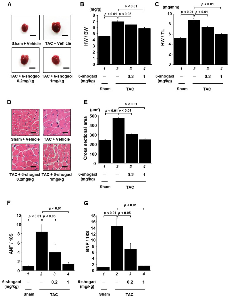
Figure 6.
LVH was significantly inhibited by 6-shogaol administration in TAC mice. (A) At 8 weeks after surgery, hearts were isolated from the sham and TAC groups. Scale bar: 5 mm. Quantified HW/BW ratio (B) and HW/TL ratio (C) of mice. Results are shown as the mean ± SEM (n = 10 mice). (D) Representative photographs of HE-stained sections of LV myocardium from sham and TAC mice. Magnification: ×400. Scale bar: 10 μm. (E) At least 70 cells in each group were used to measure the cardiomyocyte cross-sectional area. Hypertrophy-related ANF (F) and BNP (G) gene expression levels were examined by quantitative RT-PCR. Results are shown as the mean ± SEM of six individual experiments.

