This is a correction to: Maolin Zhang, Fei Jiang, Xiaochen Zhang, Shaoyi Wang, Yuqin Jin, Wenjie Zhang, Xinquan Jiang, The Effects of Platelet-Derived Growth Factor-BB on Human Dental Pulp Stem Cells Mediated Dentin-Pulp Complex Regeneration, Stem Cells Translational Medicine, Volume 6, Issue 12, December 2017, Pages 2126–2134,https://doi.org/10.1002/sctm.17-0033
In the originally published version of this article, please note the following errors, which have been corrected only in this correction notice to preserve the published version of record. The authors and journal editor have confirmed that these corrections do not affect the conclusion of this work.
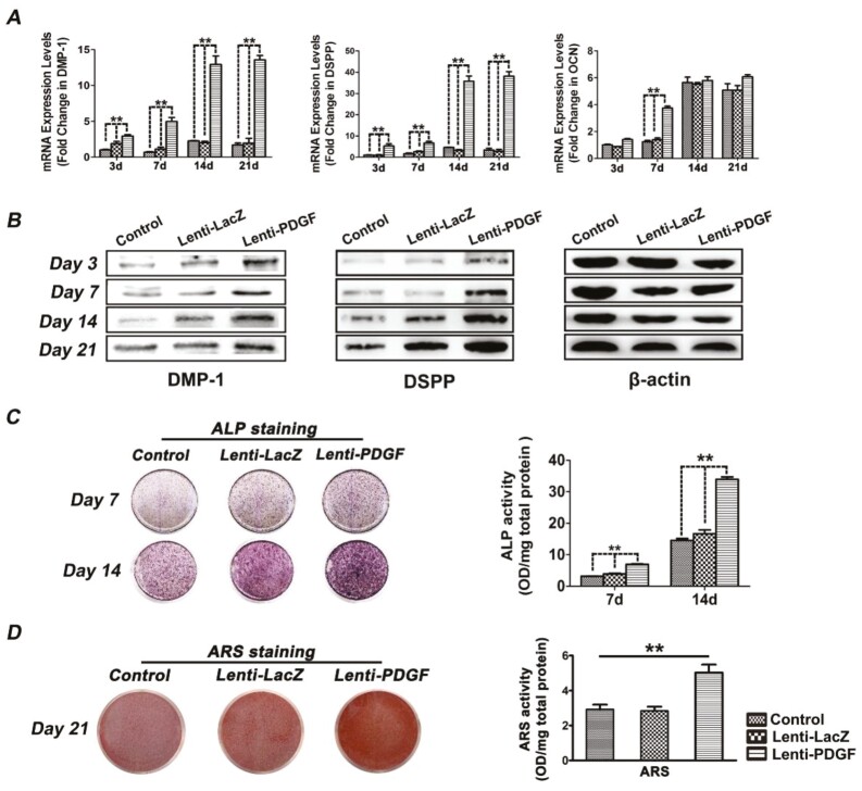
During figure assembly, the Day 21 β-actin band in Figure 2B was accidentally rotated. The corrected figure is shown here.
Corrected Figure 2B:
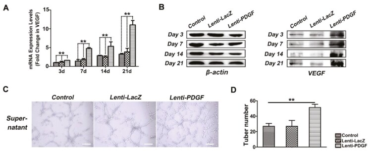
In Figure 5B, the label β-catenin was incorrectly applied. The panel should have been labeled as β-actin. β-actin was used as the internal standard to detect the expression of DMP-1 and DSPP (as shown in Figure 2B) and of VEGF (as shown in Figure 5B). The experiments were performed using the same group of samples, so the β-actin bands should appear the same in both figures.
Corrected Figure 5B:
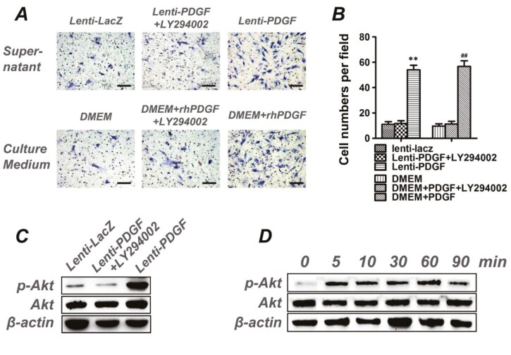
The image of Lenti-PDGF+LY294002 group in Figure 4A as originally published is incorrect and should be replaced with the following.
Corrected Figure 4A:
In the Results section under the heading “Human DPSCs Culture and Characterization”, the corrected text is as follows:
The teeth were dissected around their cervical part with a dental bur to exposure the dental pulp tissue (Fig. S1A, S1B). The isolated pulp tissue (Fig. S1C) was minced into 1-2 mm3 pieces, and these tissue pieces were then plated in a petri dish and cultured in Dulbecco’s Modified Eagle’s medium (DMEM) containing 20% fetal bovine serum (FBS). The primary hDPSCs (outgrowth cells) presented a fibroblast-like spindle shape (Fig. S1D). At passage three, hDPSCs were harvested for flow cytometric analysis. Flow cytometric analysis revealed that these cells were negative for the hematopoietic markers CD31 and CD34 (3.6% and 3.1%, respectively), and that they expressed high levels of stem cell markers CD90 and CD105 (71.9% and 96.4%, respectively), (Fig. S1E). We also tested the multidirectional differentiation potential of hDPSCs. The results show that the isolated cells could differentiate into adipocytes positive staining for lipid droplets with oil Red O (Fig. S1F), chondrocytes positive staining for cartilage proteoglycan with Alcian blue (Fig. S1G), and osteoblasts positive staining for mineral nodules with alizarin red S (Fig. S1H), respectively.


