Abstract
Discoidin Domain Receptor (DDR)-1 is activated by collagen. Nilotinib is a tyrosine kinase inhibitor that is FDA-approved for leukemia and potently inhibits DDR-1. Individuals diagnosed with mild–moderate Alzheimer’s disease (AD) treated with nilotinib (versus placebo) for 12 months showed reduction of amyloid plaque and cerebrospinal fluid (CSF) amyloid, and attenuation of hippocampal volume loss. However, the mechanisms are unclear. Here, we explored unbiased next generation whole genome miRNA sequencing from AD patients CSF and miRNAs were matched with their corresponding mRNAs using gene ontology. Changes in CSF miRNAs were confirmed via measurement of CSF DDR1 activity and plasma levels of AD biomarkers. Approximately 1050 miRNAs are detected in the CSF but only 17 miRNAs are specifically altered between baseline and 12-month treatment with nilotinib versus placebo. Treatment with nilotinib significantly reduces collagen and DDR1 gene expression (upregulated in AD brain), in association with inhibition of CSF DDR1. Pro-inflammatory cytokines, including interleukins and chemokines are reduced along with caspase-3 gene expression. Specific genes that indicate vascular fibrosis, e.g., collagen, Transforming Growth Factors (TGFs) and Tissue Inhibitors of Metalloproteases (TIMPs) are altered by DDR1 inhibition with nilotinib. Specific changes in vesicular transport, including the neurotransmitters dopamine and acetylcholine, and autophagy genes, including ATGs, indicate facilitation of autophagic flux and cellular trafficking. Inhibition of DDR1 with nilotinib may be a safe and effective adjunct treatment strategy involving an oral drug that enters the CNS and adequately engages its target. DDR1 inhibition with nilotinib exhibits multi-modal effects not only on amyloid and tau clearance but also on anti-inflammatory markers that may reduce cerebrovascular fibrosis.
Supplementary Information
The online version contains supplementary material available at 10.1186/s12974-023-02802-0.
Keywords: Discoidin domain receptor 1, Alzheimer’s disease, MicroRNAs, Collagen, Nilotinib, Cytokines, Chemokines
Background
Vascular contributions to cognitive impairment are increasingly recognized [1–6]. Small vessel disease of the brain may contribute to all dementias, including Alzheimer’s disease (AD) [5–7]. Vascular changes in AD have been attributed to the vasculo-toxic effects of β-amyloid (Aβ) [8]. Early cognitive dysfunction is associated with hippocampal capillary damage and blood–brain barrier (BBB) breakdown irrespective of Aβ and/or tau pathologies [9]. Animal studies suggest that Aβ and tau lead to blood vessel abnormalities and BBB breakdown [8, 10–12]. However, the development of neurovascular dysfunction and its relationship with Aβ and tau in the dementia continuum are not well understood.
Discoidin Domain Receptor (DDR)-1 is a non-receptor tyrosine kinase (TK) that is implicated in several fibrotic disorders. DDR1 inhibition reduces accumulation of macrophages, attenuates the inflammatory response and suppresses fibrosis in models of atherosclerosis [13, 14], and renal and lung fibrosis [15, 16]. DDR1 is expressed in the CNS and its activation increases microglial activity and matrix metalloproteases (MMPs), leading to BBB breakdown [17]. DDR1 inhibition stimulates clearance of neurotoxic proteins via autophagy in animal models of neurodegeneration [18–22] and DDR1 is upregulated in post-mortem Parkinson’s disease (PD) and AD brains [18, 19, 23–29]. Knockdown of DDR1 via shRNA reduces Aβ levels and prevents cell loss in vivo and in vitro [25]. Whole genome miRNA sequencing of CSF from patients diagnosed with PD with mild cognitive impairment (PD–MCI) and treated with the DDR1 inhibitor, nilotinib, demonstrates changes of molecular pathways that regulate angiogenesis and the BBB, neuro-inflammation, and protein clearance via autophagy [30–32]. Changes of miRNAs that control angiogenesis and the BBB are significantly correlated with motor and cognitive stability in PD–MCI patients [31].
To address the question of DDR1 effects on blood vessels and inflammation in dementia, we performed longitudinal epigenomic studies via unbiased next generation whole miRNA genome sequencing of CSF obtained from mild–moderate AD patients who were treated with nilotinib (versus placebo), which significantly reduced CSF Aβ levels and CNS amyloid burden as measured with Positron Emission Tomography (PET) [33].
Materials and methods
Study design
The primary research objective of this study was to assess longitudinal alterations in CSF microRNAs from samples previously collected in mild–moderate AD patients [33]. As previously described, AD patients [33] were randomized to placebo for 12 months (n = 20) or nilotinib, 150 mg for 6 months followed by nilotinib, 300 mg, for another 6 months (n = 17). All enrollees (N = 37) met inclusion/exclusion criteria and the study excluded cardiovascular disease, mini mental status exams (MMSE) score out of range, or a diagnosis of AD not supported by biomarker evidence. Concomitant drugs known to prolong the QTc interval and history of any cardiovascular disease, including myocardial infraction or cardiac failure, angina, arrhythmia were not permitted. Any cardiovascular or cerebrovascular event (e.g., myocardial infarction, unstable angina, or stroke), congestive heart failure, second- or third-degree atrioventricular block, sick sinus syndrome, or other serious cardiac rhythm disturbances were excluded. Significant pathological findings on brain MRI (historical in the last 12 months prior to enrollment), including cerebral microhemorrhages, cerebral macro-hemorrhages, or superficial siderosis with vasogenic edema were excluded. Other excluded MRI findings included more than 4 micro hemorrhages (defined as 10 mm or less at the greatest diameter); a single macro hemorrhage greater than 10 mm at greatest diameter; evidence of cerebral contusion, encephalomalacia, aneurysms, vascular malformations, or infective lesions; evidence of multiple lacunar infarcts or stroke involving a major vascular territory, severe small vessel, or white matter disease; space occupying lesions; or brain tumors. There was two diabetic patients in this study. Mean (SD) age of the participants was 70.7 years (6.48) and the sample included 10 men (27%) and 27 women (73%). The mean (SD) MMSE was 19.2 (3.1) in the placebo group and 19.8 (2.5) in the nilotinib group. The apolipoprotein-E (APOE) genotype in the placebo group was E4/E4 5 (25%), E3/E4 5 (25%), E2/E4, 4 (20%), E2/E2 1 (5%), and 5 (25%) inconclusive genotypes. In the nilotinib group, APOE was E4/E4 7 (41.2%), E3/E4 1 (5.9%), E2/E4 3 (17.6%), E3/E3 1 (5.9%), E2/E2 1 (5.9%), and 4 (23.5%) inconclusive genotypes. Participants and study partners were recruited, and this study was conducted in accordance with Good Clinical Practice guidelines and approved by the Institutional Review Board (IRB #2016–0351) at GUMC as well as GHUCCTS scientific review board. The study was conducted under FDA Investigational New Drug (IND) #130732 and registered in ClinicalTrials.gov (NCT02947893) [33]. All samples collected and used for experiments presented in this study were de-identified and relabeled with a study ID.
Cerebrospinal fluid microRNA sequencing and analysis
CSF was collected via lumbar puncture (LPs), which was performed on all patients between 1 and 4 h at baseline and 12 months (end of treatment) with placebo or nilotinib. Cell-free total RNA was isolated from 200 µl of CSF using the Qiagen miRNAeasy extraction kit (Qiagen, 217,184) and CSF microRNA sequencing was performed as we previously explained [32]. Samples were normalized to an input volume of 5 μl RNA eluate to prepare miRNAseq libraries using Qiagen QiaSeq miRNA-seq library preparation kit (Qiagen, 331,502). Next-generation sequencing (NGS) was performed on a NextSeq 550 Sequencing System (Illumina) using single-end (SE) 1 × 75 base pairs (bp) sequencing to a depth of 25 million raw reads per sample.
Statistical analysis
Data analysis was performed using Partek Flow software to identify differentially expressed microRNAs. An optimized workflow was used to perform quality assessment, alignment, quantification, normalization, and different analysis. Quality assessment of data in fastq format was performed in Partek flow and reads containing the adapters or low-quality bases (Qscore ≤5) were removed. The trimmed reads were then aligned to the hg38 reference genome using the Bowtie method. The aligned reads were then quantified to the miRBase reference to obtain the total miRNA read count for each sample. DESeq2 method was used for normalization of the miRNA read counts, followed by DESeq2 implementation of the likelihood ratio test (LRT) to test for multiple terms at once and interaction between the variables. The LRT for count data is conceptually similar to an analysis of variance (ANOVA) calculation, except that in the case of the Negative Binomial GLM, an analysis of deviance (ANODEV) is used, where the deviance captures the difference in likelihood between a full and a reduced model [34]. The p values obtained are then adjusted for multiple testing using the Benjamini and Hochberg method. Gene ontology (GO) pathway and functional enrichment analyses were performed on the gene targets of the significant DEMs using PANTHER Classification System. Only enriched pathways and GO terms that met Benjamini–Hochberg p < 0.05 were reported.
Measurement of CSF DDR1 activity
To estimate spectrophotometrically the levels of active or phosphorylated DDR1 (pDDR1) in CSF, we measured the level of pan-tyrosine pDDR1 via ELSA using PathScan phosphorylated DDR1 panTyrosine (Cell Signaling, KIT# 7863) and determined the absorbance (AU) change during the assay. To perform longitudinal measurement of CSF pDDR1 over time within the same patient, absorbance measurement of CSF pDDR1 collected at baseline was subtracted from absorbance (AU) at the next CSF collection and estimated the change of linear absorbance versus time of assay mixture measured at wavelength 450 nm. We plotted the linear graph with absorbance change versus time that is the absorbance change per time of the reaction (ΔA/time) and found the slope (m) that is y = mx + c using linear regression analysis as a convenient mathematical expression. Slower slope suggests less active DDR1 (less pDDR1), and faster slope is more active DDR1 (more pDDR1) over time.
Plasma biomarkers
Plasma Aβ40, Aβ42, total tau, and p-tau181 and p-tau217 were measured using Biofluid Biomarker Assay via SQUID IMR (Immuno Magnetic Reagents) Platform (MagQu Co., Ltd) that uses UMI magnetic reagents (Cat # AB0-0068, MagQu) and Aβ40, Aβ42, total tau, and p-tau 181 and p-tau217 IMR reagent (Cat. #MF-ASC-006B; MagQu).
Data availability
The data supporting the current study are available from the corresponding author on request.
Results
miRNAs that target DDR1 are significantly altered by nilotinib treatment.
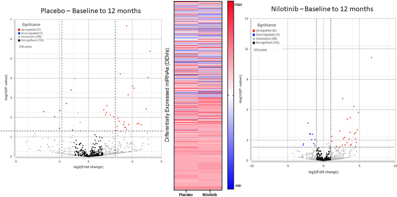
Unbiased next generation whole genome sequencing of CSF miRNA showed expression profiles of a total of 1050 miRNA (Fig. 1) between baseline and 12 months in the placebo (n = 11) and nilotinib groups (n = 12). A total of 17 microRNAs (1.6%) were significantly different between the placebo and nilotinib groups, accounting for both treatment and time. Ingenuity Pathway Analysis (IPA) microRNA Target filter tool matched associated mRNAs or targets that are experimentally validated from TarBase, miRecords, as well as highly predicted miRNA–mRNA interactions from TargetScan and revealed that these microRNAs are associated with genes that primarily control molecular pathways of autophagy, inflammation, collagen expression, and dopamine metabolism.
Fig. 1.
Unbiased next generation whole genome sequencing of CSF miRNA showed expression profiles of a total of 1050 miRNA between baseline and 12 months in the placebo (n = 11) and nilotinib groups (n = 12) (two-way ANOVA)
DDR1 is longitudinally inhibited in the CSF of AD patients treated with nilotinib
To verify the effects of nilotinib on DDR1 activity, we show that the level of CSF pDDR1 in placebo-treated AD patients (Fig. 2A) was significantly increased between baseline and end of treatment (slope = 1.003, p = 0.04). However, treatment of AD patients with the DDR1 inhibitor nilotinib (Fig. 2B) showed a remarkably slower (> 10x) and negative slope (slope = -0.1610, p = 0.7). We previously demonstrated that DDR1 inhibition with nilotinib lowers CSF Aβ levels and reduces CNS amyloid burden by PET in the same cohort of AD patients [33]. Here, we show that the ratio of plasma Aβ42/ptau217 is also significantly reduced with nilotinib compared to placebo (Fig. 3C), suggesting alterations of amyloid and tau levels, in agreement with previous findings [33]. A specific miRNA (hsa-miR-199b-5p) that targets DDR1 gene expression is reduced in the placebo versus nilotinib groups (Fig. 3), showing downregulation of DDR1 expression in the nilotinib versus placebo.
Fig. 2.
DDR1 is longitudinally inhibited in the CSF of AD patients treated with nilotinib. ELISA measurement of pan-tyrosine phosphorylated DDR1 (pDDR1) shows the slope (m) of log10 linear regression of mean difference values in the CSF of A) AD patients from baseline-end of treatment, indicating (rising slopes) an increase in active pDDR1 level over time. Nilotinib treatment B) results in slower slope (m) of log10 linear regression of mean difference values between baseline and end of treatment in AD patients treated with nilotinib, indicating reduction of pDDR1 (active). N = 12 placebo and n = 12 in nilotinib. C) measurement of plasma Aβ42 and pTau217 using Biofluid Biomarker Assay via SQUID IMR (Immuno Magnetic Reagents) Platform (MagQu Co., Ltd). N = 12 placebo and N = 10 nilotinib. Graphed as mean ± standard error of the mean. The changes in the ratio of Aβ42/pTau (217) across each group were compared using a one‐tailed unpaired t‐test with Welch's corrections. Asterisks denote actual P‐value significances (*P < 0.05) between groups and are noted in the figure. Statistical analysis was performed using GraphPad Prism, version 9.1.2 (GraphPad Software Inc.)
Fig. 3.
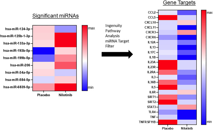
miRNAs that target inflammation are significantly reduced in nilotinib treated patients. A total of 17 microRNAs (1.6%) were significantly different between the placebo and nilotinib groups, accounting for both treatment and time. Ingenuity Pathway Analysis of microRNA matched associated mRNAs or targets that are experimentally validated from TarBase, miRecords, as well as highly predicted miRNA–mRNA interactions from TargetScan and revealed that 9 of these microRNAs are associated with genes that regulate inflammation. N = 11 placebo, n = 12 nilotinib
miRNAs that target inflammation are reduced in nilotinib treated patients
We observed changes in specific miRNAs that control interleukins (ILs) and chemokines that are collectively indicative of reduced inflammation with nilotinib treatment (Fig. 3). Specific miRNAs that target genes related to pro-inflammatory cytokines were enhanced following nilotinib treatment (Fig. 3), including but not limited to IL1β (hsa-miR-125b-1-3p), IL2 (hsa-miR-199b-5p), IL3 (hsa-miR-206), IL5 (hsa-miR-193b-5p), IL12 (hsa-miR-96-5p), IL13 (hsa-miR-125b-1-3p), IL17 (hsa-miR-206), IL23 (hsa-miR-193b-5p, hsa-miR-199b-5p), IL36 (hsa-miR-199b-5p), resulting in decreased expression, while the anti-inflammatory IL6 (hsa-miR-34a-5p) was increased. IL-6 and signal transducer and activator of transcription 3 (STAT3) (hsa-miR-124-3p), a signaling pathway in the regulation of macrophage polarization, M1-type and M2-types, are both upregulated with nilotinib, suggesting an overall change in the anti-inflammatory profile. Expression of the pro-inflammatory tissue necrosis factor (TNF) (hsa-miR-125b-1-3p) and its receptors (TNFRs) (hsa-miR-193b-5p), in addition to toll-like receptor (TLR-4) (hsa-miR-6839-5p) are also reduced in nilotinib-treated patients. There is significant regulation of CCL2 (hsa-miR-206), CCL5 (hsa-miR-193b-5p, hsa-miR-584-5p), and CXCL10 (hsa-miR-34a-5p), CXCL11 (hsa-miR-133a-3p), CXCR3 (hsa-miR-124-3p), and CXCR6 (hsa-miR-6839-5p).
miRNAs that target vascular fibrosis are reduced in nilotinib treated patients
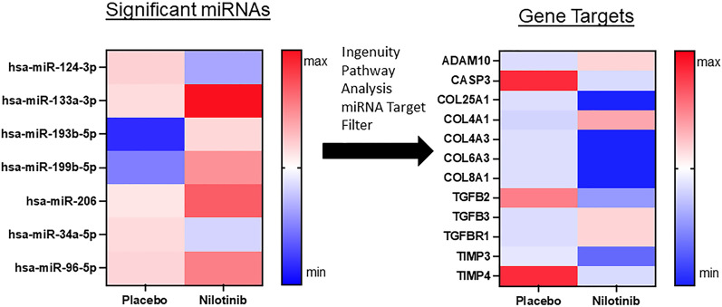
Specific miRNAs that target collagens (hsa-miR-133a-3p, hsa-miR-96-5p) were increased in the nilotinib group (Fig. 4), indicating a decrease in expression of collagens 25, 4, 6, and 8. There are differential changes between nilotinib and placebo groups in transforming growth factor-beta 2 (TGF-β2) (hsa-miR-199b-5p) and 3 (hsa-miR-34a-5p) and an increase with nilotinib in expression of the corresponding receptor (TGFBR1) (hsa-miR-34a-5p), which is a major mediator of tissue fibrosis and regulation of MMPs, suggesting reduced effects on fibrosis. Significant increases in miRNAs (hsa-miR-206, hsa-miR-193b-5p) that target Tissue Inhibitors of Metalloproteases (TIMP)3 and TIMP4 activity that regulate amyloid precursor protein (APP) cleavage and are expressed at increased levels in AD brains, indicate a normalization of TIMP expression—further supporting the hypothesis that DDR1 inhibition promotes neurovascular health. ADAM10 (A Disintegrin and Metalloproteinase), known as α-secretase that plays a critical role in APP cleavage, is downregulated in AD but is increased (hsa-miR-34a-5p) in nilotinib-treated patients. Notably, expression of critical markers of apoptosis such as caspase-3 (hsa-miR-193b-5p) and BCL2 (hsa-miR-206) are reduced with nilotinib. To validate the effects of DDR1 on collagen expression in a mouse model of vascular angiopathy (See Additional file 1), we generated DDR1 knockout mice (TgAPPtg/tg/DDR−/−) and showed that vascular collagen IV (Additional file 2: Fig. S1) was significantly reduced along the endothelial lining of blood vessels in TgAPPtg/tg/DDR−/−, resulting in increased blood vessel diameter, compared to TgAPPtg/tg/DDR+/+ littermate controls.
Fig. 4.
miRNAs that target vascular fibrosis are significantly reduced in nilotinib treated patients. A total of 17 microRNAs (1.6%) were significantly different between the placebo and nilotinib groups, accounting for both treatment and time. Ingenuity Pathway Analysis of microRNA matched associated mRNAs or targets that are experimentally validated from TarBase, miRecords, as well as highly predicted miRNA–mRNA interactions from TargetScan and revealed that 7 of these microRNAs are associated with genes that regulate collagen expression. N = 11 placebo, n = 12 nilotinib
miRNAs that target DDR1 and autophagy are significantly altered by nilotinib treatment
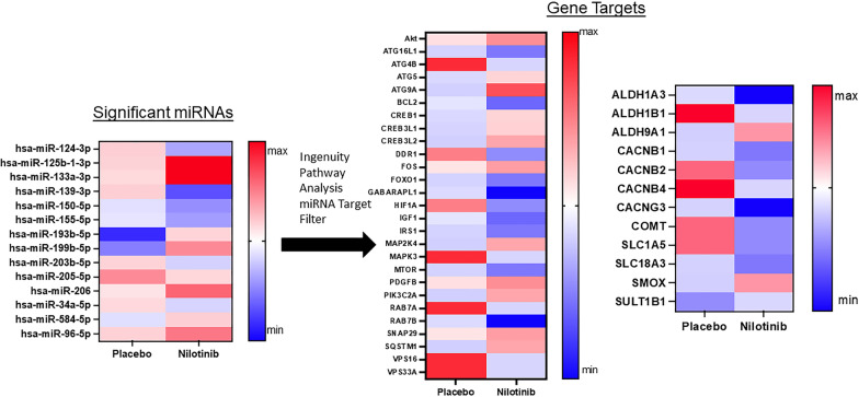
In addition, specific miRNAs that target autophagy-associated genes (ATGs) were altered to promote autophagy flux in the nilotinib group when compared to the placebo group (Fig. 5). These miRNAs and ATG changes include downregulation of ATG16L1 (hsa-miR-96-5p) that inhibits autophagosome formation, and ATG4B (hsa-miR-193b-5p), that downregulates autophagy [35]. Upregulation of AKT (hsa-miR-124-3p, hsa-miR-150-5p) is known to enhance ATG4B activity, which is a key regulator of the LC3/GABA-A Receptor associated protein GABARAPL1 (hsa-miR-133a-3p) that interacts with the family of ATG8 (also known as LC3)—indispensable for autophagosome formation and maturation [35]. ATG9A (hsa-miR-139-3p) is critical for autophagosome formation and ATG5 (hsa-miR-34a-5p) is an integral part of the ATG5–ATG12–ATG16L1 complex that catalyzes ATG8/LC3 lipidation for autophagosome formation, and maturation [35]. Interestingly, positive markers of autophagic flux in the nilotinib group are also associated with downregulation of BCL2 (hsa-miR-206), that binds to Beclin 1, thus preventing assembly of the pre-autophagosome structure and inhibiting autophagy and apoptosis. Furthermore, inhibition of the mammalian target of rapamycin (mTOR) (hsa-miR-96-5p), which is downregulated by nilotinib, is associated with induction of autophagy [35]. Several endosome-related genes, i.e., protein vesicle trafficking via RAB7A and RAB7B (hsa-miR-193b-5p, hsa-miR-133a-3p), the autophagosome cargo known as ubiquitin-binding protein p62 (SQSTM1) (hsa-miR-124-3p), and Vacuolar Sorting genes VPS16 and VPS33A (hsa-miR-193b-5p) and synaptosome associated protein (SNAP) 29 (hsa-miR-155-5p) are also differentially regulated between the placebo and nilotinib groups. Collectively, these changes are indicative of autophagosome maturation and autophagy flux, membrane trafficking and cellular transport [35].
Fig. 5.
miRNAs that target autophagy and dopamine metabolism are significantly altered between placebo and nilotinib. A total of 17 microRNAs (1.6%) were significantly different between the placebo and nilotinib groups, accounting for both treatment and time. Ingenuity Pathway Analysis of microRNA matched associated mRNAs or targets that are experimentally validated from TarBase, miRecords, as well as highly predicted miRNA–mRNA interactions from TargetScan and revealed that 14 of these microRNAs are associated with genes that control autophagy and dopamine metabolism. N = 11 placebo, n = 12 nilotinib
We also observed upregulation of the nuclear transcription factor CREB (hsa-miR-34a-5p, hsa-miR-203b-5p), that binds to cAMP response element (CRE) to regulate neuronal processes, including metabolism and survival and expression of different transcription factors and growth factors. Platelet derived growth factor (PDGF)B (hsa-miR-150-5p) is upregulated in the nilotinib group. There is also a significant decrease in insulin growth factor (IGF), (hsa-miR-206) and Insulin Receptor Signaling (IRS1), (hsa-miR-96-5p) that control main pathways of insulin signaling, such as the phosphatidylinositol 3-kinase (PI3K) (hsa-miR-124-3p), Akt (hsa-miR-150-5p), Akt substrates, Ras and mitogen-activated protein (MAP) (hsa-miR-124-3p, hsa-miR-193b-5p) kinase. Hypoxia-Inducible Factor (HIF1A) (hsa-miR-199b-5p), and the Forkhead/winged helix box gene (FOXO) (hsa-miR-96-5p) are involved in energy metabolism, oxidative stress, proteostatis, apoptosis, immunity, and inflammation, and are downregulated, whereas, expression of the immediate early gene, Fos (hsa-miR-155-5p), that is critical to learning and memory in increased with nilotinib.
miRNAs that target dopamine and acetylcholine metabolism are significantly altered between placebo and nilotinib
We previously demonstrated that AD patients treated with nilotinib exhibit a decrease in dopamine metabolism following 12 months of treatment with nilotinib versus placebo [33]. Here, we observe inhibition of Catechol-O-methyltransferase (COMT) (hsa-miR-199b-5p) which is one of several enzymes that degrade catecholamine, including dopamine (Fig. 5). Interestingly, SLC18A3 (hsa-miR-96-5p), the vesicular acetylcholine transporter, and VMAT1, the vesicular monoamine (dopamine) transporter (SLC1A5) (hsa-miR-199b-5p) are reduced by nilotinib, suggesting prolonged synaptic presence of these neurotransmitters prior to active transport into synaptic vesicles. There is an increase in sulfotransferases (SULT1B1) (hsa-miR-205-5p), which mediates sulfonation or detoxification of dopamine. There is an increase in spermine oxidase (SMOX) (hsa-miR-124-3p, hsa-miR-34a-5p) that is involved in polyamine catabolism. We also observed upregulation of the nuclear transcription factor (CREB) (hsa-miR-34a-5p, hsa-miR-203b-5p) that binds to cAMP response element (CRE), to regulate neuronal processes, including metabolism and survival and expression of different transcription factors and growth factors. Expression of several components of a calcium channel complex consisting of the ancillary subunits CACNB1 (hsa-miR-96-5p), CACNB2 (hsa-miR-199b-5p, hsa-miR-584-5p), CACNB4 (hsa-miR-193b-5p, hsa-miR-96-5p, and CACNG3 (hsa-miR-125b-1-3p) are decreased, suggesting reduced calcium-induced toxicity. Changes in expression of several subunits or isozymes of Aldehyde Dehydrogenase 1, including ALDH1A (hsa-miR-133a-3p), ALDH1B (hsa-miR-193b-5p), and ALDH9A (hsa-miR-124-3p), that are associated with detoxification, energy metabolism and Gamma-Amino Butyric Acid (GABA) metabolism were also observed.
Discussion
The current study is a continuation of our previous phase 2 investigation in mild–moderate AD patients treated with the DDR1 inhibitor, nilotinib, which significantly lowered CSF Aβ levels and amyloid burden as measured by PET [33]. Nilotinib inhibits Abelson (IC50 > 20 nM) [36] and is FDA-approved for Philadelphia chromosome Chronic Myelogenous Leukemia (CML) that results from Abelson (Abl) mutations at oral doses of 300 mg twice daily. However, nilotinib more potently inhibits DDR1 (IC50 = 1 nM) [36]. Pharmacologically, nilotinib was measured in the CSF and plasma of AD patients at the 150 mg daily dosage (Cmax: 3.46 nM and 1099 nM, respectively) and the 300 mg daily dosage (Cmax: 4.7 nM and 1410 nM, respectively), suggesting a linearly proportional increase to dose in the brain [33] that would be sufficient to inhibit DDR1 (IC50 1 nM). Here, we show that treatment with nilotinib significantly increases CSF miRNAs that inversely regulate DDR1 mRNA expression to decrease active CSF pDDR1, indicating a pharmacokinetic and pharmacodynamic relationship. Nilotinib penetrates the CNS (> 1 nM) and directly inhibits DDR1 and alters AD biomarkers, including Aβ as measured by PET, CSF Aβ42 and Aβ40, and the plasma ratio of Aβ42 to p-Tau217. Clinically, nilotinib (Tasigna) 150 mg and 300 mg, resulted in stabilization of cognitive and motor decline in PD–MCI over 27 months [31]. In AD, nilotinib, 150 mg may influence executive functions, but the higher 300 mg dose increases agitation and irritability according to caregiver reports [33]. Importantly, miRNA data demonstrate reduction of chemokines and cytokines and other inflammatory markers in the CSF of nilotinib-treated AD patients compared to placebo suggesting reduction of inflammation, in agreement with pre-clinical data that show DDR1 inhibition stimulates clearance of neurotoxic proteins via autophagy and reduces inflammation in animal models of neurodegeneration [18, 22, 25, 26, 30]. Notably, critical markers of apoptosis such as caspase-3 and BCL2 expression are reduced with nilotinib, consistent with volumetric magnetic resonance imaging (MRI) that show reduced atrophy of hippocampal volume in nilotinib-treated AD patients [33].
DDR1 plays a direct role in angiogenesis [37] and regulates the BBB [38] via interaction with collagen [39] and epithelial cell maintenance as an ECM-sensing TK [40] that controls inflammation [41]. We observed reduction of DDR1 and COL25A1, which is also known as collagen-like Alzheimer’s amyloid plaque component precursor specifically expressed in neurons and colocalized with Aβ plaques in AD brains [42, 43]. COL25A1 increases AD risk in a Swedish population [44]. COL25A1 is a type of collagen expressed by neurons that interacts with BACE1 that cleaves APP, and induces synapse damage and astrocyte activation [45]. The COL4A1 gene, in association with COL4A3, are necessary for maturation of type IV collagen, and COL4A1 mutations cause cerebral small vessel disease, i.e., intracerebral hemorrhage and shortened life-span [46, 47]. COL4A1 mutations are associated with increased thickening of capillary basement membranes [47], suggesting vascular fibrosis, in agreement with the preclinical data (Additional file 1) in APP/DDR1 mice. The differential changes in direction in COL4A1 and COL4A3 expression in the nilotinib-treated group may be due to subunits being associated with distinct processes that regulate overall collagen IV levels. Interestingly, DDR1 activation is required for Collagen IV synthesis [48] and DDR1 regulates collagen transcription and MMP production [49]; we observed alterations in subtypes of tissue TIMP3 and TIMP4 as key regulators of MMP expression [50] in the nilotinib group. We also observed reduction of TGF, which is the master regulator of fibrosis [51], as well as attenuation of inflammatory markers, including chemokines, cytokines and TNF-alpha, in the nilotinib-treated group in agreement with reduced collagen and fibrosis, in association with cytokines [52] and chemokine production [53]. COL6 genes encode the ECM protein collagen VI, and mutations in these genes are associated with muscular dystrophies [54].
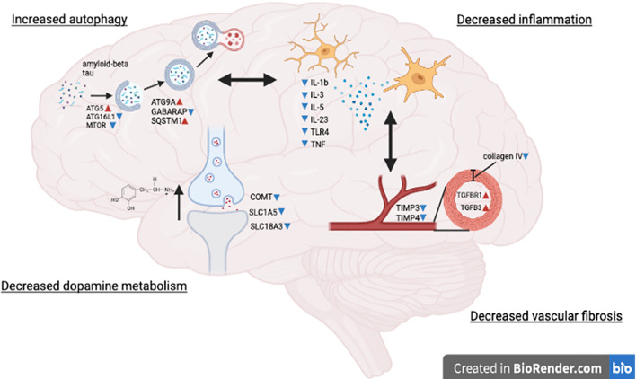
Dopamine plays an important role in executive functions via dopaminergic neurons in the mesolimbic system and this role may be particularly relevant to AD [55, 56], as nilotinib prevents loss of dopamine in the ventral tegmental area (VTA) and improves cognition in AD models [57]. Indeed, AD and PD studies support a dose-dependent increase of CNS dopamine via measurement of CSF metabolites in individuals treated with nilotinib [33, 58–60], suggesting that the increases in CNS dopamine [31] with 300 mg nilotinib-treated AD patients may lead to behavioral changes [61]. Importantly, no mood swings or behavioral changes were reported in patients treated with nilotinib, 150 mg, indicating that this dosage is more adequate to avoid behavioral adverse effects in AD [33]. Our current miRNA data is consistent with previous reports showing reduction of dopamine catabolism [33] perhaps due to downregulation of COMT expression and reduced dopamine transport, which result in increased dopamine bioavailability. The miRNA results also show an effect on acetylcholine metabolism, downstream calcium signaling and early gene expression, pointing to possible nilotinib effects on neurotransmitters, including dopamine and acetylcholine. Collectively, these data indicate that nilotinib penetrates the BBB to inhibit DDR1 and alter CNS dopamine metabolism (Fig. 6).
Fig. 6.
Tyrosine kinase DDR1 inhibition with nilotinib. Treatment of AD patients with nilotinib may induce multiple mechanisms that include activation of genes that control the execution and completion of autophagy, suggesting clearance of intracellular amyloid and tau that would perhaps reduce plaque burden and affect plasma amyloid and tau. Nilotinib treatment led to downregulation of several pro-inflammatory makers along with reduction of collagen, TIMPs and TGFs that maintain blood vessel integrity and prevent vascular fibrosis. Nilotinib treatment leads to reduction of dopamine breakdown probably via altered vesicular transport and reduction of COMT
Demonstration of miRNA changes that control genes which regulate molecular pathways involved in autophagy are indicative of autophagosome maturation, membrane trafficking and cellular transport [35] and are consistent with previous data showing the effects of nilotinib on ubiquitination and the endosomal and autophagy–lysosomal pathways [30]. Importantly, the increase in autophagy flux leads to reduction (or enhanced clearance) of intracellular or intraneuronal protein levels, including Aβ and tau, in animal models [18, 19, 62–64], suggesting that the observed reduction on Aβ PET and CSF and plasma Aβ and tau in AD patients may be due to the effect of nilotinib on neuronal autophagy (Fig. 6). Nilotinib effects on Aβ clearance provide a stark contrast with the effects of anti-amyloid therapies, e.g., Lecanemab, that targets extracellular amyloid species and only partially slows cognitive decline (27%) in early AD patients despite elimination of extracellular plaques [65]. Reduction of intracellular Aβ and tau via autophagy may also result in reduction of extracellular Aβ due to possible degradation of extracellular Aβ oligomers that internalize into endosomes/lysosomes in neurons [66] or the microglial endocytic pathway [67], suggesting that nilotinib enhances neuronal and microglial lysosome-mediated Aβ degradation.
The current miRNA analysis has several limitations due to underpowered study design that would consider more adequate inclusion and stratification of other co-morbidities of AD, such as diabetes, cerebrovascular disease, and other factors, such as sex differences and ApoE genotypes. Furthermore, evaluating extracellular vesicles in CSF following nilotinib delivery is highly desirable in future studies.
In conclusion, the tyrosine kinase inhibitor nilotinib enters the CNS, inhibits DDR1, and is associated with diverse tissue changes (Fig. 6) not only on intracellular amyloid and tau clearance but also on neuroinflammation and cerebrovascular fibrosis. Much more work is needed to prove whether diverse tissue changes are (i) reproducible, (ii) a direct or indirect effect of the drug nilotinib, and (iii) occur in significant numbers of persons. Considering that anti-amyloid antibodies, including, Lecanemab, may result in adverse effects including intracerebral hemorrhage and edema in a subset (18%) of patients [65]. Nilotinib is available on market and safe in humans, and our data support investigation of this drug as potential combination therapy with nilotinib with Lecanemab..
Supplementary Information
Additional file 1. Additional methods and results.
Additional file 2: Fig. S1. Collagen 4and amyloid-betastaining in the cortex of 5-month-old male and female A–D APPtg/tgDDR−/−mice and E–H age matched APPtg/tg DDR+/+littermate controls, and quantification of I vessel wall thickness and J vessel diameter. A–C and E–G blood vessels were imaged longitudinally, D and H blood vessels were imaged cross-sectionally. N=4 mice per group, **=0.0048, ***=0.0002, unpaired two-tailed students t test.
Acknowledgements
We wish to thank the nurses and staff of the Clinical Research Unit at GUMC, Center for Translational and Clinical Science and all staff of the research pharmacy at MedStar Georgetown University Hospital and the Genomics and Epigenomics Shared Resource at GUMC Lombardi Cancer Center.
Abbreviations
- miRNA
MicroRNA
- DDR
Discoidin domain receptor
- MMP
Matrix metalloproteases
- BBB
Blood–brain barrier
- TKI
Tyrosine kinase inhibition
- IC50
Half-maximal inhibitor concentration
- CML
Chronic myelogenous leukemia
- UMI
Unique molecular identifier
- DEM
Differentially expressed miRNA
- GO
Gene ontology
- ECM
Extracellular matrix
- VEGF
Vascular endothelial growth factor
- MCI
Mild cognitive impairment
Author contributions
MS, RV, collected and analyzed miRNA data, curated the figures, and performed statistical analysis MLH, XL, NR, and AS, collected ELISA data and generated the figures. RST and CM, obtained funding, designed experiments, and wrote the manuscript. All authors read and approved the final manuscript.
Funding
This work was supported by Georgetown University funding to C.M and grant to C.M and RST from the Alzheimer Drug Discovery Foundation (ADDF).
Availability of data and materials
The data supporting the current study are available from the corresponding author on request.
Declarations
Ethics approval and consent to participate
Participants and study partners were recruited, and this study was conducted in accordance with Good Clinical Practice guidelines and approved by the Institutional Review Board (IRB #2016–0351) at GUMC as well as GHUCCTS scientific review board. The study was conducted under FDA Investigational New Drug (IND) #130732 and registered in ClinicalTrials.gov (NCT02947893. All samples collected and used for experiments presented in this study were de-identified and relabeled with a study ID.
Consent for publication
Consent for publication was obtained as part of patient’s informed consent.
Competing interests
C.M. is an inventor on several US and international Georgetown University patents to use nilotinib and other tyrosine kinase inhibitors as a treatment for neurodegenerative diseases. Georgetown University and C.M are shareholders in KeifeRX LLC, a biopharmaceutical company, from which C.M. receives consulting fees and is a co-founder. C.M. receives consulting fees from SkyBio LLC and research grants from NIH–NIA and Sun Pharmaceuticals Advanced Research Corporation (SPARC). RST reports research support to Georgetown University from Alector, Biogen, Eisai, Janssen, Lilly, Roche/Genentech, Vaccinex, and Vivoryon. The remaining authors have no potential competing interests.
Footnotes
Publisher's Note
Springer Nature remains neutral with regard to jurisdictional claims in published maps and institutional affiliations.
References
- 1.Snyder HM, Corriveau RA, Craft S, Faber JE, Greenberg SM, Knopman D, Lamb BT, Montine TJ, Nedergaard M, Schaffer CB, et al. Vascular contributions to cognitive impairment and dementia including Alzheimer's disease. Alzheimers Dement. 2015;11:710–717. doi: 10.1016/j.jalz.2014.10.008. [DOI] [PMC free article] [PubMed] [Google Scholar]
- 2.Zlokovic BV. Neurovascular pathways to neurodegeneration in Alzheimer's disease and other disorders. Nat Rev Neurosci. 2011;12:723–738. doi: 10.1038/nrn3114. [DOI] [PMC free article] [PubMed] [Google Scholar]
- 3.Gottesman RF, Schneider AL, Zhou Y, Coresh J, Green E, Gupta N, Knopman DS, Mintz A, Rahmim A, Sharrett AR, et al. Association between midlife vascular risk factors and estimated brain amyloid deposition. JAMA. 2017;317:1443–1450. doi: 10.1001/jama.2017.3090. [DOI] [PMC free article] [PubMed] [Google Scholar]
- 4.Costa RP, Padamsey Z, D'Amour JA, Emptage NJ, Froemke RC, Vogels TP. Synaptic transmission optimization predicts expression loci of long-term plasticity. Neuron. 2017;96(177–189):e177. doi: 10.1016/j.neuron.2017.09.021. [DOI] [PMC free article] [PubMed] [Google Scholar]
- 5.Iadecola C. The neurovascular unit coming of age: a journey through neurovascular coupling in health and disease. Neuron. 2017;96:17–42. doi: 10.1016/j.neuron.2017.07.030. [DOI] [PMC free article] [PubMed] [Google Scholar]
- 6.Sweeney MD, Sagare AP, Zlokovic BV. Blood-brain barrier breakdown in Alzheimer disease and other neurodegenerative disorders. Nat Rev Neurol. 2018;14:133–150. doi: 10.1038/nrneurol.2017.188. [DOI] [PMC free article] [PubMed] [Google Scholar]
- 7.Wardlaw JM, Smith EE, Biessels GJ, Cordonnier C, Fazekas F, Frayne R, Lindley RI, O'Brien JT, Barkhof F, Benavente OR, et al. Neuroimaging standards for research into small vessel disease and its contribution to ageing and neurodegeneration. Lancet Neurol. 2013;12:822–838. doi: 10.1016/S1474-4422(13)70124-8. [DOI] [PMC free article] [PubMed] [Google Scholar]
- 8.Montagne A, Zhao Z, Zlokovic BV. Alzheimer's disease: a matter of blood-brain barrier dysfunction? J Exp Med. 2017;214:3151–3169. doi: 10.1084/jem.20171406. [DOI] [PMC free article] [PubMed] [Google Scholar]
- 9.Nation DA, Sweeney MD, Montagne A, Sagare AP, D'Orazio LM, Pachicano M, Sepehrband F, Nelson AR, Buennagel DP, Harrington MG, et al. Blood-brain barrier breakdown is an early biomarker of human cognitive dysfunction. Nat Med. 2019;25:270–276. doi: 10.1038/s41591-018-0297-y. [DOI] [PMC free article] [PubMed] [Google Scholar]
- 10.Bennett RE, Robbins AB, Hu M, Cao X, Betensky RA, Clark T, Das S, Hyman BT. Tau induces blood vessel abnormalities and angiogenesis-related gene expression in P301L transgenic mice and human Alzheimer's disease. Proc Natl Acad Sci U S A. 2018;115:E1289–E1298. doi: 10.1073/pnas.1710329115. [DOI] [PMC free article] [PubMed] [Google Scholar]
- 11.Blair LJ, Frauen HD, Zhang B, Nordhues BA, Bijan S, Lin YC, Zamudio F, Hernandez LD, Sabbagh JJ, Selenica ML, Dickey CA. Tau depletion prevents progressive blood-brain barrier damage in a mouse model of tauopathy. Acta Neuropathol Commun. 2015;3:8. doi: 10.1186/s40478-015-0186-2. [DOI] [PMC free article] [PubMed] [Google Scholar]
- 12.Jack CR, Jr, Bennett DA, Blennow K, Carrillo MC, Dunn B, Haeberlein SB, Holtzman DM, Jagust W, Jessen F, Karlawish J, et al. NIA-AA research framework: toward a biological definition of Alzheimer's disease. Alzheimers Dement. 2018;14:535–562. doi: 10.1016/j.jalz.2018.02.018. [DOI] [PMC free article] [PubMed] [Google Scholar]
- 13.Franco C, Britto K, Wong E, Hou G, Zhu SN, Chen M, Cybulsky MI, Bendeck MP. Discoidin domain receptor 1 on bone marrow-derived cells promotes macrophage accumulation during atherogenesis. Circ Res. 2009;105:1141–1148. doi: 10.1161/CIRCRESAHA.109.207357. [DOI] [PubMed] [Google Scholar]
- 14.Franco C, Hou G, Ahmad PJ, Fu EY, Koh L, Vogel WF, Bendeck MP. Discoidin domain receptor 1 (ddr1) deletion decreases atherosclerosis by accelerating matrix accumulation and reducing inflammation in low-density lipoprotein receptor-deficient mice. Circ Res. 2008;102:1202–1211. doi: 10.1161/CIRCRESAHA.107.170662. [DOI] [PubMed] [Google Scholar]
- 15.Flamant M, Placier S, Rodenas A, Curat CA, Vogel WF, Chatziantoniou C, Dussaule JC. Discoidin domain receptor 1 null mice are protected against hypertension-induced renal disease. J Am Soc Nephrol. 2006;17:3374–3381. doi: 10.1681/ASN.2006060677. [DOI] [PubMed] [Google Scholar]
- 16.Avivi-Green C, Singal M, Vogel WF. Discoidin domain receptor 1-deficient mice are resistant to bleomycin-induced lung fibrosis. Am J Respir Crit Care Med. 2006;174:420–427. doi: 10.1164/rccm.200603-333OC. [DOI] [PubMed] [Google Scholar]
- 17.Zhu M, Xing D, Lu Z, Fan Y, Hou W, Dong H, Xiong L, Dong H. DDR1 may play a key role in destruction of the blood-brain barrier after cerebral ischemia-reperfusion. Neurosci Res. 2015;96:14–19. doi: 10.1016/j.neures.2015.01.004. [DOI] [PubMed] [Google Scholar]
- 18.Hebron ML, Lonskaya I, Moussa CE. Nilotinib reverses loss of dopamine neurons and improves motor behavior via autophagic degradation of alpha-synuclein in Parkinson's disease models. Hum Mol Genet. 2013;22:3315–3328. doi: 10.1093/hmg/ddt192. [DOI] [PMC free article] [PubMed] [Google Scholar]
- 19.Lonskaya I, Hebron ML, Desforges NM, Franjie A, Moussa CE. Tyrosine kinase inhibition increases functional parkin-Beclin-1 interaction and enhances amyloid clearance and cognitive performance. EMBO Mol Med. 2013;5:1247–1262. doi: 10.1002/emmm.201302771. [DOI] [PMC free article] [PubMed] [Google Scholar]
- 20.Lonskaya I, Hebron M, Chen W, Schachter J, Moussa C. Tau deletion impairs intracellular beta-amyloid-42 clearance and leads to more extracellular plaque deposition in gene transfer models. Mol Neurodegener. 2014;9:46. doi: 10.1186/1750-1326-9-46. [DOI] [PMC free article] [PubMed] [Google Scholar]
- 21.Fowler AJ HM, Missner AA, Wang R, Gao X, Kurd-Misto B, Liu X, Moussa C E-H: Multikinase Abl/DDR/Src inhibition produces optimal effects for tyrosine kinase inhibition in neurodegeneration. Mol Neurodegener 2018. [DOI] [PMC free article] [PubMed]
- 22.Fowler AJ, Hebron M, Missner AA, Wang R, Gao X, Kurd-Misto BT, Liu X, Moussa CE. Multikinase Abl/DDR/Src inhibition produces optimal effects for tyrosine kinase inhibition in neurodegeneration. Drugs R D. 2019 doi: 10.1007/s40268-019-0266-z. [DOI] [PMC free article] [PubMed] [Google Scholar]
- 23.Schlatterer SD, Acker CM, Davies P. c-Abl in neurodegenerative disease. J Mol Neurosci. 2011;45:445–452. doi: 10.1007/s12031-011-9588-1. [DOI] [PMC free article] [PubMed] [Google Scholar]
- 24.Lee S, Kim S, Park YJ, Yun SP, Kwon S-H, Kim D, Kim DY, Shin JS, Cho DJ, Lee GY, et al. The c-Abl inhibitor, Radotinib HCl is neuroprotective in a preclinical Parkinson’s disease mouse model. Hum Mol Genet. 2018 doi: 10.1093/hmg/ddy143. [DOI] [PMC free article] [PubMed] [Google Scholar]
- 25.Hebron M, Peyton M, Liu X, Gao X, Wang R, Lonskaya I, Moussa CE. Discoidin domain receptor inhibition reduces neuropathology and attenuates inflammation in neurodegeneration models. J Neuroimmunol. 2017;311:1–9. doi: 10.1016/j.jneuroim.2017.07.009. [DOI] [PubMed] [Google Scholar]
- 26.Hebron ML, Lonskaya I, Moussa CE. Tyrosine kinase inhibition facilitates autophagic SNCA/alpha-synuclein clearance. Autophagy. 2013;9:1249–1250. doi: 10.4161/auto.25368. [DOI] [PMC free article] [PubMed] [Google Scholar]
- 27.Jing Z, Caltagarone J, Bowser R. Altered subcellular distribution of c-Abl in Alzheimer's disease. J Alzheimers Dis. 2009;17:409–422. doi: 10.3233/JAD-2009-1062. [DOI] [PMC free article] [PubMed] [Google Scholar]
- 28.Ko HS, Lee Y, Shin JH, Karuppagounder SS, Gadad BS, Koleske AJ, Pletnikova O, Troncoso JC, Dawson VL, Dawson TM. Phosphorylation by the c-Abl protein tyrosine kinase inhibits parkin's ubiquitination and protective function. Proc Natl Acad Sci U S A. 2010;107:16691–16696. doi: 10.1073/pnas.1006083107. [DOI] [PMC free article] [PubMed] [Google Scholar]
- 29.Imam SZ, Zhou Q, Yamamoto A, Valente AJ, Ali SF, Bains M, Roberts JL, Kahle PJ, Clark RA, Li S. Novel regulation of parkin function through c-Abl-mediated tyrosine phosphorylation: implications for Parkinson's disease. J Neurosci. 2011;31:157–163. doi: 10.1523/JNEUROSCI.1833-10.2011. [DOI] [PMC free article] [PubMed] [Google Scholar]
- 30.Fowler AJ, Hebron M, Balaraman K, Shi W, Missner AA, Greenzaid JD, Chiu TL, Ullman C, Weatherdon E, Duka V, et al. Discoidin domain receptor 1 is a therapeutic target for neurodegenerative diseases. Hum Mol Genet. 2020 doi: 10.1093/hmg/ddaa177. [DOI] [PMC free article] [PubMed] [Google Scholar]
- 31.Pagan FL, Wilmarth B, Torres-Yaghi Y, Hebron ML, Mulki S, Ferrante D, Matar S, Ahn J, Moussa C. Long-term safety and clinical effects of nilotinib in Parkinson's disease. Mov Disord. 2020 doi: 10.1002/mds.28389. [DOI] [PMC free article] [PubMed] [Google Scholar]
- 32.Fowler AJ, Ahn J, Hebron M, Chiu T, Ayoub R, Mulki S, Ressom H, Torres-Yaghi Y, Wilmarth B, Pagan FL, Moussa C. CSF MicroRNAs reveal impairment of angiogenesis and autophagy in parkinson disease. Neurol Genet. 2021;7:e633. doi: 10.1212/NXG.0000000000000633. [DOI] [PMC free article] [PubMed] [Google Scholar]
- 33.Turner RS, Hebron ML, Lawler A, Mundel EE, Yusuf N, Starr JN, Anjum M, Pagan F, Torres-Yaghi Y, Shi W, et al. Nilotinib effects on safety, tolerability, and biomarkers in Alzheimer's disease. Ann Neurol. 2020 doi: 10.1001/jamaneurol.2019.4200. [DOI] [PMC free article] [PubMed] [Google Scholar]
- 34.Love MI, Huber W, Anders S. Moderated estimation of fold change and dispersion for RNA-seq data with DESeq2. Genome Biol. 2014;15:550. doi: 10.1186/s13059-014-0550-8. [DOI] [PMC free article] [PubMed] [Google Scholar]
- 35.Yamamoto H, Zhang S, Mizushima N. Autophagy genes in biology and disease. Nat Rev Genet. 2023 doi: 10.1016/j.cell.2018.09.048. [DOI] [PMC free article] [PubMed] [Google Scholar]
- 36.Weisberg E, Manley PW, Breitenstein W, Bruggen J, Cowan-Jacob SW, Ray A, Huntly B, Fabbro D, Fendrich G, Hall-Meyers E, et al. Characterization of AMN107, a selective inhibitor of native and mutant Bcr-Abl. Cancer Cell. 2005;7:129–141. doi: 10.1016/j.ccr.2005.01.007. [DOI] [PubMed] [Google Scholar]
- 37.Oh S, Seo M, Choi JS, Joo CK, Lee SK. MiR-199a/b-5p inhibits lymphangiogenesis by targeting discoidin domain receptor 1 in corneal injury. Mol Cells. 2018;41:93–102. doi: 10.14348/molcells.2018.2163. [DOI] [PMC free article] [PubMed] [Google Scholar]
- 38.Stamatovic SM, Keep RF, Andjelkovic AV. Brain endothelial cell-cell junctions: how to "open" the blood brain barrier. Curr Neuropharmacol. 2008;6:179–192. doi: 10.2174/157015908785777210. [DOI] [PMC free article] [PubMed] [Google Scholar]
- 39.Leitinger B, Saltel F. Discoidin domain receptors: multitaskers for physiological and pathological processes. Cell Adh Migr. 2018;12:398–399. doi: 10.1080/19336918.2018.1491495. [DOI] [PMC free article] [PubMed] [Google Scholar]
- 40.Roberts ME, Magowan L, Hall IP, Johnson SR. Discoidin domain receptor 1 regulates bronchial epithelial repair and matrix metalloproteinase production. Eur Respir J. 2011;37:1482–1493. doi: 10.1183/09031936.00039710. [DOI] [PubMed] [Google Scholar]
- 41.Dorison A, Dussaule JC, Chatziantoniou C. The role of discoidin domain receptor 1 in inflammation. Fibrosis Renal Dis Nephron. 2017;137:212–220. doi: 10.1159/000479119. [DOI] [PubMed] [Google Scholar]
- 42.Hashimoto T, Wakabayashi T, Watanabe A, Kowa H, Hosoda R, Nakamura A, Kanazawa I, Arai T, Takio K, Mann DM, Iwatsubo T. CLAC: a novel Alzheimer amyloid plaque component derived from a transmembrane precursor. CLAC-P/collagen type XXV EMBO J. 2002;21:1524–1534. doi: 10.1093/emboj/21.7.1524. [DOI] [PMC free article] [PubMed] [Google Scholar]
- 43.Kowa H, Sakakura T, Matsuura Y, Wakabayashi T, Mann DM, Duff K, Tsuji S, Hashimoto T, Iwatsubo T. Mostly separate distributions of CLAC- versus Abeta40- or thioflavin S-reactivities in senile plaques reveal two distinct subpopulations of beta-amyloid deposits. Am J Pathol. 2004;165:273–281. doi: 10.1016/S0002-9440(10)63295-6. [DOI] [PMC free article] [PubMed] [Google Scholar]
- 44.Forsell C, Bjork BF, Lilius L, Axelman K, Fabre SF, Fratiglioni L, Winblad B, Graff C. Genetic association to the amyloid plaque associated protein gene COL25A1 in Alzheimer's disease. Neurobiol Aging. 2010;31:409–415. doi: 10.1016/j.neurobiolaging.2008.04.009. [DOI] [PubMed] [Google Scholar]
- 45.Tong Y, Xu Y, Scearce-Levie K, Ptacek LJ, Fu YH. COL25A1 triggers and promotes Alzheimer's disease-like pathology in vivo. Neurogenetics. 2010;11:41–52. doi: 10.1007/s10048-009-0201-5. [DOI] [PMC free article] [PubMed] [Google Scholar]
- 46.Lanfranconi S, Markus HS. COL4A1 mutations as a monogenic cause of cerebral small vessel disease: a systematic review. Stroke. 2010;41:e513–518. doi: 10.1161/STROKEAHA.110.581918. [DOI] [PubMed] [Google Scholar]
- 47.Lemmens R, Maugeri A, Niessen HW, Goris A, Tousseyn T, Demaerel P, Corveleyn A, Robberecht W, van der Knaap MS, Thijs VN, Zwijnenburg PJ. Novel COL4A1 mutations cause cerebral small vessel disease by haploinsufficiency. Hum Mol Genet. 2013;22:391–397. doi: 10.1093/hmg/dds436. [DOI] [PMC free article] [PubMed] [Google Scholar]
- 48.Borza CM, Su Y, Tran TL, Yu L, Steyns N, Temple KJ, Skwark MJ, Meiler J, Lindsley CW, Hicks BR, et al. Discoidin domain receptor 1 kinase activity is required for regulating collagen IV synthesis. Matrix Biol. 2017;57–58:258–271. doi: 10.1016/j.matbio.2016.11.009. [DOI] [PMC free article] [PubMed] [Google Scholar]
- 49.Chiusa M, Hu W, Liao HJ, Su Y, Borza CM, de Caestecker MP, Skrypnyk NI, Fogo AB, Pedchenko V, Li X, et al. The extracellular matrix receptor discoidin domain receptor 1 regulates collagen transcription by translocating to the nucleus. J Am Soc Nephrol. 2019;30:1605–1624. doi: 10.1681/ASN.2018111160. [DOI] [PMC free article] [PubMed] [Google Scholar]
- 50.Bode W, Fernandez-Catalan C, Grams F, Gomis-Ruth FX, Nagase H, Tschesche H, Maskos K. Insights into MMP-TIMP interactions. Ann N Y Acad Sci. 1999;878:73–91. doi: 10.1111/j.1749-6632.1999.tb07675.x. [DOI] [PubMed] [Google Scholar]
- 51.Tie Y, Tang F, Peng D, Zhang Y, Shi H. TGF-beta signal transduction: biology, function and therapy for diseases. Mol Biomed. 2022;3:45. doi: 10.1186/s43556-022-00109-9. [DOI] [PMC free article] [PubMed] [Google Scholar]
- 52.Matsuyama W, Mitsuyama H, Watanabe M, Shirahama Y, Higashimoto I, Osame M, Arimura K. Involvement of discoidin domain receptor 1 in the deterioration of pulmonary sarcoidosis. Am J Respir Cell Mol Biol. 2005;33:565–573. doi: 10.1165/rcmb.2005-0236OC. [DOI] [PubMed] [Google Scholar]
- 53.Matsuyama W, Watanabe M, Shirahama Y, Oonakahara K, Higashimoto I, Yoshimura T, Osame M, Arimura K. Activation of discoidin domain receptor 1 on CD14-positive bronchoalveolar lavage fluid cells induces chemokine production in idiopathic pulmonary fibrosis. J Immunol. 2005;174:6490–6498. doi: 10.4049/jimmunol.174.10.6490. [DOI] [PubMed] [Google Scholar]
- 54.Batra A, Lott DJ, Willcocks R, Forbes SC, Triplett W, Dastgir J, Yun P, Reghan Foley A, Bonnemann CG, Vandenborne K, Walter GA. Lower extremity muscle involvement in the intermediate and bethlem myopathy forms of COL6-related dystrophy and duchenne muscular dystrophy: a cross-sectional study. J Neuromuscul Dis. 2020;7:407–417. doi: 10.3233/JND-190457. [DOI] [PMC free article] [PubMed] [Google Scholar]
- 55.D'Amelio M, Serra L, Bozzali M. Ventral tegmental area in prodromal Alzheimer's disease: bridging the gap between mice and humans. J Alzheimers Dis. 2018;63:181–183. doi: 10.3233/JAD-180094. [DOI] [PubMed] [Google Scholar]
- 56.Nobili A, Latagliata EC, Viscomi MT, Cavallucci V, Cutuli D, Giacovazzo G, Krashia P, Rizzo FR, Marino R, Federici M, et al. Dopamine neuronal loss contributes to memory and reward dysfunction in a model of Alzheimer's disease. Nat Commun. 2017;8:14727. doi: 10.1038/ncomms14727. [DOI] [PMC free article] [PubMed] [Google Scholar]
- 57.La Barbera L, Vedele F, Nobili A, Krashia P, Spoleti E, Latagliata EC, Cutuli D, Cauzzi E, Marino R, Viscomi MT, et al. Nilotinib restores memory function by preventing dopaminergic neuron degeneration in a mouse model of Alzheimer's Disease. Prog Neurobiol. 2021 doi: 10.1016/j.pneurobio.2021.102031. [DOI] [PubMed] [Google Scholar]
- 58.Pagan FL, Hebron ML, Wilmarth B, Torres-Yaghi Y, Lawler A, Mundel EE, Yusuf N, Starr NJ, Arellano J, Howard HH, et al. Pharmacokinetics and pharmacodynamics of a single dose Nilotinib in individuals with Parkinson's disease. Pharmacol Res Perspect. 2019;7:e00470. doi: 10.1002/prp2.470. [DOI] [PMC free article] [PubMed] [Google Scholar]
- 59.Pagan F, Hebron M, Valadez EH, Torres-Yaghi Y, Huang X, Mills RR, Wilmarth BM, Howard H, Dunn C, Carlson A, et al. Nilotinib effects in parkinson's disease and dementia with lewy bodies. J Parkinsons Dis. 2016;6:503–517. doi: 10.3233/JPD-160867. [DOI] [PMC free article] [PubMed] [Google Scholar]
- 60.Pagan FL, Hebron ML, Wilmarth B, Torres-Yaghi Y, Lawler A, Mundel EE, Yusuf N, Starr NJ, Anjum M, Arellano J, et al. Nilotinib effects on safety, tolerability, and potential biomarkers in parkinson disease: a phase 2 randomized clinical trial. JAMA Neurol. 2019 doi: 10.1001/jamaneurol.2020.4725. [DOI] [PMC free article] [PubMed] [Google Scholar]
- 61.Ashok AH, Marques TR, Jauhar S, Nour MM, Goodwin GM, Young AH, Howes OD. The dopamine hypothesis of bipolar affective disorder: the state of the art and implications for treatment. Mol Psychiatry. 2017;22:666–679. doi: 10.1038/mp.2017.16. [DOI] [PMC free article] [PubMed] [Google Scholar]
- 62.Lonskaya I, Desforges NM, Hebron ML, Moussa CE. Ubiquitination increases parkin activity to promote autophagic alpha-synuclein clearance. PLoS ONE. 2013;8:e83914. doi: 10.1371/journal.pone.0083914. [DOI] [PMC free article] [PubMed] [Google Scholar]
- 63.Lonskaya I, Hebron M, Desforges NM, Schachter JB, Moussa CE. Nilotinib-induced autophagic changes increase endogenous parkin level and ubiquitination, leading to amyloid clearance. J Mol Med. 2014;92:373–386. doi: 10.1007/s00109-013-1112-3. [DOI] [PMC free article] [PubMed] [Google Scholar]
- 64.Hebron ML, Javidnia M, Moussa CE. Tau clearance improves astrocytic function and brain glutamate-glutamine cycle. J Neurol Sci. 2018;391:90–99. doi: 10.1016/j.jns.2018.06.005. [DOI] [PubMed] [Google Scholar]
- 65.van Dyck CH, Swanson CJ, Aisen P, Bateman RJ, Chen C, Gee M, Kanekiyo M, Li D, Reyderman L, Cohen S, et al. Lecanemab in early Alzheimer's disease. N Engl J Med. 2023;388:9–21. doi: 10.1056/NEJMoa2212948. [DOI] [PubMed] [Google Scholar]
- 66.Yang WN, Ma KG, Qian YH, Zhang JS, Feng GF, Shi LL, Zhang ZC, Liu ZH. Mitogen-activated protein kinase signaling pathways promote low-density lipoprotein receptor-related protein 1-mediated internalization of beta-amyloid protein in primary cortical neurons. Int J Biochem Cell Biol. 2015;64:252–264. doi: 10.1016/j.biocel.2015.04.013. [DOI] [PubMed] [Google Scholar]
- 67.Sole-Domenech S, Cruz DL, Capetillo-Zarate E, Maxfield FR. The endocytic pathway in microglia during health, aging and Alzheimer's disease. Ageing Res Rev. 2016;32:89–103. doi: 10.1016/j.arr.2016.07.002. [DOI] [PMC free article] [PubMed] [Google Scholar]
Associated Data
This section collects any data citations, data availability statements, or supplementary materials included in this article.
Supplementary Materials
Additional file 1. Additional methods and results.
Additional file 2: Fig. S1. Collagen 4and amyloid-betastaining in the cortex of 5-month-old male and female A–D APPtg/tgDDR−/−mice and E–H age matched APPtg/tg DDR+/+littermate controls, and quantification of I vessel wall thickness and J vessel diameter. A–C and E–G blood vessels were imaged longitudinally, D and H blood vessels were imaged cross-sectionally. N=4 mice per group, **=0.0048, ***=0.0002, unpaired two-tailed students t test.
Data Availability Statement
The data supporting the current study are available from the corresponding author on request.
The data supporting the current study are available from the corresponding author on request.


