SUMMARY:
PET with amino acid tracers provides additional insight beyond MR imaging into the biology of gliomas that can be used for initial diagnosis, delineation of tumor margins, planning of surgical and radiation therapy, assessment of residual tumor, and evaluation of posttreatment response. Hybrid PET MR imaging allows the simultaneous acquisition of various PET and MR imaging parameters in a single investigation with reduced scanning time and improved anatomic localization. This review aimed to provide neuroradiologists with a concise overview of the various amino acid tracers and a practical understanding of the clinical applications of amino acid PET MR imaging in glioma management. Future perspectives in newer advances, novel radiotracers, radiomics, and cost-effectiveness are also outlined.
Gliomas represent approximately 80% of malignant brain tumors, with an annual incidence rate of 5.6 cases per 100,000 individuals worldwide. Glioblastoma multiforme (GBM) is the most common primary malignant brain tumor, representing approximately 50% of all gliomas and 16% of all brain tumors.1 Depending on the size and extent of these tumors, the standard of care for newly diagnosed GBMs usually includes maximal surgical resection followed by radiation and chemotherapy. Despite substantial development in managing high-grade gliomas (HGGs), the median survival is <15 months, with 1- and 5-year survival rates of 40% and 5.5%, respectively.2
Imaging is crucial for diagnosing, guiding biopsy, surgical planning, and distinguishing treatment-related changes (TRC) from recurrence in glioma management. MR imaging is the primary imaging technique; however, it lacks specificity to distinguish between viable neoplastic tissue and tumor-free areas. Advanced MR imaging techniques such as PWI, DWI, DTI, MRS, and molecular imaging (PET) facilitate visualization and quantification of different metabolic processes and improve overall diagnostic performance in brain tumors. Hybrid PET MR imaging with novel radiotracers provides a noninvasive, simultaneous assessment of brain tumor morphologic, functional, metabolic, and molecular parameters.3,4
This review aimed to provide neuroradiologists with a concise overview of the various amino acid tracers (AATs) and a practical understanding of the clinical applications of amino acid (AA) PET MR imaging in glioma management. We will review the current literature regarding AA-PET MR imaging in glioma treatment and discuss its role in the initial diagnosis, delineation of tumor margin, planning of radiation therapy, assessment of residual tumor, and evaluation of treatment response. We will sum up with future perspectives on newer advances, novel radiotracers, radiomics, and cost-effectiveness.
Radiopharmaceuticals
The Joint European Association of Nuclear Medicine (EANM)/European Association of Neuro-Oncology (EANO)/Response Assessment in Neuro-Oncology (RANO)/Society of Nuclear Medicine and Molecular Imaging (SNMMI) guidelines provide the performance, interpretation of molecular imaging, and clinical application of several PET radiotracers (Online Supplemental Data), including imaging of glucose metabolism FDG and the L-amino acid transport system 11C-methionine (MET), O-(2-[18F]fluoroethyl)-L-tyrosine (FET), and 3,4-dihydroxy-6-[18F]fluoro-L-phenylalanine (FDOPA).3
Why Are AATs Supplanting FDG?
The radiotracer most comprehensively explored and evaluated for oncology is FDG. Tumors overexpress GLUT1 with enhanced hexokinase phosphorylation, resulting in increased FDG uptake. FDG has physiologic uptake in the brain parenchyma, resulting in a poor tumor-to-background ratio (TBR). A recent review showed higher sensitivity of AATs than FDG in differentiating tumor progression and TRC in HGGs. FDG (12 studies, 171 lesions), FET (7 studies, 172 lesions), and MET (8 studies, 151 lesions) showed a pooled sensitivity of 84%, 90%, 93% and specificity of 84%, 85%, 82%, respectively.5 In a meta-analysis (33 studies, 1734 patients), FET-PET has shown a higher sensitivity (0.88) and lower specificity (0.78) than FDG PET (sensitivity, 0.78; specificity, 0.87) for glioma recurrence. MET and FDOPA-PET also offer good sensitivity (0.92 and 0.85) with moderate specificity (0.78 and 0.70).6 Despite the drawbacks, FDG is commonly used due to the limited availability of AATs.3 FDG is a ubiquitous PET tracer and the most widely used tracer in oncology. It has a long half-life (110 minutes) and is easily transportable to a distant lab if the center does not have an onsite cyclotron. At the same time, the 11C-labeled tracer has a short half-life (20 minutes 4 seconds), which limits the remote transport and scheduling of multiple patients.3
What Are Amino Acids, and What Are Their Functions?
AAs are the building blocks of proteins, including enzymes, hormones, membrane channels, and transporters. They are essential for growth regulation, signaling pathways, and energy production. There are 2 main groups of AA transporters: the large amino acid transporter (LAT) and Na+-independent transporters (alanine-serine-cysteine transporter [ASCT]). The active intracellular absorption of radiolabeled AATs by the LAT and ASCT is the basis for AAT-PET. L-DOPA, L-tyrosine, and methionine are specific substrates for the LAT1. FET and [18F] fluciclovine resemble their corresponding AAs, tyrosine and L-leucine.7
Why AATs Are an Excellent Choice for Glioma Metabolism Imaging.
HGGs show a strong correlation between the uptake of MET and FDOPA with elevated LAT1 expression.8,9 LAT1 and ASCT2 transporters are overexpressed in gliomas more than in normal brain cells, resulting in high TBR. FDOPA exhibits physiologic uptake in the basal ganglia, which underestimates basal ganglia region malignancies.10
Hybrid PET MR Imaging Acquisition Protocol
Two types of hybrid PET MR imaging scanners are sequential and integrated. A sequential system uses a single bed for both MR imaging and PET scans, which lessens misregistration. An integrated system simultaneously acquires PET and MR imaging data. Brain AAT-PET MR imaging is performed immediately after tracer injection for 20–25 minutes, during which the advanced MR imaging sequences are performed with continuous PET acquisition (Fig 1).3 Dynamic [18F] FET-PET imaging studies the temporal distribution of tracers within the tumor and healthy brain. It generates the time-activity curves (TACs) to characterize the pattern of FET kinetics in gliomas. Among the various AATs, FET has an established added clinical value for dynamic acquisition.3 HGGs may be distinguished from low-grade gliomas (LGGs) on the basis of their early time-to-peak (0–20 minutes) and the late-phase analysis (20–40 minutes) of plateaued or declining uptake.11-13 A steady increase in uptake up to 40 minutes after the injection suggests grade I and II gliomas and TRC.3
FIG 1.
Simultaneous AAT-PET MR imaging acquisition protocol. Min indicates minute; UTE, ultrashort echo time; vol, volume; SUV, standardized uptake value.
Clinical Indications for PET MR Imaging in Glioma Management
Recommendations from the Joint EANM/EANO/RANO/SNMMI guidelines for using AATs in clinical imaging fall into 4 categories: 1) diagnosis and grading, 2) noninvasive tumor genotyping, 3) tumor margin delineation for radiation therapy planning, and 4) disease and treatment monitoring.3 These are discussed in more detail in the following sections.
Initial Diagnosis, Sampling, and Grading.
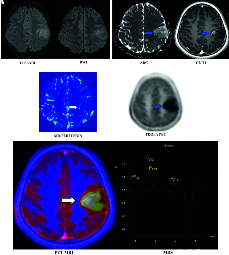
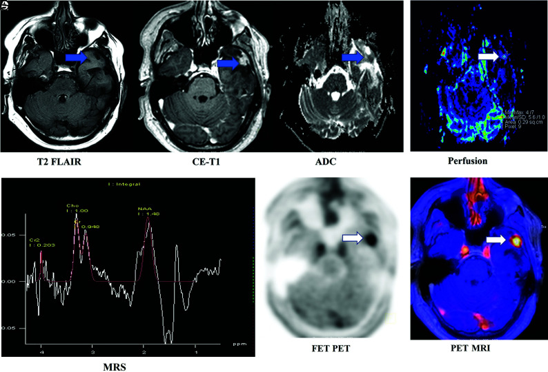
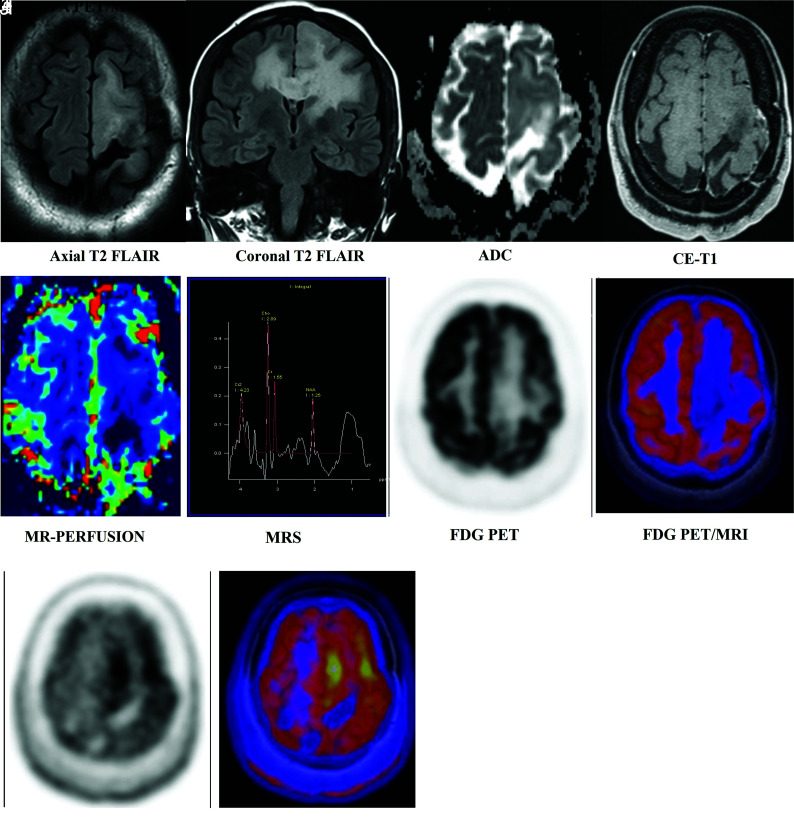
It is crucial to distinguish benign from malignant brain lesions to avoid invasive biopsy (Fig 2). The high specificity and negative predictive value of FET-PET MR imaging help to rule out malignancy with an accuracy of 85% and a change in management in 33% of the untreated equivocal lesions.14 FET-PET offers supplemental information on tumor extent and biopsy target selection compared with MR imaging, which further improves with dynamic PET. Dual time-point FET-PET-based targeted biopsies from non-contrast-enhanced areas have shown that FET uptake corresponded to HGGs as far as 3 cm from contrast enhancement.15 FET-PET-based TBR and PWI-determined relative CBV (rCBV) provide congruent and complementary information on glioma biology, with a moderate overlap of the tumor volumes.16 FET-PET may improve the outcome of surgical planning in both newly diagnosed and recurrent tumors (Fig 3). AAT-PET biologic tumor volume (BTV) often differs from contrast-enhanced MR imaging BTV (Fig 4). Patients with residual FET uptake have worse outcomes than those with complete FET-PET BTV resection (median overall survival, 13.7 versus 19.3 months, P = .007). The results were consistent regardless of age, MGMT, IDH mutation, promoter, or adjuvant therapy regimens.17
FIG 2.
A 12-year-old boy presented with right focal seizures for a month. Electroencephalography was noncontributory. Imaging-based diagnosis of a tuberculoma was made on the initial contrast-enhanced MR imaging in November 2019. Antitubercular treatment was started empirically. Follow-up MR imaging in February 2022 showed an interval increase of the mass from approximately 1.5 × 1.3 cm to 3.8 × 3.6 cm (images not shown), which led to further work-up, and the patient underwent AA-PET MR imaging and FDOPA-PET MR imaging. T2 FLAIR (A) demonstrates a hyperintense left posterior frontal mass with equivocal diffusion restriction on the diffusion-weighted image (B and C, blue arrow). A predominantly nonenhancing mass with small peripheral nodular enhancement is seen on postcontrast T1-weighted image (D, blue arrow). No apparent increased regional CBV is seen on DSC PWI (E, white arrow). On FDOPA-PET (F, blue arrow) and fused FDOPA-PET MR imaging (G, white arrow), the lesion showed uniformly increased DOPA uptake throughout with a high maximum standard uptake value of 3.62 (lesion/striatum ratio of 1.81 versus <1.0 as normal) and TBR. Multivoxel MRS (H) showed Cho/NAA and Cho/Cr ratios of 2.78 and 1.4, respectively, with a prominent lactate peak. In this patient, FDOPA-PET MR imaging confirmed the precise diagnosis of neoplastic etiology, which, on biopsy, was revealed to be an anaplastic astrocytoma. CE indicates contrast-enhanced.
FIG 3.
A 43-year-old man treated for a left anterior temporal lobe glioblastoma (IDH wild-type) status post resection (positive for generalized paroxysmal fast activity, negative for p53 and IDH1, MIB1 labeling index = 10%–12%, fluorescence in site hybridization epidermal growth factor receptor amplification, and no loss of 1p19q) and chemoradiation. He underwent follow-up FET-PET MR imaging after 3 years. T2 FLAIR (A, blue arrow) image shows postsurgical changes in the left anterior temporal lobe with peripheral nodular enhancement on the postcontrast T1-weighted image (B), mild increased diffusion restriction (C), and increased perfusion (rCBV of 6.7). Multivoxel MRS (E) shows noisy spectra with mildly raised choline and an inverted lactate peak. Corresponding increased FET uptake (TBR = 2.8; maximum standard uptake value = 3.7) on FET-PET (F) and fused FET-PET MR imaging (G, white arrow). The patient underwent resection, and pathology showed a recurrent tumor. This case highlights the congruent findings on contrast-enhanced MR imaging and FET-PET with a larger TBR leading to less interobserver variability and improving diagnostic performance for differentiating recurrence from TRC. CE indicates contrast-enhanced.
FIG 4.
A 35-year-old man was initially diagnosed with left frontal lobe oligodendroglioma grade II, status post resection and chemoradiation in 2008. He underwent a follow-up [18F] DOPA-PET MR imaging. T2 FLAIR axial (A) and coronal (B) images demonstrate a large cortical and subcortical area of abnormal T2-FLAIR hyperintensity in the left parasagittal frontal lobe extending to the left gangliocapsular area and the corpus callosum and across the midline in the right parietal region without apparent diffusion restriction (C), enhancement (D), and increased rCBV perfusion (E). Multivoxel MRS (F) shows increased Cho/Cr and Cho/NAA ratios (1.86 and 2.31, respectively). FDG-PET MR imaging (G and H) shows no appreciable FDG uptake. FDOPA-PET MR imaging shows areas of significant DOPA tracer uptake (maximum standard uptake value = 1.54 versus <1.0 as normal) more prominently in the left paramedian frontal region. FDOPA-avid, FDG-nonavid nonenhancing lesion in the left frontal region involving the corpus callosum with positive MR imaging correlates suggests active underlying residual/recurrent disease. This case again highlights the superiority of AATs over FDG and the importance of multiparametric MR imaging over individual sequences. AAT uptake in the absence of contrast enhancement and increased perfusion helped with the planning of surgery and radiation. CE indicates contrast-enhanced.
High- and low-grade subregions may coexist in gliomas. HGGs demonstrate higher uptake of AATs than LGGs. In a recent meta-analysis (7 studies, 219 patients), FDOPA-PET sensitivity and specificity for glioma grading were 0.88 and 0.73, respectively.18 The diagnostic accuracy of FET-PET and contrast PWI to discriminate LGGs from HGGs were similar, with an area under the curve TBR maximum of FET-PET uptake and rCBV being 0.83 and 0.81, respectively.19 Similar trends were observed with combined FDOPA-PET and multiparametric MR imaging.20
Noninvasive Tumor Genetic Profile and Molecular Markers.
Gliomas with IDH1 mutations have better chemoradiation responses and longer survival.21 AAT-PET MR imaging has the potential to serve as an alternative to invasive tissue characterization. In 52 patients with gliomas, FET-PET with PWI differentiated 1p/19q codeletion oligodendrogliomas with IDH mutations from IDH wild-type glioblastomas.22 FDOPA-PET and MR imaging metrics predicted the IDH mutation and 1p/19q codeletion with sensitivities of 73% and 76% and specificities of 100% and 94%, respectively.23 Oligodendrogliomas with the 1p/19q codeletion had MET uptake as high as that of IDH wild-type gliomas regardless of IDH1 mutation status. Therefore, MET-PET seems more useful for glioma grading in IDH wild-type gliomas.24 The FET-PET and the DWI-derived TBR/ADC ratio showed higher diagnostic accuracy than the individual technique for HGGs and IDH1, human telomerase reverse transcriptase, and epidermal growth factor receptor–mutated gliomas. Tumor regions with a human telomerase reverse transcriptase mutation had higher TBR and lower ADC values, while tumor protein P53 mutation showed lower TBR and higher ADC values. The 1p/19q codeletion and epidermal growth factor receptor mutations had lower ADC, and IDH1 mutations had higher TBR mean values.25 FET-PET MR imaging predicted ATRX, MGMT, IDH1, and 1p19q mutations in 85%, 76%, 89%, and 98%, respectively.26
Tumor Margin Delineation and Defining Tumor Extent for Radiation Therapy Planning.
In radiation planning, accurate tumor delineation is essential to provide the maximum tumor dose and minimize treatment-related damage to uninvolved regions. Enhancing components on MR imaging are the usual target for target-volume definition, whereas a tumor may extend beyond areas of enhancement. Conventional MR imaging inadequately distinguishes edema, contrast-enhancing, non-contrast-enhancing, and infiltrating tumors. PET hotspots represent high tumor cell densities. In a recent biopsy-validated study, FET-PET revealed precise glioma extent, allowing personalized treatment planning.27 FDOPA-PET–guided dose-escalated radiation therapy significantly improved the overall survival in methylated and progression-free survival in unmethylated GBMs.28 In 30 patients with HGGs, rCBV and the permeability (K2) map correlated with enhancing tumor volumes. FET-PET provided complementary information, suggesting that contrast-enhancing MR imaging underestimates the metabolically active tumor volume.29 Tumor volumes were larger in FET-PET than in rCBV maps (P < .001), with low spatial similarity of both imaging parameters.30 Similar results by Lohmann et al31 demonstrated larger metabolically active tumor volume by FET-PET than by contrast enhancement (P < .001) and lower spatial similarity. FET-PET resulted in a mean increase of 27% from clinical target volumes to biologic tumor volumes.32
Immediate Postsurgical Residual Tumor Evaluation.
The assessment of the early postoperative resection status in HGGs is necessary for surgical re-evaluation in the large residual disease. Early postoperative MR imaging may be ambiguous for residual tumors. In 25 patients with HGGs, FET-PET, MR imaging, and intraoperative assessment consistently showed complete resection in 48% of cases and residual disease in 24%.33 A Prospective PET/MRI study correlated the MET accumulation in GBM patients before postoperative chemoradiation with time to recurrence. The median time to recurrence was significantly shorter in MET-positive than MET-negative patients (6.3 and 19 months, P < .001).34 Rosen et al35 evaluated the prognostic value of dynamic FET-PET in partially resected IDH wild-type astrocytic gliomas with minimal or absent contrast enhancement. Smaller pre-irradiation FET-PET tumor volumes correlated with a favorable progression-free survival (7.9 versus 4.2 months; P = .012) and overall survival (16.6 versus 9.0 months; P = .002). In contrast, the mean TBR and time-to-peak values were associated with only a longer progression-free survival (P = .048 and P = .045, respectively).
Disease and Treatment Monitoring: Differentiation between Tumor Recurrence and TRC.
After initial glioma management, TRC are common and may mimic or coexist with tumor recurrence. RANO criteria based on T2-weighted, FLAIR, and contrast-enhancement changes are affected by BBB damage, which fails to differentiate tumor recurrence from treatment-related changes. In 20%–30% of patients, early postirradiation MR imaging exhibited increased enhancement, mimicking progression. However, it gradually disappeared without intervention.36 TRC can appear as new sites of enhancement or an increased extent of enhancement. Multiparametric MRI has certain limitations in cases of pseudoprogression within the first 12 weeks of therapy and in cases of radionecrosis after 12 weeks of treatment.37 The recent RANO recommendation for AAT-PET also includes assessing the response to radiation in gliomas apart from target delineation, prognostication, and re-irradiation.38 MET-PET was moderately specific (74%) and highly sensitive (97%) in detecting recurrence.39 The sensitivity remained high (97%) and the specificity rose to 93% when MET-PET MR imaging was used.39,40 A large meta-analysis (33 studies, 1734 patients) evaluated FET, MET, and FDOPA tracers for tumor recurrence with a sensitivity of 0.88, 0.92, and 0.85 and a specificity of 0.78, 0.78, and 0.70, respectively.6 The meta-analysis supports the incorporation of FET and MET in the treatment evaluation of HGGs. The amount of literature on the efficacy of 3’-deoxy3’-18F-fluorothymidine [18F-FLT] and FDOPA is insufficient for a conclusion.5
The FET-PET TBR and PWI-rCBV indicators performed moderately well in distinguishing progression from TRC in 104 patients (P <.01). A criterion of rCBV maximum of >2.85 allowed a correct diagnosis of progression in 44 patients with a positive predictive value of 100%. In the remaining 60 patients, progression and TRC were discriminated in 78% of patients.41 FET-PET is of significant clinical importance in diagnosing pseudoprogression related to chemoradiation. FET-PET performed a mean of 10 (SD, 7) days after the equivocal MR imaging findings showed a high accuracy of 87% to identify pseudoprogression, with an improved specificity of 100%.42 A combined analysis of arterial spin-labeled CBF and FDOPA uptake allowed high diagnostic performance in differentiating progression and pseudoprogression in treated gliomas.43 Given the high sensitivity for identifying tumor progression, FET-PET MR imaging could change clinical management in nearly one-half of patients.14
Monitoring Treatment Response to Newer Drugs in Recurrent HGGs.
AAT-PET can predict patient survival in both HGGs and LGGs treated with temozolomide. AAT-PET imaging reveals metabolic alterations in response to temozolomide earlier than morphologic alterations on MR imaging. Dynamic TAC patterns are helpful for diagnostic and prognostic purposes, particularly in distinguishing progression from TRC.44 Bevacizumab is an antiangiogenic chemotherapy drug targeting circulating vascular endothelial growth factor and lowering cerebrovascular permeability. It is an adjunctive therapy in recurrent HGGs and significantly reduces contrast enhancement, underestimating residual tumor evaluation. FET, MET, and FDOPA-PET combined with multiparametric MRI have shown promising results for improving accuracy in diagnosing tumor recurrence and detecting early treatment failure and TRC in patients with recurrent HGGs treated with bevacizumab.9 In a recurrent HGG in a patient on bevacizumab, the relationship between response to therapy on FET-PET and improved overall survival or progression-free survival is not well-understood. Compared with MR imaging, FET-PET could determine the failure of bevacizumab therapy 9–10.5 weeks earlier and a change in diagnosis or treatment planning in one-third of patients.45
The response assessment after immunotherapy in gliomas continues to be a significant challenge due to the rising occurrence of pseudoprogression. In patients with recurrent HGGs treated with regorafenib (multikinase inhibitor), FET and DWI-ADC metrics can predict the overall survival, and could serve as semiquantitative independent biomarkers of response to treatment.46 A recent study analyzed data from patients with GBM who received autologous dendritic cell vaccination therapy. In MR imaging for suspicion for GBM recurrence, FET-PET showed congruent tumor progression in 3/5 patients and TRC in the remaining 2/5.47 AAT-PET may help to identify the personalized bevacizumab treatment dose to improve therapeutic efficacy. Tumor volumetric and ADC analyses of serial MR imaging scans from 67 patients and serial FET-TBRs from 31 patients revealed overall survival benefits from bevacizumab plus radiation therapy compared with radiation alone. A high FET-TBR of nonenhancing tumor portions during bevacizumab therapy was associated with an inferior overall survival on multivariate analysis (hazard ratio, 5.97; 95% CI, 1.16–30.8).48
Role in Nonenhancing Tumors.
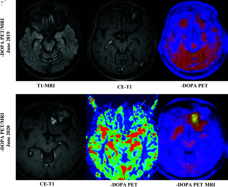
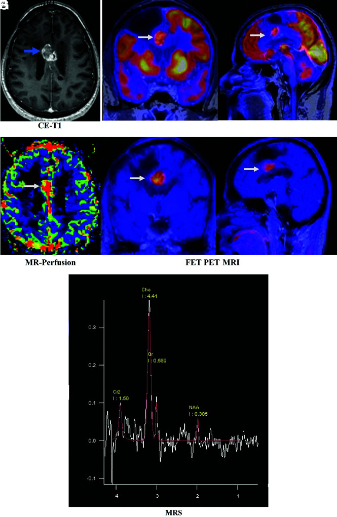
Most LGGs do not grossly disrupt BBBs, and many HGGs have nonenhancing regions suboptimally evaluated on conventional MR imaging. In GBM, FLAIR changes adjacent to an enhancement are presumed to be related to edema, which alters radiation protocols. The Radiation Therapy Oncology Group protocol suggests the possibility of microscopic disease in this region and includes it in the target volume with a lower prescribed radiation dose. In contrast, the European Society for Radiation Therapy and Oncology protocols do not specifically target this region. However, identifying this zone to precisely cover the target volume with the appropriate radiation dose is essential for treatment. AA-PET could image the nonenhancing areas better than conventional MR imaging or FDG (Fig 4).49 In a biopsy-validated analysis, combined FDOPA-PET MR imaging detected high-grade subregions with an accuracy of 58% compared with 42% with contrast-enhanced MR imaging (P = .03). The hybrid technique leads to larger delineation volumes and better accuracy for detecting high-grade subregions.20 In GBM, AA-PET MR imaging helps to clarify the nature of the suspected nonenhancing region (Figs 5 and 6). It improves delineation of the radiation therapy target, thus reducing undertreatment.32
FIG 5.
A 35-year-old man was treated for left frontal glioma (grade II), status post surgical resection and chemoradiation in 2009. He underwent a reoperation in February 2018 for tumor recurrence. In June 2019, [18F] DOPA PET MR imaging (A–C) showed a postresection surgical cavity in the left anterior parasagittal basifrontal region without nodular enhancement or any increased focal FDOPA uptake. There was no evidence of recurrence. In June 2020, follow-up [18F]-DOPA PET MR imaging (D and E) showed a new focal nodular enhancing lesion in the left basifrontal area (arrow) on the postcontrast T1-weighted image (D), with mildly increased rCBV perfusion (E) and corresponding increased FDOPA uptake on fused FET-PET MR imaging (F) (maximum standard uptake value = 3.9; lesion/striatal ratio; 1.38; <1.0 as normal), suggesting recurrence. The patient underwent a reoperation with recurrence found. This case also highlights the congruent findings on contrast-enhanced MR imaging and FDOPA-PET in differentiating TRC from recurrence. CE indicates contrast-enhanced.
FIG 6.
A 37-year-old man with a known diagnosis of oligodendroglioma, status post resection and chemoradiation. Postcontrast T1-weighted image (A, blue arrow) shows a recurrent enhancing lesion along the inferomedial aspect of the resection cavity of the right frontal region involving the body of the corpus callosum. Fused-PET MR images (B and C) show intralesional increased FDG slightly higher than in the white matter and lower than in the gray matter with increased rCBV perfusion (D, white arrows). Fused FET-PET MR images (E and F) show a relatively larger volume of a recurrent lesion, allowing better estimates of the extent of the lesion. Multivoxel MRS (G) shows an increased choline peak and decreased NAA peak. The pathologic diagnosis was a recurrence. This case highlights the superiority of AAT over FDG, congruent findings on MR imaging and FET-PET, and an excellent TBR, which were helpful in radiation therapy planning and re-surgical resection. CE indicates contrast-enhanced.
Future Perspectives: Newer Advances and Novel Radiotracers
Numerous non-AA tracers demonstrate uptake in gliomas, but the PET signal is frequently not solely attributed to the tumor cells. Glutamine-increased concentration in gliomas correlates with tumor proliferation and treatment resistance. The glutamine fluoro-analog, 4-[18F]-(2S,4R)-fluoroglutamine distinguishes proliferating gliomas from stable tumors.50 The extracellular matrix, stromal cells, immune cells, and blood vessels comprise the tumor microenvironment contributing to tumorigenesis. New therapeutic approaches target tumor microenvironment cells, necessitating the discovery of relevant imaging biomarkers to identify individuals who benefit from such therapy.51 [18F]DPA-714(TSPO)-PET MR imaging is used to image the glioma-associated immunosuppressive tumor microenvironment for targeting immunotherapy, drug target engagement, and clinical response assessment.52
Intratumoral hypoxia is associated with resistance to treatment and entails radiation to hypoxic subregions of tumors. [18F] fluoromisonidazole, a nitroimidazole derivative that images viable hypoxic cells, a biomarker of glioblastoma that correlates with prolonged overall survival53 and distinguishes pseudoprogression from recurrence in patients with HGGs treated with pembrolizumab54 [18F]-GE-180 (a novel TSPO ligand), is an imaging biomarker of tumor heterogeneity with improved binding affinity and a high TBR in glioblastoma. TSPO-PET can differentiate potentially aggressive forms of gliomas, graded according to the World Health Organization (WHO) classification, with a positive rate on PET of 100% among the HGGs.55 The Arg-Gly-Asp peptide is an angiogenesis-targeting radiotracer that binds αvβ3 integrins and monitors antiangiogenic therapies.56 The fibroblast activation protein gallium 68-labeled fibroblast activation protein inhibitor [68Ga]FAPI shows high accumulation in IDH wild-type WHO grade IV gliomas and WHO grade III and IV gliomas compared with WHO grade II gliomas.57 Although non-AA tracers are not specific for tumor cell imaging, they might still be used to better delineate tumor extent and, more important, as radiotheranostic agents. However, the results are very preliminary, and validation studies are required.
Application of Feature-Based PET MR Imaging Radiomics in Patients with Gliomas
Radiomics allows the voxel-level extraction of quantitative features from various diagnostic imaging integrated with clinical, histopathologic, and molecular factors to produce diagnostic, prognostic, or predictive mathematic models.58 Several PET radiomics analyses have been performed in neuro-oncology with reasonable accuracy to predict the glioma grade and identify the IDH mutation and the patients with a high risk of progression after receiving first-line therapy. FET-PET MR imaging combined with textural analysis predicts IDH mutations at 93%.59 The proposed machine learning model on MET-PET can predict grades of disease.60 FET-radiomic features discriminated significantly between tumor and nontumor components of 32 patients with recurrent GBM. The texture feature showed the best performance for predicting time to progression and overall survival and localizing the site of recurrence. The authors postulated that FET-PET radiomics might help with prognostic evaluation and selecting patients with recurrent GBM who would benefit from re-irradiation.61 Further studies are needed to improve radiomics algorithms to personalize predictive and prognostic models and potentially support the medical decision process.
Cost-Effectiveness of Treatment Monitoring of Gliomas Using AAT-PET
It is crucial to ensure that financial resources are used as efficiently as possible, given the limited resources available for health care. Imaging techniques require substantial investment. They should preferably be used only when the extra value appears to justify the expense. The known benefit of AAT-PET in patients with glioma is to prevent unnecessary treatment and its side effects. Joint recommendations state that AAT-PET MR imaging is appropriate for clinical use and FET-PET is the most useful for predicting treatment responses of glioblastomas.62,63 Despite the ubiquitous use of PET MR imaging, the FDA requires the treating institution to obtain an Investigational New Drug Application, which has prevented the mainstream adoption of AAT-PET into the neuro-oncology clinical algorithm in the United States.
Heinzel et al64 evaluated the cost-effectiveness of FET-PET MR imaging–guided biopsy for diagnosing gliomas. FET-PET MR imaging resulted in an increase of 18.5% in the likelihood of a correct diagnosis. The incremental cost-effectiveness ratio for 1 additional accurate diagnosis was €6405 for the baseline scenario and €9114 for the scenario based on higher disease severity. Heinzel et al65 investigated the cost-effectiveness of recurrent HGGs treated with bevacizumab and irinotecan. The authors suggested that the additional use of FET-PET in managing patients may be cost-effective. Baguet et al66 evaluated the cost-effectiveness of a follow-up PET scan performed on patients with glioblastoma postsurgery and before temozolomide. The decision tree based on overall survival demonstrated that the number of nonresponders identified using PET was 57.14% higher than that with conventional MR imaging.
AATs need to be FDA-approved to become widely available and approved by the Centers for Medicare and Medicaid Services to be reimbursable. Cost-effective analyses of hybrid PET MR imaging scanners are still pending. Further work is needed to update treatment guidelines and to include more PET agents when appropriate, encouraging insurance companies to reimburse for these potentially valuable agents.67 A simple-but-effective solution to the limited availability, use, and cost-effectiveness of the hybrid PET MR imaging remains to standardize the protocol for separate PET and MR imaging acquisitions.
Limitations of AATs
Combining PET and MR imaging is more patient-friendly than separate examinations and avoids the limitations of each imaging technique. With the development of PET MR imaging, it is now possible to swiftly and effectively study a variety of PET and MR imaging parameters. Significant hurdles include accessibility, the cost of PET MR imaging examinations, and the limited availability of the AATs. Uncertainty exists regarding the cost-effectiveness of PET MR imaging and the patients who will benefit from combined PET MR imaging. AATs have known limitations, with false-positive results in inflammation, infection, postsurgical regions, and postchemoradiation. Small tumor volumes may lead to false-negative results due to the partial volume effect. Acquisition of a baseline scan helps to compare pre- and posttreatment imaging findings. The estimation of TBR mean, TBR maximum, and BTV with AATs relies on reference brain parenchymal uptake. A decreased uptake due to atrophy, trauma, infarcts, and ischemia may lead to overestimating parameters. Initial dynamic FET images may show a reasonably high blood-pool uptake, and TAC in vascular structures may resemble tumor uptake. An increasing TAC may indicate inflammatory lesions, while a decreasing TAC may be seen in WHO grade II oligodendroglial tumors.3 A significant proportion (30%) of grade II IDH-mutated gliomas do not exhibit considerable AAT uptake. A negative finding on AA-PET is insufficient to exclude LGGs.68,69
CONCLUSIONS
In the current era of precision and personalized medicine, AAT-PET can provide additional insight beyond MR imaging for the clinical management of brain tumors (Online Supplemental Data). Furthermore, sufficient literature is available to demonstrate the utility of AAT-PET MR imaging in distinguishing recurrence from TRC in gliomas. There are several limitations in the existing literature, which may have impacted the diagnostic efficacy of the radionuclide tracers. Prospective validation studies of PET imaging criteria are needed to get beyond these restrictions and allow comparisons of direct results. Even though hybrid PET MR imaging is more patient-friendly and offers practical advantages, the cost-effectiveness and accessibility of these systems must be weighed against the additional effort involved in sequential examinations. As a result, it is projected that more effective therapy monitoring will be available in the upcoming years, which may be beneficial for glioma management.
ABBREVIATIONS:
- AAT
amino acid tracer
- AA
amino acid
- ASCT
alanine-serine-cysteine transporter
- BTV
biologic tumor volume
- GBM
glioblastoma multiforme
- FDOPA
3,4-dihydroxy-6-[18F]fluoro-L-phenylalanine
- FET
O-(2-[18F]fluoroethyl)-L-tyrosine
- LAT
large amino acid transporter
- HGG
high-grade glioma
- LGG
low-grade glioma
- MET
11C-methionine
- rCBV
relative CBV
- TAC
time-activity curve
- TBR
tumor-to-background ratio
- TRC
treatment-related changes
- TSPO
translocator protein
- WHO
World Health Organization
Footnotes
Neetu Soni and Manish Ora are first authors.
Disclosure forms provided by the authors are available with the full text and PDF of this article at www.ajnr.org.
References
- 1.Lin D, Wang M, Chen Y, et al. Trends in intracranial glioma incidence and mortality in the United States, 1975-2018. Front Oncol 2021;11:748061 10.3389/fonc.2021.748061 [DOI] [PMC free article] [PubMed] [Google Scholar]
- 2.Ostrom QT, Gittleman H, Liao P, et al. CBTRUS Statistical Report: primary brain and other central nervous system tumors diagnosed in the United States in 2010-2014. Neuro Oncol 2017;19:v1–88 10.1093/neuonc/nox158 [DOI] [PMC free article] [PubMed] [Google Scholar]
- 3.Law I, Albert NL, Arbizu J, et al. Joint EANM/EANO/RANO practice guidelines/SNMMI procedure standards for imaging of gliomas using PET with radiolabelled amino acids and [(18)F]FDG: version 1.0. Eur J Nucl Med Mol Imaging 2019;46:540–57 10.1007/s00259-018-4207-9 [DOI] [PMC free article] [PubMed] [Google Scholar]
- 4.Soni N, Ora M, Mohindra N, et al. Diagnostic performance of PET and perfusion-weighted imaging in differentiating tumor recurrence or progression from radiation necrosis in posttreatment gliomas: a review of literature. AJNR Am J Neuroradiol 2020;41:1550–57 10.3174/ajnr.A6685 [DOI] [PMC free article] [PubMed] [Google Scholar]
- 5.de Zwart PL, van Dijken BR, Holtman GA, et al. Diagnostic accuracy of PET tracers for the differentiation of tumor progression from treatment-related changes in high-grade glioma: a systematic review and metaanalysis. J Nucl Med 2020;61:498–504 10.2967/jnumed.119.233809 [DOI] [PubMed] [Google Scholar]
- 6.Cui M, Zorrilla-Veloz RI, Hu J, et al. Diagnostic accuracy of PET for differentiating true glioma progression from post treatment-related changes: a systematic review and meta-analysis. Front Neurol 2021;12:671867 10.3389/fneur.2021.671867 [DOI] [PMC free article] [PubMed] [Google Scholar]
- 7.Moreau A, Febvey O, Mognetti T, et al. Contribution of different positron emission tomography tracers in glioma management: focus on glioblastoma. Front Oncol 2019;9:9 10.3389/fonc.2019.01134 [DOI] [PMC free article] [PubMed] [Google Scholar]
- 8.Lopes C, Pereira C, Medeiros R. ASCT2 and LAT1 contribution to the hallmarks of cancer: from a molecular perspective to clinical translation. Cancers (Basel) 2021;13:203 10.3390/cancers13020203 [DOI] [PMC free article] [PubMed] [Google Scholar]
- 9.Hughes KL, O’Neal CM, Andrews BJ, et al. A systematic review of the utility of amino acid PET in assessing treatment response to bevacizumab in recurrent high-grade glioma. Neurooncol Adv 2021;3:vdab003 10.1093/noajnl/vdab003 [DOI] [PMC free article] [PubMed] [Google Scholar]
- 10.Fuchs BC, Bode BP. Amino acid transporters ASCT2 and LAT1 in cancer: partners in crime? Semin Cancer Biol 2005;15:254–66 10.1016/j.semcancer.2005.04.005 [DOI] [PubMed] [Google Scholar]
- 11.Jansen NL, Suchorska B, Wenter V, et al. Dynamic 18F-FET PET in newly diagnosed astrocytic low-grade glioma identifies high-risk patients. J Nucl Med 2014;55:198–203 10.2967/jnumed.113.122333 [DOI] [PubMed] [Google Scholar]
- 12.Jansen NL, Graute V, Armbruster L, et al. MRI-suspected low-grade glioma: is there a need to perform dynamic FET PET? Eur J Nucl Med Mol Imaging 2012;39:1021–29 10.1007/s00259-012-2109-9 [DOI] [PubMed] [Google Scholar]
- 13.Jansen NL, Schwartz C, Graute V, et al. Prediction of oligodendroglial histology and LOH 1p/19q using dynamic [(18)F]FET-PET imaging in intracranial WHO grade II and III gliomas. Neuro Oncol 2012;14:1473–80 10.1093/neuonc/nos259 [DOI] [PMC free article] [PubMed] [Google Scholar]
- 14.Brendle C, Maier C, Bender B, et al. Impact of (18)F-FET PET/MRI on clinical management of brain tumor patients. J Nucl Med 2022;63:522–27 10.2967/jnumed.121.262051 [DOI] [PMC free article] [PubMed] [Google Scholar]
- 15.Furtak J, Rakowska J, Szylberg T, et al. Glioma biopsy based on hybrid dual time-point FET-PET/MRI: a proof of concept study. Front Neurol 2021;12:634609 10.3389/fneur.2021.634609 [DOI] [PMC free article] [PubMed] [Google Scholar]
- 16.Göttler J, Lukas M, Kluge A, et al. Intra-lesional spatial correlation of static and dynamic FET-PET parameters with MRI-based cerebral blood volume in patients with untreated glioma. Eur J Nucl Med Mol Imaging 2017;44:392–97 10.1007/s00259-016-3585-0 [DOI] [PubMed] [Google Scholar]
- 17.Ort J, Hamou HA, Kernbach JM, et al. (18)F-FET-PET-guided gross total resection improves overall survival in patients with WHO grade III/IV glioma: moving towards a multimodal imaging-guided resection. J Neurooncol 2021;155:71–80 10.1007/s11060-021-03844-1 [DOI] [PMC free article] [PubMed] [Google Scholar]
- 18.Xiao J, Jin Y, Nie J, et al. Diagnostic and grading accuracy of (18)F-FDOPA PET and PET/CT in patients with gliomas: a systematic review and meta-analysis. BMC Cancer 2019;19:767 10.1186/s12885-019-5938-0 [DOI] [PMC free article] [PubMed] [Google Scholar]
- 19.Verger A, Filss CP, Lohmann P, et al. Comparison of (18)F-FET PET and perfusion-weighted MRI for glioma grading: a hybrid PET/MR study. Eur J Nucl Med Mol Imaging 2017;44:2257–65 10.1007/s00259-017-3812-3 [DOI] [PubMed] [Google Scholar]
- 20.Girard A, Le Reste PJ, Metais A, et al. Combining (18)F-DOPA PET and MRI with perfusion-weighted imaging improves delineation of high-grade subregions in enhancing and non-enhancing gliomas prior treatment: a biopsy-controlled study. J Neurooncol 2021;155:287–95 10.1007/s11060-021-03873-w [DOI] [PubMed] [Google Scholar]
- 21.Osborn AG, Louis DN, Poussaint TY, et al. The 2021 World Health Organization Classification of Tumors of the Central Nervous System: what neuroradiologists need to know. AJNR Am J Neuroradiol 2022;43:928–37 10.3174/ajnr.A7462 [DOI] [PMC free article] [PubMed] [Google Scholar]
- 22.Song S, Wang L, Yang H, et al. Static (18)F-FET PET and DSC-PWI based on hybrid PET/MR for the prediction of gliomas defined by IDH and 1p/19q status. Eur Radiol 2021;31:4087–96 10.1007/s00330-020-07470-9 [DOI] [PubMed] [Google Scholar]
- 23.Tatekawa H, Yao J, Oughourlian TC, et al. Maximum uptake and hypermetabolic volume of 18F-FDOPA PET estimate molecular status and overall survival in low-grade gliomas: a PET and MRI study. Clin Nucl Med 2020;45:e505–11 10.1097/RLU.0000000000003318 [DOI] [PMC free article] [PubMed] [Google Scholar]
- 24.Kim D, Chun JH, Kim SH, et al. Re-evaluation of the diagnostic performance of (11)C-methionine PET/CT according to the 2016 WHO classification of cerebral gliomas. Eur J Nucl Med Mol Imaging 2019;46:1678–84 10.1007/s00259-019-04337-0 [DOI] [PubMed] [Google Scholar]
- 25.Cheng Y, Song S, Wei Y, et al. Glioma imaging by O-(2-18F-fluoroethyl)-L-tyrosine PET and diffusion-weighted MRI and correlation with molecular phenotypes, validated by PET/MR-guided biopsies. Front Oncol 2021;11:743655 10.3389/fonc.2021.743655 [DOI] [PMC free article] [PubMed] [Google Scholar]
- 26.Haubold J, Demircioglu A, Gratz M, et al. Non-invasive tumor decoding and phenotyping of cerebral gliomas utilizing multiparametric (18)F-FET PET-MRI and MR fingerprinting. Eur J Nucl Med Mol Imaging 2020;47:1435–45 10.1007/s00259-019-04602-2 [DOI] [PubMed] [Google Scholar]
- 27.Meyer HS, Liesche-Starnecker F, Mustafa M, et al. (18)F FET PET uptake indicates high tumor and low necrosis content in brain metastasis. Cancers (Basel) 2021;13:355 10.3390/cancers13020355 [DOI] [PMC free article] [PubMed] [Google Scholar]
- 28.Laack NN, Pafundi D, Anderson SK, et al. Initial results of a Phase 2 trial of (18)F-DOPA PET-guided dose-escalated radiation therapy for glioblastoma. Int J Radiat Oncol Biol Phys 2021;110:1383–95 10.1016/j.ijrobp.2021.03.032 [DOI] [PubMed] [Google Scholar]
- 29.Dissaux G, Dissaux B, Kabbaj OE, et al. Radiotherapy target volume definition in newly diagnosed high grade glioma using (18)F-FET PET imaging and multiparametric perfusion MRI: a prospective study (IMAGG). Radiother Oncol 2020;150:164–71 10.1016/j.radonc.2020.06.025 [DOI] [PubMed] [Google Scholar]
- 30.Filss CP, Galldiks N, Stoffels G, et al. Comparison of 18F-FET PET and perfusion-weighted MR imaging: a PET/MR imaging hybrid study in patients with brain tumors. J Nucl Med 2014;55:540–45 10.2967/jnumed.113.129007 [DOI] [PubMed] [Google Scholar]
- 31.Lohmann P, Stavrinou P, Lipke K, et al. FET PET reveals considerable spatial differences in tumour burden compared to conventional MRI in newly diagnosed glioblastoma. Eur J Nucl Med Mol Imaging 2019;46:591–602 10.1007/s00259-018-4188-8 [DOI] [PubMed] [Google Scholar]
- 32.Hayes AR, Jayamanne D, Hsiao E, et al. Utilizing 18F-fluoroethyltyrosine (FET) positron emission tomography (PET) to define suspected nonenhancing tumor for radiation therapy planning of glioblastoma. Pract Radiat Oncol 2018;8:230–38 10.1016/j.prro.2018.01.006 [DOI] [PubMed] [Google Scholar]
- 33.Kläsner B, Buchmann N, Gempt J, et al. Early [18F]FET-PET in gliomas after surgical resection: comparison with MRI and histopathology. PLoS One 2015;10:e0141153 10.1371/journal.pone.0141153 [DOI] [PMC free article] [PubMed] [Google Scholar]
- 34.Seidlitz A, Beuthien-Baumann B, Löck S, et al. Final results of the Prospective Biomarker Trial PETra: [(11)C]-MET-accumulation in postoperative PET/MRI predicts outcome after radiochemotherapy in glioblastoma. Clin Cancer Res 2021;27:1351–60 10.1158/1078-0432.CCR-20-1775 [DOI] [PubMed] [Google Scholar]
- 35.Rosen J, Stoffels G, Lohmann P, et al. Prognostic value of pre-irradiation FET PET in patients with not completely resectable IDH-wildtype glioma and minimal or absent contrast enhancement. Sci Rep 2021;11:20828 10.1038/s41598-021-00193-x [DOI] [PMC free article] [PubMed] [Google Scholar]
- 36.Ellingson BM, Chung C, Pope WB, et al. Pseudoprogression, radionecrosis, inflammation or true tumor progression? Challenges associated with glioblastoma response assessment in an evolving therapeutic landscape. J Neurooncol 2017;134:495–504 10.1007/s11060-017-2375-2 [DOI] [PMC free article] [PubMed] [Google Scholar]
- 37.Albert NL, Weller M, Suchorska B, et al. Response Assessment in Neuro-Oncology Working Group and European Association for Neuro-Oncology recommendations for the clinical use of PET imaging in gliomas. Neuro Oncol 2016;18:1199–208 10.1093/neuonc/now058 [DOI] [PMC free article] [PubMed] [Google Scholar]
- 38.Galldiks N, Niyazi M, Grosu AL, et al. Contribution of PET imaging to radiotherapy planning and monitoring in glioma patients: a report of the PET/RANO group. Neuro Oncol 2021;23:881–93 10.1093/neuonc/noab013 [DOI] [PMC free article] [PubMed] [Google Scholar]
- 39.Deuschl C, Kirchner J, Poeppel TD, et al. 11C-MET PET/MRI for detection of recurrent glioma. Eur J Nucl Med Mol Imaging 2018;45:593–601 10.1007/s00259-017-3916-9 [DOI] [PubMed] [Google Scholar]
- 40.D’Souza MM, Sharma R, Jaimini A, et al. 11C-MET PET/CT and advanced MRI in the evaluation of tumor recurrence in high-grade gliomas. Clin Nucl Med 2014;39:791–98 10.1097/RLU.0000000000000532 [DOI] [PubMed] [Google Scholar]
- 41.Steidl E, Langen KJ, Hmeidan SA, et al. Sequential implementation of DSC-MR perfusion and dynamic [(18)F]FET PET allows efficient differentiation of glioma progression from treatment-related changes. Eur J Nucl Med Mol Imaging 2021;48:1956–65 10.1007/s00259-020-05114-0 [DOI] [PMC free article] [PubMed] [Google Scholar]
- 42.Werner JM, Weller J, Ceccon G, et al. Diagnosis of pseudoprogression following lomustine-temozolomide chemoradiation in newly diagnosed glioblastoma patients using FET-PET. Clin Cancer Res 2021;27:3704–13 10.1158/1078-0432.CCR-21-0471 [DOI] [PubMed] [Google Scholar]
- 43.Pellerin A, Khalifé M, Sanson M, et al. Simultaneously acquired PET and ASL imaging biomarkers may be helpful in differentiating progression from pseudo-progression in treated gliomas. Eur Radiol 2021;31:7395–405 10.1007/s00330-021-07732-0 [DOI] [PubMed] [Google Scholar]
- 44.Prather KY, O’Neal CM, Westrup AM, et al. A systematic review of amino acid PET in assessing treatment response to temozolomide in glioma. Neurooncol Adv 2022;4:vdac008 10.1093/noajnl/vdac008 [DOI] [PMC free article] [PubMed] [Google Scholar]
- 45.Harris RJ, Cloughesy TF, Pope WB, et al. 18F-FDOPA and 18F-FLT positron emission tomography parametric response maps predict response in recurrent malignant gliomas treated with bevacizumab. Neuro Oncol 2012;14:1079–89 10.1093/neuonc/nos141 [DOI] [PMC free article] [PubMed] [Google Scholar]
- 46.Lombardi G, Spimpolo A, Berti S, et al. PET/MR in recurrent glioblastoma patients treated with regorafenib: [(18)F]FET and DWI-ADC for response assessment and survival prediction. Br J Radiol 2022;95:20211018 10.1259/bjr.20211018 [DOI] [PMC free article] [PubMed] [Google Scholar]
- 47.Kristin Schmitz A, Sorg RV, Stoffels G, et al. Diagnostic impact of additional O-(2-[18F]fluoroethyl)-L-tyrosine ((18)F-FET) PET following immunotherapy with dendritic cell vaccination in glioblastoma patients. Br J Neurosurg 2021;35:736–42 10.1080/02688697.2019.1639615 [DOI] [PubMed] [Google Scholar]
- 48.Wirsching HG, Roelcke U, Weller J, et al. MRI and (18)FET-PET predict survival benefit from bevacizumab plus radiotherapy in patients with isocitrate dehydrogenase wild-type glioblastoma: results from the randomized ARTE Trial. Clin Cancer Res 2021;27:179–88 10.1158/1078-0432.CCR-20-2096 [DOI] [PubMed] [Google Scholar]
- 49.Niyazi M, Brada M, Chalmers AJ, et al. ESTRO-ACROP guideline “target delineation of glioblastomas.” Radiother Oncol 2016;118:35–42 10.1016/j.radonc.2015.12.003 [DOI] [PubMed] [Google Scholar]
- 50.Ekici S, Nye JA, Neill SG, et al. Glutamine imaging: a new avenue for glioma management. AJNR Am J Neuroradiol 2022;43:11–18 10.3174/ajnr.A7333 [DOI] [PMC free article] [PubMed] [Google Scholar]
- 51.Anderson NM, Simon MC. The tumor microenvironment. Curr Biol 2020;30:R921–25 10.1016/j.cub.2020.06.081 [DOI] [PMC free article] [PubMed] [Google Scholar]
- 52.Zinnhardt B, Müther M, Roll W, et al. TSPO imaging-guided characterization of the immunosuppressive myeloid tumor microenvironment in patients with malignant glioma. Neuro Oncol 2020;22:1030–43 10.1093/neuonc/noaa023 [DOI] [PMC free article] [PubMed] [Google Scholar]
- 53.Leimgruber A, Hickson K, Lee ST, et al. Spatial and quantitative mapping of glycolysis and hypoxia in glioblastoma as a predictor of radiotherapy response and sites of relapse. Eur J Nucl Med Mol Imaging 2020;47:1476–85 10.1007/s00259-020-04706-0 [DOI] [PubMed] [Google Scholar]
- 54.Barajas RF Jr, Ambady P, Link J, et al. [(18)F]-fluoromisonidazole (FMISO) PET/MRI hypoxic fraction distinguishes neuroinflammatory pseudoprogression from recurrent glioblastoma in patients treated with pembrolizumab. Neurooncol Pract 2022;9:246–50 10.1093/nop/npac021 [DOI] [PMC free article] [PubMed] [Google Scholar]
- 55.Unterrainer M, Fleischmann DF, Vettermann F, et al. TSPO PET, tumour grading and molecular genetics in histologically verified glioma: a correlative (18)F-GE-180 PET study. Eur J Nucl Med Mol Imaging 2020;47:1368–80 10.1007/s00259-019-04491-5 [DOI] [PubMed] [Google Scholar]
- 56.Echavidre W, Picco V, Faraggi M, et al. Integrin-αvβ3 as a therapeutic target in glioblastoma: back to the future? Pharmaceutics 2022;14:1054 10.3390/pharmaceutics14051053 [DOI] [PMC free article] [PubMed] [Google Scholar]
- 57.Rohrich M, Loktev A, Wefers AK, et al. IDH-wildtype glioblastomas and grade III/IV IDH-mutant gliomas show elevated tracer uptake in fibroblast activation protein-specific PET/CT. Eur J Nucl Med Mol Imaging 2019;46:2569–80 10.1007/s00259-019-04444-y [DOI] [PubMed] [Google Scholar]
- 58.Lohmann P, Meißner AK, Kocher M, et al. Feature-based PET/MRI radiomics in patients with brain tumors. Neurooncol Adv 2020;2:iv15–21 10.1093/noajnl/vdaa118 [DOI] [PMC free article] [PubMed] [Google Scholar]
- 59.Lohmann P, Lerche C, Bauer EK, et al. Predicting IDH genotype in gliomas using FET PET radiomics. Sci Rep 2018;8:13328 10.1038/s41598-018-31806-7 [DOI] [PMC free article] [PubMed] [Google Scholar]
- 60.Russo G, Stefano A, Alongi P, et al. Feasibility on the use of radiomics features of 11[C]-MET PET/CT in central nervous system tumours: preliminary results on potential grading discrimination using a machine learning model. Curr Oncol 2021;28:5318–31 10.3390/curroncol28060444 [DOI] [PMC free article] [PubMed] [Google Scholar]
- 61.Carles M, Popp I, Starke MM, et al. FET-PET radiomics in recurrent glioblastoma: prognostic value for outcome after re-irradiation? Radiat Oncol 2021;16:46 10.1186/s13014-020-01744-8 [DOI] [PMC free article] [PubMed] [Google Scholar]
- 62.Langen KJ, Galldiks N. Update on amino acid pet of brain tumours. Curr Opin Neurol 2018;31:354–61 10.1097/WCO.0000000000000574 [DOI] [PubMed] [Google Scholar]
- 63.Langen K-J, Heinzel A, Lohmann P, et al. Advantages and limitations of amino acid PET for tracking therapy response in glioma patients. Expert Rev Neurother.2020;20:137–46 10.1080/14737175.2020.1704256 [DOI] [PubMed] [Google Scholar]
- 64.Heinzel A, Stock S, Langen K-J, et al. Cost-effectiveness analysis of FET PET-guided target selection for the diagnosis of gliomas. Eur J Nucl Med Mol Imaging 2012;39:1089–96 10.1007/s00259-012-2093-0 [DOI] [PubMed] [Google Scholar]
- 65.Heinzel A, Müller D, Langen KJ, et al. The use of O-(2-18F-fluoroethyl)-L-tyrosine PET for treatment management of bevacizumab and irinotecan in patients with recurrent high-grade glioma: a cost-effectiveness analysis. J Nucl Med 2013;54:1217–22 10.2967/jnumed.113.120089 [DOI] [PubMed] [Google Scholar]
- 66.Baguet T, Verhoeven J, De Vos F, et al. Cost-effectiveness of [18F] fluoroethyl-L-tyrosine for temozolomide therapy assessment in patients with glioblastoma. Front Oncol 2019;9:9 10.3389/fonc.2019.00814 [DOI] [PMC free article] [PubMed] [Google Scholar]
- 67.Ehman EC, Johnson GB, Villanueva-Meyer JE, et al. PET/MRI: Where might it replace PET/CT? J Magn Reson Imaging 2017;46:1247–62 10.1002/jmri.25711 [DOI] [PMC free article] [PubMed] [Google Scholar]
- 68.Suchorska B, Giese A, Biczok A, et al. Identification of time-to-peak on dynamic 18F-FET-PET as a prognostic marker specifically in IDH1/2 mutant diffuse astrocytoma. Neuro Oncol 2018;20:279–88 10.1093/neuonc/nox153 [DOI] [PMC free article] [PubMed] [Google Scholar]
- 69.Zhang-Yin JT, Girard A, Bertaux M. What does PET imaging bring to neuro-oncology in 2022? a review. Cancers (Basel) 2022;14:879 10.3390/cancers14040879 [DOI] [PMC free article] [PubMed] [Google Scholar]


