

Abstract
Background: A spontaneous cerebrospinal leak from Sternberg’s canal with meningoencephalocele is a very rare clinical entity. Endoscopic repair of the defect is challenging and crucial in identifying the defect. The aim of this case report is to highlight the presence and management with endoscopic surgery in repairing Sternberg canal. Case: 40-year-old woman presents with spontaneous CSF rhinorrhea with no predisposing factors. CT imaging and MRI showed osteodural defect in the lateral recess of sphenoid with meningoencephalocoele lateral to the foramen rotundum. Endoscopic transethmoidal - transphenoidal - transpterygoid approach was used to repair the defect, and patient is well post-operative with least complication from the intervention surgery. Conclusion: Endoscopic approach proved to be the best and safest method in localizing the defect and closure of the leak. Angled scopes and image guided system were used to identify the precise location of the leak.
Supplementary Information
The online version contains supplementary material available at 10.1007/s12070-022-03347-z.
Keywords: Sternberg’s Canal, Lateral Craniopharyngeal Canal, Cerebrospinal Fluid, Rhinorrhoea, Lateral Sphenoid Recess
Introduction
Cerebrospinal fluid (CSF) rhinorrhea, a phenomenon where there is leakage of CSF through nasal cavity, often occurs due to skull base fracture after a massive head injury or as a result of complication from skull base surgery [1]. Spontaneous CSF leak has also been identified in patient without previous history of trauma, surgery, tumor or radiation therapy [2]. This phenomenon is rare and its actual cause still remains doubtful [3]. It can be associated with or without cephalocele. Pre-existing dural sac weakness and herniation of intracranial meninges or brain tissues through a skull base defect has slowly been recognized as the possible causes of this condition [3, 4]. CSF rhinorrhea has a low incidence of approximately 1 in 35,000 population, and are common in the anterior skull base [4]. Intrasphenoidal encephalocele is uncommon and subdivided into medial, perisellar and lateral sphenoid recess types, depends on whether it is an acquired or congenital anatomical defect [4, 5].
Sternberg canal is a congenital defect of the lateral craniopharyngeal wall with a channel coursing through and ends blindly into the sphenoid sinus. This weak defect allows the herniation of temporal lobe brain tissue into the lateral recess of sphenoid sinus. Certain literatures have doubted the existence of the Sternberg Canal and was reported only in 4% of adults [6].
We are presenting a case report of a 40-year-old lady who presented with spontaneous CSF rhinorrhea and was diagnosed to have meningocele through the Sternberg canal and was treated with endoscopic endonasal surgery.
Case Report
A healthy 40 - year - old lady, presented to us with three months history of right nasal rhinorrhoea which was persistent throughout the day and aggravated by straining and stooping. The rhinorrhoea was associated with headache. Otherwise, patient does not have symptoms of meningitis or encephalitis, and previous history of trauma or any skull base surgeries. Clinical examination revealed no neurological deficit. A rigid nasoendoscope revealed clear, glistening discharge at right sphenoid ostia, which was dripping to nasopharynx. The clear nasal discharge was collected and sent for biochemical analysis which then yielded non-infectious CSF fluid. Computed tomography (CT) and Magnetic Resonance Imaging (MRI) scan showed a bony dehiscent of the lateral recess of sphenoid bone with homeogenous opacity in sphenoid sinus (Fig. 1).
Fig. 1.
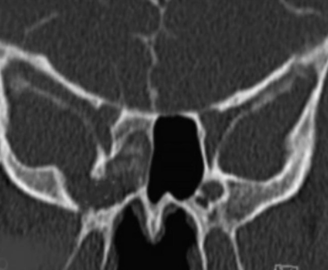
Coronal section of CT scan which shows a bony defect on the right cribriform plate. (pre rotundum pneumatisation)
Patient had Endoscopic Endonasal Transethmoidal Transphenoidal Trans-pterygoid surgery performed for Lateral Recess of Sphenoid Sinus (Sternberg Canal) defect repair with neurosurgery team. Prior to the surgery, a lumbar drain was inserted to reduce the intracranial pressure. Surgery started with medial maxillectomy followed by ethmoidectomy and sphenoidotomy to approach the sphenoid sinus. Hadad nasoseptal flap was raised from contralateral septal mucosa. Pterygoid base was drilled to visualize the foramen rotundum. With the assistance of image guided surgical (IGS) system and angled scope, the lateral recess of lateral sphenoid sinus (LRSS) defect was identified and meningonencephalocele was noted (Fig. 2). The meningoencephalocele was ablated using bipolar cautery and the mucosa was completely removed. Valsalva manoeuvre resulted in frank leak of CSF from the Sternberg canal, further confirming the presence of defect Multi-layered skull base reconstruction was performed with artificial dura (Duragen), fat, bioglue and Hadad flap. The repair was covered by surgicell and supported with nasal packing.
Fig. 2.

Endoscopic operative view of the defect (MC-meningocoele)with pooling of cerebrospinal fluid (CSF)
Nasal packing removed on day five postoperatively. Repeated nasoendoscope two weeks postoperative showed well taken flap with no evidence of CSF leak. Subsequent follow-up reviewed uneventful recovery from her problem, and she was discharged from the clinic after a year of follow up (Fig. 3).
Fig. 3.

Three months outcome post endoscopic repair reveals a well epithelised flap with no evidence of CSF leakage
Discussion
The sphenoid bone develops from independent cartilaginous precursors, which are presphenoid and postsphenoid/basisphenoid centers, orbitosphenoid, alisphenoid and the lateral pterygoid process [4]. The ossification of three cartilaginous precursors starts at third to fourth month of foetal life, and achieves full ossification in adolescences [7]. The shape and size of sphenoid bone depends on the variation of pneumatization along the growth of the skull base which ideally limits the pneumatization in the direction from anterior posteriorly [4, 7]. Most literatures explained VR line, which is an imaginary line connecting the medial wall of anterior opening of vidian canal to extracranial end of foramen rotundum.[8] Excessive pneumatization across the VR line forming lateral recess of sphenoid sinus, extending into either greater wing of sphenoid or pterygoid process [8].
Maximillian Sternberg proposed Sternberg canal, also known as “canalis cranio-pharyngeus lateralis” in year 1888. It is a lateral craniopharyngeal fistula coursing through inferior of rim of the orbit, extending to lateral root of orbital wing [9]. The canal may appear in children, but only demonstrated in 4% of adult population [9].
Patients with this congenital defect are usually asymptomatic. The defect canal may form a channel of herniation of brain substance when there is persistent intracranial hypertension. CSF rhinorrhoea due to rupture of dura overlying the meningoencephalocele is the common first presentation. Obesity is a known risk of spontaneous CSF rhinorrhoea, mainly due to increasing intracranial pressure secondary to increase intrathoracic and intraabdominal pressure in this group of people [10]. Other associated symptoms include orthostatic throbbing headache, nausea, vomiting and blurring vision with diplopia [11]. Fever, neck discomfort and altered behaviour should raise a high index of suspicion of meningoencephalitis [4, 11]. Even though spontaneous CSF rhinorrhoea due to Sternberg’s canal was observed, it is not a common phenomenon. Samuel et al. collected data in 17 patients with spontaneous CSF rhinorrhoea, only 1 patient had anatomical defect near to description to what is described in Stenberg’s canal [6].
Imaging plays an undisputable role in diagnosing the presence of Sternberg canal by identifying the site of bone defect in sphenoid sinus in thin-sliced computed tomography (CT). CT cisternography, if possible, should be obtained as it offers more advantage in identifying the morphology, size and quantity of CSF rhinorrhoea by visualizing the flow of the intrathecally injected contrast at the site of dura [12]. Sinus magnetic resonance imaging (MRI) gives synergistic information for soft tissue, especially the severity of meningoencephalocele. Both CT imaging and MRI also assist surgeons in planning the surgery to be performed. A 3D model can be reconstructed showing the pathway of the Sternberg canal as well as for the purpose of image guided surgery (IGS) during the repairing surgery [12, 13].
Intrasphenoidal cephalocele with spontaneous CSF, if not well treated, may promote ascending infection intracranially [4]. Traditionally, a lateral sphenoid sinus recess cephalocele is repaired by extracranial approach via the pterygoid route through the middle cranial fossa [4]. But the traction of the temporal lobe along the pathway in reaching the lateral recess of sphenoid adding the risk of intracranial contusion or hematoma intra- or post-operatively. The sterility of using transnasal endoscopy method of exposing surgical skull base to nasal cavity using this approach is questionable. In our case, intra- nasal and oral cavity were flushed with povidone prior to surgery and prophylactic antibiotic Ceftriaxone was administered. Combination of endoscopy transnasal and transpterygoid approach provide a wide surgical field for a proper direct vision to the lateral recess of sphenoid sinus. After the removal of the meningocele, the repair was done in multi-layered by using the contralateral nasoseptal flap and secured by the biology adhesive glue. Endoscopic endonasal surgery is considered less destructive in removing the cephalocele and repairing the lateral recess of sphenoid sinus. It is also cosmetically favourable to the patient as it avoids external prominent scar. Paolo et al. had treated 15 patients with Sternberg canal via endoscopic transnasal surgery and the average hospital stay were 1 week with no major complication [14]. Our patient has a short hospital stay post-operative without any post-surgery complication She is now well and asymptomatic for 2 years after the surgery.
Conclusion
Sternberg canal is a rare lateral recess of sphenoid sinus. It usually presents as incidentalfinding when patient presented with spontaneous CSF rhinorrhoea. To identify CSF leak from Sternberg canal, it requires a vast knowledge of the anatomical region. With the advancement of technology in endoscopic transnasal surgery, it is a procedure worth considering in managing Sternberg canal defect as it is not associated with major complication and cosmetically acceptable.
Electronic Supplementary Material
Below is the link to the electronic supplementary material.
Funding
There is no funding declared in this case report.
Originality statement: We are declaring that this manuscript is our own work, it is not under consideration by another journal, and this material has not been previously published.
Declarations
This article has not been sent to any journal neither being published, and compliance to ethical standard.
Competing Interests
The authors declare that they have no competing interests.
Informed Consent
The patient has been consented. This manuscript has gotten ethics approval from Clinical Research Center of Sarawak General Hospital.
Footnotes
Publisher’s Note
Springer Nature remains neutral with regard to jurisdictional claims in published maps and institutional affiliations.
References
- 1.Jayaprakash N, Kumar RR, Devanand B, Vaishnavi VA. Sternberg’s canal - A rare cause of spontaneous cerebrospinal fluid rhinorrhea. Neurol India. 2017;65(6):1429–1430. doi: 10.4103/0028-3886.217954. [DOI] [PubMed] [Google Scholar]
- 2.Tomaszewska M, Brożek-Mądry E, Krzeski A (2015) Spontaneous sphenoid sinus cerebrospinal fluid leak and meningoencephalocele - are they due to patent Sternberg’s canal? Wideochirurgia i inne techniki maloinwazyjne = videosurgery and other miniinvasive techniques. 10:347–358. 10.5114/wiitm.2014.47097. 2 [DOI] [PMC free article] [PubMed]
- 3.Mokri B. Spontaneous low pressure, low CSF volume headaches: spontaneous CSF leaks. Headache. 2013;53(7):1034–1053. doi: 10.1111/head.12149. [DOI] [PubMed] [Google Scholar]
- 4.Bendersky DC, Landriel FA, Ajler PM, Hem SM, Carrizo AG. Sternberg’s canal as a cause of encephalocele within the lateral recess of the sphenoid sinus: a report of two cases. Surg Neurol Int. 2011;2:171. doi: 10.4103/2152-7806.90034. [DOI] [PMC free article] [PubMed] [Google Scholar]
- 5.Lai SY, Kennedy DW, Bolger WE. Sphenoid encephaloceles: disease management and identification of lesions within the lateral recess of the sphenoid sinus. Laryngoscope. 2002;112(10):1800–1805. doi: 10.1097/00005537-200210000-00018. [DOI] [PubMed] [Google Scholar]
- 6.Barañano CF, Cure J, Palmer JN, Woodworth BA. Sternberg’s Canal: fact or fiction? Am J Rhinol Allergy. 2009;23(2):167–171. doi: 10.2500/ajra.2009.23.3290. [DOI] [PubMed] [Google Scholar]
- 7.Schick B, Brors D, Prescher A. Sternberg’s canal–cause of congenital sphenoidal meningocele. European archives of oto-rhino-laryngology: official journal of the European Federation of Oto-Rhino-Laryngological Societies (EUFOS) : affiliated with the german society for Oto-Rhino-Laryngology. Head and Neck Surgery. 2000;257(8):430–432. doi: 10.1007/s004050000235. [DOI] [PubMed] [Google Scholar]
- 8.Wang J, Bidari S, Inoue K, Yang H, Rhoton A. Jr. Extensions of the sphenoid sinus: a new classification. Neurosurgery. 2010;66(4):797–816. doi: 10.1227/01.neu.0000367619.24800.b1. [DOI] [PubMed] [Google Scholar]
- 9.Thakur JD, Manzi B, Savardekar AR, Singh MP, Menger R, Nanda A. Commentary: Maximilian Sternberg (1863–1934): the Man behind Sternberg’s Canal and his contribution to the Modern-Day Skull Base anatomy and neuroscience-historical vignette. Neurosurgery. 2018;83(3):E120–e4. doi: 10.1093/neuros/nyy242. [DOI] [PubMed] [Google Scholar]
- 10.Hanwate R, Thorawade V, Jagade M, Attakil A, Parelkar K, Pandare M, et al. CSF rhinorrhoea with encephalocele through Sternberg’s canal: our experience. Int J Otolaryngol Head Neck Surg. 2015;04:50–54. doi: 10.4236/ijohns.2015.41009. [DOI] [Google Scholar]
- 11.Mokri B (2013) Spontaneous Low Pressure, Low CSF Volume Headaches: Spontaneous CSF Leaks. Headache: The Journal of Head and Face Pain. ;53(7):1034-53. doi: 10.1111/head.12149 [DOI] [PubMed]
- 12.Chen GY, Ma L, Xu ML, Zhang JN, He ZD, He CY et al (2018) Spontaneous cerebrospinal fluid rhinorrhea: a case report and analysis.Medicine. ; 97(5) [DOI] [PMC free article] [PubMed]
- 13.Connor SE. Imaging of skull-base cephalocoeles and cerebrospinal fluid leaks. Clin Radiol. 2010;65(10):832–841. doi: 10.1016/j.crad.2010.05.002. [DOI] [PubMed] [Google Scholar]
- 14.Castelnuovo P, Dallan I, Pistochini A, Battaglia P, Locatelli D, Bignami M. Endonasal endoscopic repair of Sternberg’s canal cerebrospinal fluid leaks. Laryngoscope. 2007;117(2):345–349. doi: 10.1097/01.mlg.0000251452.90657.3a. [DOI] [PubMed] [Google Scholar]
Associated Data
This section collects any data citations, data availability statements, or supplementary materials included in this article.