. 2023 Apr 27;43(5):e220105. doi: 10.1148/rg.220105

Figure 1.

Diagram of the binary classification model for predicting progressive versus stable (nonprogressive) IPF from three-dimensional lung high-resolution CT (HRCT) images. Patients were imaged at total lung capacity (TLC) in the prone position using standard diffuse lung disease CT protocols, and a few patients were imaged in the supine position owing to their severe symptoms. A notable data shift in this example is the change in the proportion of supine images for patients with progressive IPF. The model shows performance drop due to data shift.

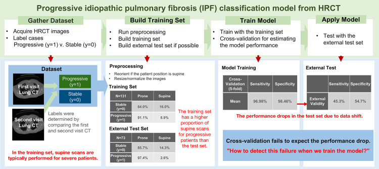
Diagram of the binary classification model for predicting progressive versus stable (nonprogressive) IPF from three-dimensional lung high-resolution CT (HRCT) images. Patients were imaged at total lung capacity (TLC) in the prone position using standard diffuse lung disease CT protocols, and a few patients were imaged in the supine position owing to their severe symptoms. A notable data shift in this example is the change in the proportion of supine images for patients with progressive IPF. The model shows performance drop due to data shift.