PLOS ONE logoLink to PLOS ONE
. 2023 May 18;18(5):e0285251. doi: 10.1371/journal.pone.0285251

HER2 inhibition increases non-muscle myosin IIA to promote tumorigenesis in HER2+ breast cancers

Samar M Alanazi 1, Wasim Feroz 1, Rosalin Mishra 1, Mary Kate Kilroy 1, Hima Patel 1, Long Yuan 1, Sarah J Storr 2, Joan T Garrett 1,*
Editor: Mohammed Soutto3
PMCID: PMC10194889  PMID: 37200287

Abstract

HER2 is over-expressed in around 15% to 20% of breast cancers. HER3 plays a critical role in HER2 mediated tumorigenesis. Increased HER3 transcription and protein levels occur upon inhibition of HER2. We aimed to identify proteins that bound to HER3 upon inhibition of the HER family with the pan-HER inhibitor neratinib in HER2+ breast cancer cells. Immunoprecipitation of HER3 followed by mass spectrometry experiments found non-muscle myosin IIA (NMIIA) increased upon neratinib treatment relative to vehicle DMSO treatment. MYH9 is the gene that encodes for the heavy chain of NMIIA. Breast cancer patients with high MYH9 were significantly associated with a shorter disease specific survival compared to patients with low MYH9 expression from the METABRIC cohort of patients. In addition, high MYH9 expression was associated with HER2+ tumors from this cohort. Immunoblots of whole cell lysates of BT474 and MDA-MB-453 HER2+ breast cancer cells demonstrated elevated HER3 and NMIIA protein levels upon neratinib treatment for 24 hours. To examine the role of NMIIA in HER2+ breast cancer, we modulated NMIIA levels in BT474 and MDA-MB-453 cells using doxycycline inducible shRNA targeting MYH9. MYH9 knockdown reduces HER3 protein levels and concomitant reduction in downstream P-Akt. In addition, loss of MYH9 suppresses cell growth, proliferation, migration, and invasion. Our data reveals that NMIIA regulates HER3 and loss of NMIIA reduces HER2+ breast cancer growth.

Introduction

HER2+ breast cancer is characterized by overexpression of human epidermal growth factor receptor 2 (HER2, HER2/neu, erbB-2). HER2 is an oncogene located on chromosome 17q12 and is amplified in about 20% of all breast cancers [1]. The HER family consists of four members: EGFR (ErbB1/HER1), ErbB2 (HER2), ErbB3 (HER3), and ErbB4 (HER4). HER2 always maintains an open confirmation with no known high affinity ligand and has potent kinase activity, making it the ideal partner for dimerization [2]. HER3 plays a central role in HER2+ breast cancer cells. Studies have shown that blocking the kinase activity of HER2 results in upregulation of HER3 [35].

Approved HER2 targeted therapies include monoclonal antibodies that target the extracellular domain of HER2: trastuzumab, pertuzumab, and margetuximab (Margenza, margetuximab-cmkb), engineered for increased binding to activating Fcγ receptor IIIA (CD16A) and decreased binding to inhibitory Fcγ receptor IIB (CD32B) relative to trastuzumab [6, 7]. HER2 antibody-drug conjugates is another class of drugs targeting HER2 including trastuzumab emtansine (T-DM1), a trastuzumab-cytotoxic drug conjugate [8] and fam-trastuzumab deruxtecan-nxki (Enhertu), which delivers a topoisomerase I inhibitor [9]. Small molecules targeting the intracellular tyrosine kinase (TK) domain of HER2 that are FDA approved to treat HER2+ breast cancer are lapatinib, neratinib [10], and tucatinib [11]. These therapies have significantly improved the outcome of HER2+ breast cancer patients [10]. However, most HER2+ metastatic breast cancer patients will eventually develop resistance to HER2 targeted therapies [12]. Neratinib is one of the FDA approved HER2 inhibitors for extended adjuvant treatment of HER2+ breast cancer patients following adjuvant trastuzumab therapy [13].

Non-muscle myosin class II, (NMII) plays a variety of functions as a skeleton protein, such as involvement in cell motility, polarity, and adhesion. They also have a role in maintaining cell shape, and signal transduction. Non-muscle myosin IIA (NMIIA) is the myosin isoform consisting of non-muscle myosin heavy chain IIA (NMHCIIA), encoded by the MYH9 gene, and regulatory and essential light chains that are shared with other NMII isoforms [1417].The MYH9 gene is localized on chromosome 22q12.3. NMIIA belongs to a superfamily of motor proteins that promote mechanical forces when they bind to actin through hydrolysis of ATP. NMIIA is a hexameric molecule composed of a dimer of heavy chains, two regulatory light chains (RLC) and two essential light chains (ELC).

Several studies have shown that MYH9 has a critical role in cancer and demonstrated that NMIIA expression in cancer cells associated with progression of various types of cancers [18]. A study in MDA-MB-231 breast cancer cells showed that depletion of NMIIA with small-interfering RNA resulted in decreased MDA-MB-231 cell migration but enhanced lamellar spreading [19]. The overexpression of NMIIA was strongly correlated with esophageal squamous cancer and non-small cell lung cancer (NSCLC) progression and was associated with worse overall survival [20, 21]. In addition, NMIIA overexpression promotes invasion, lymph node metastasis in gastric cancer [22, 23], increases proliferation and migration in colorectal cancer (CRC) cells and pancreatic cancer [24, 25], and associated with poor prognosis in hepatocellular carcinoma (HCC) [26]. Collectively, the data of these studies indicate that overexpression of NMIIA could contribute to cancer progression and poor prognosis. In this study, we aim to investigate the role of NMIIA in HER2+ breast cancer cells. Our data indicate that NMIIA controls HER3 downstream signaling and promotes HER2+ breast cancer growth, proliferation, migration, and invasion.

Materials and methods

Cell culture and inhibitors

Human HER2+ breast cancer cells (BT474 and MDA-MB-453) were obtained from American Type Culture Collection, ATCC (Manassas, VA). Neratinib was obtained from LC Laboratories. BT474 cells were maintained in RPMI with 10% fetal bovine serum (FBS) at 37°C in 5% CO2 in a humidified incubator. MDA-MB-453 cells were maintained in DMEM with 10% fetal bovine serum at 37°C in 5% CO2. Blebbestatin was obtained from SelleckChem (Cat # S7099). Doxycycline was obtained from MP Biomedicals, LLC (Cat # 198955)

Immunoprecipitation

BT474 and MDA-MB-453 cells were grown to 70% confluency and lysed in 1% NP40 lysis buffer containing 20 mM Tris, pH 7.4, 150 mM NaCl, 10% glycerol, 1 mM EDTA, 1 mM EGTA, 5 mM NaPyro-PO4, 50 mM NaF, 10 mM β-glycerol-PO4. 0.7–1.4 milligrams (mg) of cleared lysate was incubated with 2 <g of HER3 (2F12) antibody (Invitrogen, Cat# MA5-12675), 2.3 <g IgG antibody (Invitrogen, Cat# MA1-10418), 1 <g NMIIA antibody (Abcam, Cat# ab238131) overnight at 4° C. The next day, 5 <l Protein A magnetic beads (Millipore) were added per 1 <g of antibody and incubated for 1 hr at 4°C. Resuspended beads were washed with lysis buffer and boiled in SDS-PAGE loading buffer. The levels of HER3 and NMIIA from immunoprecipitated samples were analyzed by western blotting. For HER3 immunoprecipitates the recovered proteins were eluted and analyzed by silver staining, mass spectrometry, and western blotting.

Silver staining, mass spectrometry

HER3 immunoprecipitated samples from BT474 treated with DMSO or neratinib for 24 hours were loaded in 4–20% SDS-polyacrylamide gel. The gel was stained using Pierce Silver Stain Kit (Thermo Scientific Cat# 24612). Briefly, gel was washed 2 × 5 minutes in ultrapure water, then fixed for 15 minutes in 30% ethanol:10% acetic acid solution. After fixation, gel was washed 2 × 5 minutes in 10% ethanol, then 2 × 5 minutes in ultrapure water. Gel sensitized for 1 minute, then washed 2 × 1 minute with water. Gel was stained for 30 minutes, then washed 2 × 20 seconds with ultrapure water. After staining and washing, the gel was developed for 2–3 minutes until appearance of bands. The selected bands were excised from the gel, pooled with like bands (resulting in 1 sample from the 24 hr neratinib and 1 sample from the DMSO), reduced with DTT, alkylated with IAA, and digested with trypsin. The peptides were extracted, dried by speed vac, resuspended in 10 ul of 0.1% FA and desalted using a micro C18 zip tip. The samples were mixed with alpha cyano 4-hydroxy cinnamic acid (HCCA) matrix and spotted for analysis by MALDI TOF-TOF. A MASCOT search of all entries was used to identify the proteins.

Immunoblotting

BT474, MDA-MB-453 Cells were lysed in RIPA buffer (Thermo Fisher Scientific, Waltham, MA, USA, Cat#BP-115) supplemented with protease and phosphatase inhibitors (Thermo Fisher Scientific, Cat#88669). Immunoblotting was performed using specific primary antibodies (Cell Signaling Technology) for P-HER3 (Y1289) (Cat#4791), HER3 (Cat#12708), NMIIA (Cat#3403), P-Akt (T308) (Cat#13038), AKT (Cat#4691), P-Erk1/2 (Cat#4370), Erk1/2 (Cat#4695), and α- Tubulin (Cat#2125). Anti-rabbit IgG-horse radish peroxidase (HRP) was used as the secondary antibody (Santa cruz biotechnology sc-2357). α- Tubulin served as loading controls.

MHY9 expression in breast cancer patients

Gene expression microarray data from the Molecular Taxonomy of Breast Cancer International Consortium (METABRIC) breast cancer cohort were obtained from the CBioPortal for Cancer Genomics (n = 1903), with detailed description found in the original publication [27]. The gene expression data for MHY9 (mRNA expression z-values relative to diploid samples) were available from 1904 patients, with one breast sarcoma patient excluded from analysis (n = 1903). Statistical analysis was performed using IBM SPSS Statistics (version 28) and all differences were deemed statistically significant at the level of P≤0.05. X-tile was used to determine cut-points for assessment using breast cancer specific survival [28]. Survival curves were plotted according to the Kaplan-Meier method with significance determined using the log-rank test. The Pearson χ2 test of association was used to determine the relationship between categorised protein expression and clinicopathological variables. Gene set enrichement analysis (GSEA) was performed using GSEA software [29, 30], with samples divided into MHY9 low and MHY9 high expression (https://www.gsea-msigdb.org/gsea/index.jsp) and gene enrichment determined in the curated hallmarks of cancer gene sets (h.all.v2022.1.Hs.symbols.gmt) [31, 32].

qPCR analysis

Total RNA was extracted from BT474 and MDA-MB-453 cell lines using the RNA extraction mini kit (Qiagen, 74104, USA) according to the manufacturer’s instruction. Two-step Real Time-qPCR was performed to assess the mRNA level of ERBB3 and MYH9. First strand cDNA was synthesized using iScriptTM cDNA Synthesis Kit (Biorad, 1708890, USA). qPCR was set up using CFX96 Real-Time System (Biorad, USA). GAPDH was used as an internal control. Relative ERBB3 and MYH9 mRNA expression was presented by 2−ΔΔCT method. Paired primer sequences used for ERBB3: 5ˋ-GGGTTAGAGGAAGAGGATGTCAAC-3ˋ (forward) and 5ˋ-GGGAGGAGGGAGTACCTTTGAG-3ˋ (reverse); for MYH9: 5ˋ-GTGAGAAGGAGACAAAGGCG-3ˋ (forward) and 5ˋ-GACTTCTCCAGCTCGTGGAC-3ˋ (reverse); and for GAPDH: 5ˋ-AAGAAGGTGGTGAAGCAG-3ˋ (forward); 5ˋ-TCATACCAGGAAATG AGC-3ˋ.

Generation of inducible MYH9-deficient cells

To ablate MYH9 expression, we transduced BT474 and MDA-MB-453 with doxycycline-dependent inducible lentiviruses expressing shRNAs that target MYH9 expression (GE Dharmacon, Lafayette, CO). A non-targeting shRNA lentivirus was used to generate control cell lines. The inducible lentiviral shRNA vector, which uses the Tet-On inducible system, only allows the expression of either MYH9-targeting shRNA (Construct (1) AGTGAGGACGAGCTACTCT, Construct (2) ATTGATACCCAAGAGATGG, Construct (3) TGTTGGGAGAAGCCACTGG or a nontargeting shRNA when cells are treated with doxycycline. The expression of GFP is driven by the same tetO promoter so that the transduction and shRNA expression upon doxycycline treatment can be visually tracked by green fluorescence. Briefly, BT474 and MDA-MB-453 cells were seeded in 96-well tissue culture plates. The following day, serum free media containing viral particles was added and incubated for 16 h; then, the medium containing viral particles was replaced with fresh media supplemented with 10% FBS. To select transduced cells, medium containing 1–2 μg/ml puromycin was added, and the cells were maintained for three days. Because the inducible lentiviral vector contains a puromycin-resistance gene, transduced cells survive the selection by puromycin, while cells without the integration of lentiviral DNA are killed by puromycin. To evaluate whether puromycin selection was complete, 500 ng/ml doxycycline (MP Biomedicals, LLC) was added for 48 h to induce the expression of shRNAs and GFP. The efficiency of inhibiting MYH9 expression by shRNA was determined by immunoblotting.

Matrigel colony formation

Three-dimensional (3D) growth assays were performed in growth factor-reduced matrigel (BD Biosciences, San Jose, CA, USA) where 96 well plates were coated with 80 <L of matrigel/well. BT474, MDA-MB-453 shCTRL or shMYH9 cells (1 × 10⁴/well) were plated and incubated at 37°C for 24 h. Cells were treated with vehicle (DMSO), 500 nM doxycycline or 200 nM neratinib or combination of both every alternate day for doxycycline. After 10 days of incubation, colonies were visualized, and photographs were captured from 3 random fields under microscope at 10× magnification. The areas of the colonies were measured by ImageJ and represented as mean areas normalized to DMSO control.

MTT cell proliferation assay

Both BT474 and MDA-MB- 453 shCTRL or shMYH9 cells (2 × 10⁴/well) were plated in 96 well plates in triplicate and incubated at 37°C for 24 h. Cells were treated with vehicle (DMSO), 500 nM doxycycline or 200 nM neratinib or combination of both. After 72 hr, the media containing the drug was replaced with 5 mg/mL MTT (3-(4,5-dimethylthiazol- 2-yl)-2,5-diphenyltetrazolium bromide) dissolved in cell line specific media and incubated for 4 hr. After 4 hr the media was aspirated, and crystals were dissolved with isopropanol (Molecular Grade, Fisher BioReagents). The absorbance was read at 570 nm using a microplate reader (SPECTR Amax PLUS Microplate Spectrophotometer Plate Reader, Molecular Devices Corporation). Data is represented as the mean of at least two independent experiments ± SEM.

Spheroid cultures and PrestoBlue assay

Spheroids were established by seeding cells in a 96-well Ultra-Low Attachment plate (Corning) in 200 <L volume per well. Extracellular matrix (ECM)-rich Cultrex (Trevigen Inc.) was included at a final concentration of 100 <g/mL to aid in the formation of spheroids. Cells were treated upon plating and every 48 hours with 500 nM doxycycline or vehicle. After centrifugation for at least 5 min at 300 Relative Centrifugal Field (RCF), the cultures were maintained in a cell culture incubator at 37°C with 5% CO2 in a humidified atmosphere. Spheroids were incubated with 10% (v/v) PrestoBlue (Thermo Fisher Scientific) for 30 min in the cell culture incubator, and the relative fluorescence (RFU) at different time points and measured utilizing a plate reader (Fluostar Omega, BMG Labtech; FlexStation II, Molecular Devices) at excitation and emission wavelengths of 544 nm and 590 nm, respectively. Data shown is normalized to the values for vehicle treatment.

Cell migration and invasion assay

BT474 and MDA-MB- 453 shCTRL or shMYH9 cells were seeded at a density of 5 × 10⁴ cells/well in 6 well plates and treated vehicle (DMSO), 500 nM doxycycline or 200 nM neratinib or combination of both. After 24 h, cells were counted and 2 × 10⁴ cells/well were added to the upper chamber (for invasion the champers were coated with matrigel) of transwell and incubated for 48 h. After 48 h, migrated and invaded cells in the lower chamber were stained with 0.5% crystal violet and images were captured from three areas under phase contrast microscope. The intensities from three areas collected from three independent experiments were measured using ImageJ and expressed as % of control and represented graphically.

Small-interfering RNA

4x10⁶ BT474 and MDA-MB-453 cells were transfected with siRNAs specifically targeting HER3 (siHER3) or control siRNA (siCon). To knock down HER3 expression, siRNA against a HER3 target sequence ACCACGGTATCTGGTCATAAA was used (Dharmacon International). Mismatched siRNA with a target sequence of GGAAGC AGACTCACTCTTATA was used as a negative control.

Statistical analysis

Data are shown as the mean ± standard error of mean (SEM) and representative of at least three independent experiments unless indicated otherwise. Statistical analysis was performed by two sample paired ‘t’-test using ANOVA (Graph Pad Prism 7). The data was considered statistically significant if p<0.05.

Results

Enhanced NMIIA–HER3 levels in response to HER2 inhibition in HER2+ breast cancer cells

Previous studies have shown that inhibition of HER2 results in upregulation of HER3 [3]. We sought to identify HER3 binding partners in the presence or absence of pharmacological inhibition of the HER family using the irreversible pan HER inhibitor neratinib. Mass spectrometry experiments were performed to identify HER3 binding proteins in HER2+ BT474 breast cancer cells. NMIIA was increased upon inhibition of HER2 with neratinib relative to DMSO control treatment from HER3 immunoprecipitates (Fig 1A). Immunoprecipitation experiments were performed in BT474 and MDA-MB-453 breast cancer cells using a HER3, IgG control, or NMIIA antibody. Immunoblots performed using antibodies against NMIIA and HER3 showed increased NMIIA and HER3 levels upon treatment with 200nM neratinib for 24 hours in HER3 and NMIIA immunoprecipitates. 200 nM of neratinib was chosen for this and subsequent experiments as this is a clinically achievable concentration in patients [33]. We observed 24 hours of neratinib treatment resulted in enhanced NMIIA bound to HER3 compared to vehicle treated cells (Fig 1B and 1C).

Fig 1. Enhanced NMIIA–HER3 levels upon HER2 inhibition in HER2+ breast cancer.

Fig 1

(A) BT474 cells were treated with DMSO or 500 nM neratinib for 24 hours. Cells were lysed and an immunoprecipitation for HER3 was performed. Triplicate samples are shown. Bands in red represent NMIIA as determined by mass spectrometry. (B) BT474 cells were treated with 200 nM neratinib or vehicle DMSO for 24 hours. The cells were lysed using NP-40 buffer and immunoprecipitated using a HER3, IgG, or NMIIA antibody. The products were analyzed by 10% SDS-PAGE followed by NMIIA and HER3 immunoblots. (C) MDA-MB-453 cells were treated with 200 nM neratinib and DMSO for 24 hours. The cells were lysed using NP-40 buffer and immunoprecipitated using a HER3, IgG, or NMIIA antibody. The products were analyzed by 10% SDSPAGE followed by NMIIA and HER3 immunoblots.

Significance of NMIIA in breast cancer

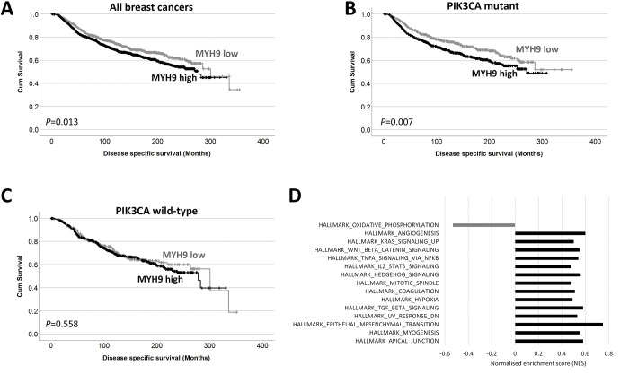
We examined the gene expression of MHY9 in the METABRIC cohort and observed that patients with high MHY9 expression have shorter survival to patients with low MHY9 levels (Fig 2A). We next examined if MHY9 expression was associated with survival in patient subgroups; high MHY9 expression was significantly associated with adverse survival of patients with mutations in P13KCA mutations (P = 0.007) but was not associated with survival of patients with PI3KCA wild type tumors (P = 0.558) (Fig 2B and 2C). PI3KCA is the most frequently mutated gene in the METABRIC cohort, and of those patients with matched gene expression data, 41.8% of cases are PI3KCA mutant (795/1903).

Fig 2. MYH9 is associated with breast cancer survival.

Fig 2

Kaplan-Meier analysis of MHY9 expression where the impact of low (grey line) and high (black line) expression are shown (A). The impact of low (grey line) and high (black line) MHY9 expression are shown in PI3KCA mutant tumors (B) and in wild type tumors (C). GSEA results showing normalized enrichment scores for the gene sets enriched as a FDR of 1% in MHY9 low samples (gray) and MHY9 high samples (black).

High MHY9 expression was associated with progesterone receptor negative tumors (χ2 = 19.970, d.f. = 1, P<0.001), HER2 positive tumors (χ2 = 12.440, d.f. = 1, P<0.001), ER negative tumors (χ2 = 41.608, d.f. = 1, P<0.001), patients under 40 years (χ2 = 4.885, d.f. = 1, P = 0.027), and tumors with p53 mutations (χ2 = 23.007, d.f. = 1, P<0.001); data is shown in Table 1. GSEA was used to explore METABRIC microarray data for enrichment of genes in the curated hallmarks of cancer gene sets in high MHY9 tumors. Normalized enrichment scores (NES) with a false discovery rate of 1% identified 14/50 enriched gene sets in MHY9 tumors, compared with 1/50 in MHY9 low tumors (Fig 2D).

Table 1. Associations between MHY9 expression and clinicopathological variables.

    MHY9 expression
Variable Low High P value
P53 mutation status Wild type 487 (25.7%) 754 (39.7%) <0.001
  Mutant 185 (9.7%) 472 (24.9%)
PgR status Negative 269 (14.2%) 622 (32.8%) <0.001
  Positive 403 (21.2%) 604 (31.8%)
HER2 status Negative 613 (32.3%) 1050 (55.3%) <0.001
  Positive 59 (3.1%) 176 (9.3%)
ER status Negative 100 (5.3%) 343 (18.1%) <0.001
  Positive 572 (30.1%) 883 (46.5%)
Patient age 40 years or less 30 (1.6%) 86 (4.5%) 0.027
  Greater than 40 years 639 (33.8%) 1136 (60.1)
Tumor size 2cm or less 297 (15.8%) 521 (27.7%) 0.616
  Greater than 2cm 373 (19.9%) 687 (36.6)

The P values are resultant from Pearson χ2 test of association.

HER3 and NMIIA protein levels are increased upon inhibition of HER2 in HER2+ breast cancer

We next sought to examine overall levels of HER3 and MYH9 mRNA and protein upon treatment with neratinib in HER2+ breast cancer cell lines. Levels of HER3 and MYH9 mRNA in BT474 and MDA-MB-453 were assessed using qRT-PCR. We did not observe that mRNA levels of HER3 or MYH9 consistently changed upon HER2 inhibition with neratinib (data not shown). We found that 24 hours of neratinib treatment reduced P-HER2 and concomitant reduction in total HER2 protein as has been reported [34, 35]. We found that neratinib treatment increased HER3 and NMIIA protein levels (Fig 3).

Fig 3. HER3 and NMIIA protein levels are increased upon inhibition of HER2 in HER2+ breast cancer cells.

Fig 3

BT474 and MDA-MB-453 cells were treated with vehicle or neratinib (200 nM) for 24 hours. Cells were lysed and products analyzed by 10% SDS-PAGE followed by probing immunoblots with antibodies for p-HER2, HER2, NMIIA, HER3 and α-tubulin.

Inducible MYH9 deficient HER2+ breast cancer cells demonstrate that NMIIA controls HER3 signaling pathway

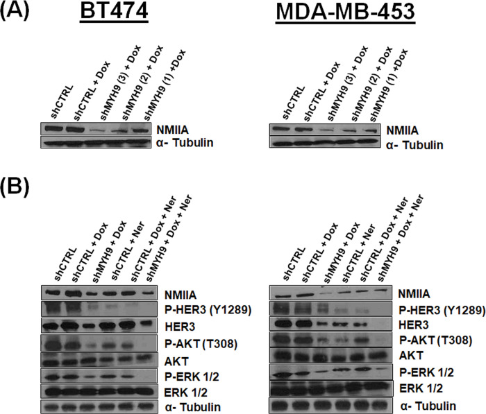
Three different MYH9-targeting shRNAs were designed and evaluated for their abilities to inhibit MYH9 expression. shMYH9 (1) was found to have the highest inhibitory efficiency in both cell lines and was used to generate MYH9 deficient BT474 and MDA-MB-453 cells (Fig 4A). We next examined the effect NMIIA loss has on HER3 signaling in the absence or presence of neratinib. Lentivirally transduced MDA-MB-453 and BT474 cells with shMYH9 and doxycycline induction demonstrate a reduction in HER3 protein levels compared to cells lentivirally transduced with a control sequence and doxycycline induction (Fig 4B). Strikingly, we observed a concomitant reduction in P-HER3 (Y1289) and downstream P-Akt (T308) and P-Erk1/2. Y1289 of HER3 resides within a YXXM motif and participates in signaling to PI3K upon phosphorylation, activating downstream signaling [36]. Neratinib alone reduced P-HER3, P-Akt and P-Erk1/2. The combination of doxycycline to induce MYH9 knockdown and neratinib treatment resulted in further reduction in P-HER3, P-Akt and P-Erk1/2. Furthermore, we investigated the effect of NMIIA siRNA in the presence or absence of neratinib in BT474 and MDA-MB-453 cells (S1 Fig). Notably, we observed NMIIA RNA and proteins levels are increased upon neratinib treatment in cells transfected with control siRNA. Cells transfected with MYH9 siRNA in the absence of neratinib treatment had decreased MYH9 mRNA and protein levels. MYH9 siRNA reduced HER3 mRNA and proteins levels in the absence of neratinib treatment, indicating that mechanism(s) exist for NMIIA to modulate HER3 mRNA and protein levels. Furthermore, in cells transfected with MYH9 siRNA, neratinib treatment increased both HER3 and NMIIA RNA and protein levels compared to vehicle DMSO treatment. Additionally, we used the pharmacological inhibitor for NMII blebbistatin to evaluate the mRNA and protein levels of NMIIA and HER3. Blebbistatin inhibits myosin ATPase activity [37]. It binds halfway between the nucleotide binding pocket and the actin binding cleft of myosin, predominantly in an actin detached conformation [38]. It has been shown that blebbistatin reduces NMIIA protein levels [39]. Blebbistatin treatment results in reduced HER3 mRNA and protein levels (S2 Fig).

Fig 4. Generation of HER2+ breast cancer cells lentivirally transduced with shRNA targeting MYH9: Loss of NMIIA inhibits HER3 signaling axis.

Fig 4

(A) Western blot showing MYH9 expression in transduced and selected BT474 and MDA-MB-453 cells after 48-hour of 500 nM doxycycline induction. (B) Western blot using the indicated antibodies in transduced and selected BT474 and MDA-MB-453 cells after 48-hour of 500 nM doxycycline induction and/or 24-hour of 200 nM neratinib treatment as indicated.

MYH9 knockdown and neratinib treatment inhibit matrigel colony formation, and proliferation in HER2+ breast cancer cells

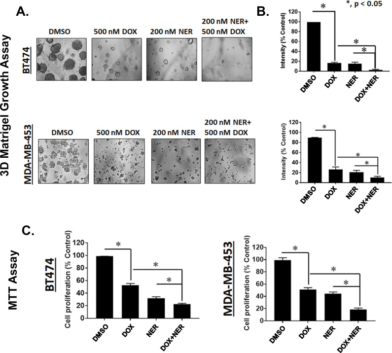
Examination of BT474 and MDA-MB-453 cells transduced with a doxycycline inducible construct expressing shRNA targeting MYH9 grown on a basement membrane of matrigel indicated that combination doxycycline (500 nM) and neratinib (200 nM) inhibit colony formation better than either single knockdown of MYH9 or neratinib treatment. Our data showed that the combination of doxycycline with neratinib significantly suppressed colony formation on matrigel compared to individual treatment or DMSO control in BT474 and MDA-MB-453 cells transduced with shMYH9. BT474 cells were more sensitive to the combination of doxycycline and neratinib marked by significantly reduced colonies versus single treatment of doxycycline, neratinib, or vehicle DMSO (Fig 5A and 5B). We speculated that the combination of doxycycline-induced MYH9 knockdown and neratinib could effectively inhibit proliferation of BT474 and MDA-MB-453 cells transduced shMYH9. Accordingly, BT474 and MDA-MB-453 cells transduced with shRNA targeting MYH9 were treated with doxycycline (500 nM) and neratinib (200 nM) for 72 hr. After 72 hr, the media containing the drug was replaced with 5 mg/mL MTT (3-(4,5-dimethylthiazol-2-yl)-2,5-diphenyltetrazolium bromide) dissolved in cell line specific media and incubated for 4 hr. After 4 hr the media was aspirated, and crystals were dissolved with isopropanol. We observed that the combination of neratinib and doxycycline to deplete MYH9 in BT474 and MDA-MB-453 cells resulted in a statistically significant reduction in proliferation compared to single treatment doxycycline or neratinib in both cell lines (Fig 5C). Parallel experiments in BT474 and MDA-MB-453 cells expressing control non-targeting shRNA were performed. For cells grown on a basement membrane of Matrigel, doxycycline added to neratinib did not result in a further reduction of colony size (S3A and S3B Fig). In BT474 cells transduced with control shRNA we observed a further reduction in proliferation for the combination of doxycycline and neratinib compared to neratinib treatment alone which is unexpected as we have found that cells stably expressing lentivirus tolerate 500 nM doxycycline (S3C Fig). We investigated the growth of BT474 cells expressing shCTRL or shMYH9 in spheroids by plating 1000 cells in ultra-low attachment plates. Spheroids can recapitulate the basic 3D structure of tumors and more closely mimic cancer cells in vivo. On day 1, 3 and 5 cells were treated with vehicle or doxycycline. Images of spheroids taken on day 3, 5 and 7 revealed BT474 cells expressing shMYH9 treated with doxycycline were smaller compared to vehicle treatment. Furthermore, assessing cell viability using PrestoBlue reagent revealed statistical reductions in cell viability on days 3, 5 and 7 in doxycycline treated cells versus vehicle treated cells (S4 Fig). Doxycycline treatment had no effect in BT474 cells expressing shCTRL.

Fig 5. MYH9 knockdown and neratinib treatment inhibit colony formation on Matrigel and proliferation in HER2+ breast cancer cells.

Fig 5

(A) BT474 and MDA-MB-453 cells transduced with MYH9 shRNA were seeded on a basement membrane of matrigel at a density of 2 × 10⁴ cells/well. Cells treated with vehicle (DMSO), doxycycline (500 nM)/48 h, neratinib (200 nM) once, and combination. Phase contrast images of acini for all cell lines were captured at 10× magnification and the average size of each cellular structure was quantified and expressed as mean of areas ± SEM, n = 5 random fields * p <0.05 for indicated comparison (B). (C) BT474 and MDA-MB-453 cells transduced with a doxycycline inducible shRNA targeting MYH9 were treated with doxycycline (500 nM) and neratinib (200 nM) for 72 hr. After 72 hr, the media containing the drug was replaced with 5 mg/mL MTT dissolved in cell line specific media and incubated for 4 hr. After 4 hr the media was aspirated, and crystals were dissolved with isopropanol. The bar graphs are represented as mean; Error bars: SEM, n = 3 * p <0.05 for indicated comparison.

MYH9 knockdown and neratinib treatment suppress HER2+ breast cancer cell migration and invasion

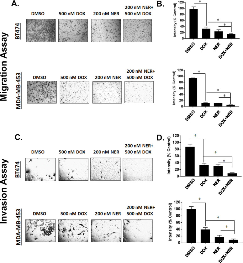
Knockdown of MYH9 in BT474 and MDA-MB-453 cells reduced migration and invasion of cells. We assessed migration by plating cells in the upper chamber of transwell plates using 10% FBS as a chemoattractant in the lower chamber. BT474 and MDA-MB-453 cells transduced with shMYH9 and treated with doxycycline (500 nM) and neratinib (200 nM) had a significantly lower number of migrated cells compared with single agent doxycycline or neratinib in both cell lines (Fig 6A and 6B). We next assessed invasion via coating the upper chamber with a layer of matrigel requiring cells to travel through the matrigel to enter the lower chamber. We observed that BT474 and MDA-MB-453 cells containing shRNA targeting MYH9 treated with doxycycline (500 nM) and neratinib (200 nM) had a significantly lower number of invading cells compared with single agent doxycycline or neratinib in both cell lines (Fig 6C and 6D). Neratinib treatment reduced migration and invasion of BT474 and MDA-MB-453 cells expressing control non-targeting shRNA (S5 Fig). Doxycycline alone or added to neratinib did not affect migration or invasion of cells expressing non-targeting shRNA.

Fig 6. MYH9 knockdown and neratinib treatment inhibit migration and invasion in HER2+ breast cancer cells.

Fig 6

(A) BT474 and MDA-MB-453 (5 × 10⁴ cells/well) stable cells transduced with MYH9 shRNA were seeded in six well plates and treated with vehicle (DMSO), doxycycline (500 nM), neratinib (200 nM) and indicated combination in serum free media for 24 h. Post 24 hr treatment, these treated cells (2 × 10⁴ cells/well) were added to the upper chamber of transwell plates with 10% FBS as chemoattractant in the lower chamber. After 24h, the migrated cells were stained and captured. (B) The intensities of the migrated cells were measured using ImageJ and expressed as mean of % control and represented as bar graph. Error bars: SEM, n = 4, * p <0.05 for indicated comparison. (C) Invasion assays were performed where each chamber was coated with 20 <l matrigel mixed with 80 <l serum free media and incubated at 37° for 30 mins. Stable cells (BT474 and MDA-MB-453) expressing MYH9 shRNA were seeded at a density of 5 × 10⁴ cells/well and treated and incubated as above. After 24h, the invaded cells were stained and photographed (D) The intensities of the invaded cells were measured using ImageJ and expressed as mean % of control. Error bars: SEM, n = 3* p <0.05 for indicated comparison.

Discussion

Previous studies indicate that inhibition of HER2 TK activity results in HER3 upregulation [40]. In our study we sought to identify HER3 binding proteins upon inhibition of HER2 by the irreversible HER inhibitor neratinib. We identified NMIIA encoded by MYH9 as a HER3 binding protein upon pharmacological inhibition of HER2 in HER2+ breast cancer cells (Fig 1).

NMIIA is known to be involved in many cellular functions including cell adhesion, cytokinesis, polarization, division, and migration [4143]. It is also a key regulator in human diseases such as cancer [18, 44]. NMIIA can have an oncogenic role in different types of cancer such as triple negative breast cancer, esophageal squamous cancer, NSCLC, gastric cancer, CRC, pancreatic cancer, HCC and nasopharyngeal carcinoma [1926, 45]. Recent work to delineate how NMII isoforms function upon a stiffening extracellular matrix in cancer cells found that NMIIA aides the establishment of cell polarity through actin polarization at the cell leading edge, lamellipodia formation and focal adhesion turnover [46].

Conversely, other studies have shown that NMIIA can act as a tumor suppressor. Repression of MYH9 predisposed mice to squamous cell carcinoma (SCC), as MYH9 was identified from an RNA interference screen [47]. The mechanism by which NMIIA acts as a tumor suppressor was via a noncanonical role in nuclear retention of activated p53. When NMIIA is defective, even though the p53 pathway can be induced in response to DNA damage, it did not occur because of an inability of p53 to remain and/or accumulate in the nucleus. Additional work found that low MYH9 expression is associated with decreased survival in patients with head and neck cancer harboring stratified low-risk mutant p53 but not high-risk mutant p53 [48]. More than half of all human cancers contain a TP53 mutation. Notably BT474 cells have been reported to have a p53 mutation and MDA-MB-453 cells have a deletion in p53 [49]. As the breast cancer models in our study may not depend on functional p53, the role of NMIIA in nuclear retention of activated p53 may be irrelevant for the breast cancer models in our study. Mice with homozygous deletion of NMIIA consistently developed SCC of the tongue [50]. Interestingly genetic deletion of NMIIA did not affect the stability of p53 but was ascribed to NMIIA maintaining mitotic stability during cell division. An additional study found that genetically knocking down MYH9 in mouse melanoma cells increased growth and metastasis in vivo in mouse models which was attributed to increased epithelial to mesenchymal (EMT) markers and potentially increased angiogenesis and inflammatory cell recruitment [51]. These studies point to the context in which NMIIA may be needed throughout development and the interplay with the tumor microenvironment in determining if NMIIA permits or hampers tumor growth.

Blebbistatin reversibly inhibits NMII by blocking its ATPase activity [37, 38, 52, 53]. Due to blebbistatin’s non-specificity and cytotoxicity [54], it may not be a good candidate to target NMIIA in HER2+ breast cancer. Recently however blebbistatin has been used in vivo in tumorgenicity studies demonstrating reduced tumor volume in a dose-dependent manner in a NSCLC model [39]. Toxicity or weight loss with blebbistatin in mice was not reported although the lack of specificity for blebbistatin is a major consideration. Blebbistatin has been reported to inhibit NMIIA and NMIIB along with several striated muscle myosins and data indicate that blebbistatin inhibits arterial smooth muscle tonic contractions [55, 56]. There is a strong rationale for the development of more specific inhibitors of NMIIA that could be used in cancer treatment, potentially in HER2+ breast cancers from data presented in this study.

Recently, Orgaz et al. demonstrated that myosin II activity increases during melanoma adaptation to MAPK pathway inhibition. High myosin II activity correlated with targeted and immunotherapy resistant melanomas [57]. Rho-kinase (ROCK) is upstream of NMIIA. ROCK inactivates the myosin light chain 2 (MLC2) phosphatase, which leads to increased phosphorylation of MLC2 (p-MLC2) and myosin II activity. ROCK inhibitors improve the efficacy of MAPK inhibitors and immunotherapies in melanoma models [57]. The utility of ROCK inhibitors should be considered in combination with HER2 inhibition in HER2+ breast cancers given the paucity of NMIIA specific inhibitors.

We observed a bidirectional relationship between NMIIA and the HER family. Figs 1 and 3 demonstrate that inhibition of HER signaling with neratinib treatment increases NMIIA protein levels. Future studies will define the mechanistic details of neratinib induced MYH9 upregulation. There are limitations to our work. This study does not determine mechanistically how HER3 and NMIIA interact nor the mechanism(s) by which loss of NMIIA reduces HER3 protein levels or how the loss of HER3 reduces NMIIA protein levels. The immunoprecipitation experiments shown in Fig 1B and 1C are challenging to interpret. The immunoprecipitation of HER3 followed by immunoblotting of HER3 reveals unequal amounts of HER3 between lanes, similarly for immunoprecipitation of NMIIA followed by immunoblotting of NMIIA. As shown in Fig 3, 24 hours of neratinib treatment results in increased NMIIA and HER3, correlating with the increased amount of HER3 and NMIIA detected from the immunoprecipitation and immunoblotting experiments shown in Fig 1B and 1C. Future studies will mechanistically decipher which compartment(s) of the cell NMIIA and HER3 interact and if blebbistatin can inhibit the interaction. Future studies will delineate if loss of MYH9 affects the half-life of HER3 protein and examine the effects of overexpression of NMIIA.

We have found NMIIA is increased upon HER2 kinase inhibition. HER2 inhibition in the clinic is very rarely curative to women with HER2+ metastatic breast cancer. As such this work aims to determine if there is potential validity in co-targeting NMIIA and HER2. Cumulatively, we found that BT474 and MDA-MB-453 cells containing shRNA targeting MYH9 treated with doxycycline and neratinib had a statistically significant: (1) reduction in the size of colonies grown on a basement membrane of Matrigel, (2) reduction in proliferation, (3) reduction in migrating cells and (4) lower number of invading cells compared with single agent doxycycline or neratinib in both cell lines. These experiments support further examination of potentially co-targeting NMIIA and HER2 in HER2+ breast cancer. Future studies in HER2+ breast cancers would determine conditional loss of MYH9 in HER2+ breast cancer xenografts in vivo and additionally examine the efficacy of inhibitors of ROCK, upstream of NMIIA, in combination with HER2 inhibition.

Conclusions

This study shows a bidirectional relationship between NMIIA and the HER family in HER2+ breast cancer cells. HER kinase inhibition increases NMIIA. Loss of NMIIA reduces HER3 protein and concomitant reductions in PI3K/Akt and MAPK signaling. Loss of NMIIA in combination with HER kinase inhibition results in reduction in HER2+ breast cancer cell proliferation, growth on a basement membrane, migration and invasion. This study identifies NMIIA as a potential therapeutic target in HER2+ breast cancers.

Supporting information

S1 Fig. NMIIA knockdown reduces HER3 and NMIIA expression that is rescued by inhibition of HER2 with 24h treatment of neratinib in HER2+ breast cancer cell lines.

BT474 and MDA-MB-453 were seeded in 60 mm plates (2x 10⁶ cells/plate), and forward and reverse transfection were performed using siRNA targeting MYH9 or scrambled sequence (control) and lipofectamine RNAmax mixture for 48 hours. (A) BT474 and MDA-MB-453 cells were then treated with 200 nM neratinib or DMSO for 24 hours. RNA was isolated and MYH9 and HER3 RNA levels determined using real time PCR. (B) BT474 and MDAMB-453 cells were treated with neratinib (200 nM) for 0–24 hours. Cells were lysed and products analyzed by 10% SDS-PAGE followed by probing immunoblots with antibodies for NMIIA, HER3 and α-tubulin.

(TIF)

S2 Fig. Reduced total HER3 levels as a result of pharmacological inhibition of NMIIA in breast cancer cell lines.

(A) BT474 and MDA-MB-453 cells were treated with DMSO, neratinib, blebbistatin, and combination therapy of neratinib and blebbistatin for 24 hours. RNA was isolated and MYH9 and HER3 RNA levels determined using real time PCR. Graphs were blotted in Prism 7 (GraphPad) (B) BT474 and MDA-MB-453 cells were treated with DMSO, neratinib, blebbistatin, and combination therapy of neratinib and blebbistatin for 24 hours. Cells were lysed in RIPA buffer and the products were analyzed by 10% SDS-PAGE followed by probing immunoblots with antibodies against NMIIA, HER3, and α-tubulin.

(TIF)

S3 Fig. Evaluation of colony formation and proliferation with doxycycline and neratinib treatment in HER2+ breast cancer cells expressing shRNA non-targeting control.

(A) BT474 and MDA-MB-453 cells transduced with non-targeting control shRNA were seeded on a basement membrane of matrigel at a density of 2 × 10⁴ cells/well. Cells treated with vehicle (DMSO), doxycycline (500 nM)/48 h, neratinib (200 nM) once, and combination. Phase contrast images of acini for cell lines were captured at 10× magnification and the average size of each cellular structure was quantified and expressed as mean of areas ± SEM, n = 5 random fields (B). (C) BT474 and MDA-MB-453 cells transduced with a doxycycline inducible shRNA targeting sh-control (shCTRL) were treated with doxycycline (500 nM) and neratinib (200 nM) for 72 hr. After 72 hr, the media containing the drug was replaced with 5 mg/mL MTT dissolved in cell line specific media and incubated for 4 hr. After 4 hr the media was aspirated, and crystals were dissolved with isopropanol. The bar graphs are represented as mean; Error bars: SEM, n = 3.

(TIF)

S4 Fig. MYH9 knockdown inhibits spheroid growth of BT474 cells.

1000 cells expressing shRNA targeting MYH9 or cell expressing non-targeting control (shCTRL) were plated in 96-well Ultra-Low Attachment plates. Cells were treated upon plating and every 48 hours with 500 nM doxycycline or vehicle. Images of spheroids were taken 10x magnification (left panel). PrestoBlue reagent was added on days 3, 5, and 7 to assess cell viability. Data shown is normalized to the values for vehicle treatment. The bar graphs are represented as mean; Error bars: SEM, n = 3. * p <0.05 for comparison to vehicle (right panel).

(TIF)

S5 Fig. Evaluation of migration and invasion with doxycycline and neratinib treatment in HER2+ breast cancer cells expressing shRNA non-targeting control.

(A) BT474 and MDA-MB-453 (5 × 10⁴ cells/well) stable cells transduced with non-targeting control shRNA were seeded in six well plates and treated with vehicle (DMSO), doxycycline (500 nM), neratinib (200 nM) and indicated combination in serum free media for 24 h. Post 24 hr treatment, these treated cells (2 × 10⁴ cells/well) were added to the upper chamber of transwell plates with 10% FBS as chemoattractant in the lower chamber. After 24h, the migrated cells were stained and captured. (B) The intensities of the migrated cells were measured using ImageJ and expressed as mean of % control and represented as bar graph. Error bars: SEM, n = 4 (C) Invasion assays were performed where each chamber was coated with 20 <l matrigel mixed with 80 <l serum free media and incubated at 37° for 30 mins. Stable cells (BT474 and MDA-MB-453) expressing non-targeting control shRNA were seeded at a density of 5 × 10⁴ cells/well and treated and incubated as above. After 24h, the invaded cells were stained and photographed (D) The intensities of the invaded cells were measured using ImageJ and expressed as mean % of control. Error bars: SEM, n = 3.

(TIF)

S1 Raw images

(PDF)

Acknowledgments

The protein identification by mass spectrometry was done in the UC Proteomics Laboratory under the direction of Ken Greis, PhD with subsidized recharge rates through support from the University of Cincinnati College of Medicine, Office of Research.

Data Availability

All relevant data are within the paper and its Supporting Information files.

Funding Statement

The author(s) received no specific funding for this work.

References

  • 1.Goutsouliak K, Veeraraghavan J, Sethunath V, De Angelis C, Osborne CK, Rimawi MF, et al. Towards personalized treatment for early stage HER2-positive breast cancer. Nature Reviews Clinical Oncology. 2020;17(4):233–50. doi: 10.1038/s41571-019-0299-9 [DOI] [PMC free article] [PubMed] [Google Scholar]
  • 2.Karamouzis MV, Dalagiorgou G, Georgopoulou U, Nonni A, Kontos M, Papavassiliou AG. HER-3 targeting alters the dimerization pattern of ErbB protein family members in breast carcinomas. Oncotarget. 2016;7(5):5576. doi: 10.18632/oncotarget.6762 [DOI] [PMC free article] [PubMed] [Google Scholar]
  • 3.Mishra R, Patel H, Alanazi S, Yuan L, Garrett JT. HER3 signaling and targeted therapy in cancer. Oncology reviews. 2018;12(1). doi: 10.4081/oncol.2018.355 [DOI] [PMC free article] [PubMed] [Google Scholar]
  • 4.Garrett JT, Olivares MG, Rinehart C, Granja-Ingram ND, Sánchez V, Chakrabarty A, et al. Transcriptional and posttranslational up-regulation of HER3 (ErbB3) compensates for inhibition of the HER2 tyrosine kinase. Proceedings of the National Academy of Sciences. 2011;108(12):5021–6. doi: 10.1073/pnas.1016140108 [DOI] [PMC free article] [PubMed] [Google Scholar]
  • 5.Chakrabarty A, Sánchez V, Kuba MG, Rinehart C, Arteaga CL. Feedback upregulation of HER3 (ErbB3) expression and activity attenuates antitumor effect of PI3K inhibitors. Proceedings of the National Academy of Sciences. 2012;109(8):2718–23. doi: 10.1073/pnas.1018001108 [DOI] [PMC free article] [PubMed] [Google Scholar]
  • 6.Swain SM, Baselga J, Kim S-B, Ro J, Semiglazov V, Campone M, et al. Pertuzumab, trastuzumab, and docetaxel in HER2-positive metastatic breast cancer. New England Journal of Medicine. 2015;372(8):724–34. doi: 10.1056/NEJMoa1413513 [DOI] [PMC free article] [PubMed] [Google Scholar]
  • 7.Markham A. Margetuximab: First Approval. Drugs. 2021;81(5):599–604. doi: 10.1007/s40265-021-01485-2 . [DOI] [PubMed] [Google Scholar]
  • 8.Müller P, Kreuzaler M, Khan T, Thommen DS, Martin K, Glatz K, et al. Trastuzumab emtansine (T-DM1) renders HER2+ breast cancer highly susceptible to CTLA-4/PD-1 blockade. Science translational medicine. 2015;7(315):315ra188–315ra188. doi: 10.1126/scitranslmed.aac4925 [DOI] [PubMed] [Google Scholar]
  • 9.Ishii T, Shitara K. Trastuzumab deruxtecan and other HER2-targeting agents for the treatment of HER2-positive gastric cancer. Expert Rev Anticancer Ther. 2021;21(11):1193–201. doi: 10.1080/14737140.2021.1982698 . [DOI] [PubMed] [Google Scholar]
  • 10.Segovia-Mendoza M, González-González ME, Barrera D, Díaz L, García-Becerra R. Efficacy and mechanism of action of the tyrosine kinase inhibitors gefitinib, lapatinib and neratinib in the treatment of HER2-positive breast cancer: preclinical and clinical evidence. American journal of cancer research. 2015;5(9):2531. [PMC free article] [PubMed] [Google Scholar]
  • 11.Shah M, Wedam S, Cheng J, Fiero MH, Xia H, Li F, et al. FDA approval summary: tucatinib for the treatment of patients with advanced or metastatic HER2-positive breast cancer. Clinical Cancer Research. 2021;27(5):1220–6. doi: 10.1158/1078-0432.CCR-20-2701 [DOI] [PubMed] [Google Scholar]
  • 12.Krasniqi E, Barchiesi G, Pizzuti L, Mazzotta M, Venuti A, Maugeri-Saccà M, et al. Immunotherapy in HER2-positive breast cancer: state of the art and future perspectives. Journal of hematology & oncology. 2019;12(1):1–26. [DOI] [PMC free article] [PubMed] [Google Scholar]
  • 13.Food and Drug Adminstration. FDA approves neratinib for extended adjuvant treatment of early stage HER2-positive breast cancer 2017, July 17. Available from: https://www.fda.gov/drugs/resources-information-approved-drugs/fda-approves-neratinib-extended-adjuvant-treatment-early-stage-her2-positive-breast-cancer. [DOI] [PubMed] [Google Scholar]
  • 14.Kolega J. Cytoplasmic dynamics of myosin IIA and IIB: spatial ’sorting’ of isoforms in locomoting cells. J Cell Sci. 1998;111 (Pt 15):2085–95. doi: 10.1242/jcs.111.15.2085 . [DOI] [PubMed] [Google Scholar]
  • 15.Saez CG, Myers JC, Shows TB, Leinwand LA. Human nonmuscle myosin heavy chain mRNA: generation of diversity through alternative polyadenylylation. Proc Natl Acad Sci U S A. 1990;87(3):1164–8. doi: 10.1073/pnas.87.3.1164 ; PubMed Central PMCID: PMC53431. [DOI] [PMC free article] [PubMed] [Google Scholar]
  • 16.Simons M, Wang M, McBride OW, Kawamoto S, Yamakawa K, Gdula D, et al. Human nonmuscle myosin heavy chains are encoded by two genes located on different chromosomes. Circ Res. 1991;69(2):530–9. doi: 10.1161/01.res.69.2.530 . [DOI] [PubMed] [Google Scholar]
  • 17.Toothaker LE, Gonzalez DA, Tung N, Lemons RS, Le Beau MM, Arnaout MA, et al. Cellular myosin heavy chain in human leukocytes: isolation of 5’ cDNA clones, characterization of the protein, chromosomal localization, and upregulation during myeloid differentiation. Blood. 1991;78(7):1826–33. . [PubMed] [Google Scholar]
  • 18.Pecci A, Ma X, Savoia A, Adelstein RS. MYH9: Structure, functions and role of non-muscle myosin IIA in human disease. Gene. 2018;664:152–67. doi: 10.1016/j.gene.2018.04.048 [DOI] [PMC free article] [PubMed] [Google Scholar]
  • 19.Betapudi V, Licate LS, Egelhoff TT. Distinct roles of nonmuscle myosin II isoforms in the regulation of MDA-MB-231 breast cancer cell spreading and migration. Cancer research. 2006;66(9):4725–33. doi: 10.1158/0008-5472.CAN-05-4236 [DOI] [PubMed] [Google Scholar]
  • 20.Xia Z, Yuan Y, Yin N, Yin B, Tan Z, Hu Y. Nonmuscle myosin IIA is associated with poor prognosis of esophageal squamous cancer. Diseases of the Esophagus. 2012;25(5):427–36. doi: 10.1111/j.1442-2050.2011.01261.x [DOI] [PubMed] [Google Scholar]
  • 21.Katono K, Sato Y, Jiang S-X, Kobayashi M, Nagashio R, Ryuge S, et al. Prognostic significance of MYH9 expression in resected non-small cell lung cancer. PLoS One. 2015;10(3):e0121460. doi: 10.1371/journal.pone.0121460 [DOI] [PMC free article] [PubMed] [Google Scholar]
  • 22.Li F, Shi J, Xu Z, Yao X, Mou T, Yu J, et al. S100A4-MYH9 axis promote migration and invasion of gastric cancer cells by inducing TGF-β-mediated epithelial-mesenchymal transition. Journal of Cancer. 2018;9(21):3839. [DOI] [PMC free article] [PubMed] [Google Scholar]
  • 23.Ye G, Huang K, Yu J, Zhao L, Zhu X, Yang Q, et al. MicroRNA-647 targets SRF-MYH9 axis to suppress invasion and metastasis of gastric cancer. Theranostics. 2017;7(13):3338. doi: 10.7150/thno.20512 [DOI] [PMC free article] [PubMed] [Google Scholar]
  • 24.Wang B, Qi X, Liu J, Zhou R, Lin C, Shangguan J, et al. MYH9 promotes growth and metastasis via activation of MAPK/AKT signaling in colorectal cancer. Journal of Cancer. 2019;10(4):874. doi: 10.7150/jca.27635 [DOI] [PMC free article] [PubMed] [Google Scholar]
  • 25.Zhou P, Li Y, Li B, Zhang M, Liu Y, Yao Y, et al. NMIIA promotes tumor growth and metastasis by activating the Wnt/β-catenin signaling pathway and EMT in pancreatic cancer. Oncogene. 2019;38(27):5500–15. [DOI] [PubMed] [Google Scholar]
  • 26.Lin X, Li A-m, Li Y-H, Luo R-C, Zou Y-J, Liu Y-Y, et al. Silencing MYH9 blocks HBx-induced GSK3β ubiquitination and degradation to inhibit tumor stemness in hepatocellular carcinoma. Signal transduction and targeted therapy. 2020;5(1):1–12. [DOI] [PMC free article] [PubMed] [Google Scholar]
  • 27.Curtis C, Shah SP, Chin SF, Turashvili G, Rueda OM, Dunning MJ, et al. The genomic and transcriptomic architecture of 2,000 breast tumours reveals novel subgroups. Nature. 2012;486(7403):346–52. Epub 2012/04/24. doi: 10.1038/nature10983 ; PubMed Central PMCID: PMC3440846. [DOI] [PMC free article] [PubMed] [Google Scholar]
  • 28.Camp RL, Dolled-Filhart M, Rimm DL. X-tile: a new bio-informatics tool for biomarker assessment and outcome-based cut-point optimization. Clin Cancer Res. 2004;10(21):7252–9. Epub 2004/11/10. doi: 10.1158/1078-0432.CCR-04-0713 . [DOI] [PubMed] [Google Scholar]
  • 29.Mootha VK, Lindgren CM, Eriksson KF, Subramanian A, Sihag S, Lehar J, et al. PGC-1alpha-responsive genes involved in oxidative phosphorylation are coordinately downregulated in human diabetes. Nat Genet. 2003;34(3):267–73. Epub 2003/06/17. doi: 10.1038/ng1180 . [DOI] [PubMed] [Google Scholar]
  • 30.Subramanian A, Tamayo P, Mootha VK, Mukherjee S, Ebert BL, Gillette MA, et al. Gene set enrichment analysis: a knowledge-based approach for interpreting genome-wide expression profiles. Proc Natl Acad Sci U S A. 2005;102(43):15545–50. Epub 2005/10/04. doi: 10.1073/pnas.0506580102 ; PubMed Central PMCID: PMC1239896. [DOI] [PMC free article] [PubMed] [Google Scholar]
  • 31.Liberzon A, Birger C, Thorvaldsdottir H, Ghandi M, Mesirov JP, Tamayo P. The Molecular Signatures Database (MSigDB) hallmark gene set collection. Cell Syst. 2015;1(6):417–25. Epub 2016/01/16. doi: 10.1016/j.cels.2015.12.004 ; PubMed Central PMCID: PMC4707969. [DOI] [PMC free article] [PubMed] [Google Scholar]
  • 32.Liberzon A, Subramanian A, Pinchback R, Thorvaldsdottir H, Tamayo P, Mesirov JP. Molecular signatures database (MSigDB) 3.0. Bioinformatics. 2011;27(12):1739–40. Epub 2011/05/07. doi: 10.1093/bioinformatics/btr260 ; PubMed Central PMCID: PMC3106198. [DOI] [PMC free article] [PubMed] [Google Scholar]
  • 33.Burstein HJ, Sun Y, Dirix LY, Jiang Z, Paridaens R, Tan AR, et al. Neratinib, an irreversible ErbB receptor tyrosine kinase inhibitor, in patients with advanced ErbB2-positive breast cancer. J Clin Oncol. 2010;28(8):1301–7. doi: 10.1200/JCO.2009.25.8707 . [DOI] [PubMed] [Google Scholar]
  • 34.Collins DM, Gately K, Hughes C, Edwards C, Davies A, Madden SF, et al. Tyrosine kinase inhibitors as modulators of trastuzumab-mediated antibody-dependent cell-mediated cytotoxicity in breast cancer cell lines. Cell Immunol. 2017;319:35–42. Epub 2017/07/25. doi: 10.1016/j.cellimm.2017.07.005 . [DOI] [PubMed] [Google Scholar]
  • 35.Collins DM, Conlon NT, Kannan S, Verma CS, Eli LD, Lalani AS, et al. Preclinical Characteristics of the Irreversible Pan-HER Kinase Inhibitor Neratinib Compared with Lapatinib: Implications for the Treatment of HER2-Positive and HER2- Mutated Breast Cancer. Cancers (Basel). 2019;11(6). Epub 2019/05/31. doi: 10.3390/cancers11060737 ; PubMed Central PMCID: PMC6628314. [DOI] [PMC free article] [PubMed] [Google Scholar]
  • 36.Kim HH, Sierke SL, Koland JG. Epidermal growth factor-dependent association of phosphatidylinositol 3-kinase with the erbB3 gene product. Journal of Biological Chemistry. 1994;269(40):24747–55. doi: 10.1016/S0021-9258(17)31455-2 [DOI] [PubMed] [Google Scholar]
  • 37.Kovacs M, Toth J, Hetenyi C, Malnasi-Csizmadia A, Sellers JR. Mechanism of blebbistatin inhibition of myosin II. J Biol Chem. 2004;279(34):35557–63. doi: 10.1074/jbc.M405319200 . [DOI] [PubMed] [Google Scholar]
  • 38.Allingham JS, Smith R, Rayment I. The structural basis of blebbistatin inhibition and specificity for myosin II. Nat Struct Mol Biol. 2005;12(4):378–9. doi: 10.1038/nsmb908 . [DOI] [PubMed] [Google Scholar]
  • 39.Chen M, Sun LX, Yu L, Liu J, Sun LC, Yang ZH, et al. MYH9 is crucial for stem cell-like properties in non-small cell lung cancer by activating mTOR signaling. Cell Death Discov. 2021;7(1):282. Epub 2021/10/13. doi: 10.1038/s41420-021-00681-z ; PubMed Central PMCID: PMC8505404. [DOI] [PMC free article] [PubMed] [Google Scholar]
  • 40.D’Amato V, Raimondo L, Formisano L, Giuliano M, De Placido S, Rosa R, et al. Mechanisms of lapatinib resistance in HER2-driven breast cancer. Cancer treatment reviews. 2015;41(10):877–83. doi: 10.1016/j.ctrv.2015.08.001 [DOI] [PubMed] [Google Scholar]
  • 41.Brito C, Sousa S. Non-Muscle Myosin 2A (NM2A): Structure, Regulation and Function. Cells. 2020;9(7):1590. doi: 10.3390/cells9071590 [DOI] [PMC free article] [PubMed] [Google Scholar]
  • 42.Vicente-Manzanares M, Ma X, Adelstein RS, Horwitz AR. Non-muscle myosin II takes centre stage in cell adhesion and migration. Nature reviews Molecular cell biology. 2009;10(11):778–90. doi: 10.1038/nrm2786 [DOI] [PMC free article] [PubMed] [Google Scholar]
  • 43.Asensio-Juárez G, Llorente-González C, Vicente-Manzanares M. Linking the landscape of MYH9-related diseases to the molecular mechanisms that control non-muscle myosin II-A function in cells. Cells. 2020;9(6):1458. doi: 10.3390/cells9061458 [DOI] [PMC free article] [PubMed] [Google Scholar]
  • 44.Newell-Litwa KA, Horwitz R, Lamers ML. Non-muscle myosin II in disease: mechanisms and therapeutic opportunities. Disease models & mechanisms. 2015;8(12):1495–515. doi: 10.1242/dmm.022103 [DOI] [PMC free article] [PubMed] [Google Scholar]
  • 45.Li Y, Liu X, Lin X, Zhao M, Xiao Y, Liu C, et al. Chemical compound cinobufotalin potently induces FOXO1-stimulated cisplatin sensitivity by antagonizing its binding partner MYH9. Signal transduction and targeted therapy. 2019;4(1):1–12. doi: 10.1038/s41392-019-0084-3 [DOI] [PMC free article] [PubMed] [Google Scholar]
  • 46.Peng Y, Chen Z, He Y, Li P, Chen Y, Chen X, et al. Non-muscle myosin II isoforms orchestrate substrate stiffness sensing to promote cancer cell contractility and migration. Cancer Lett. 2022;524:245–58. Epub 2021/10/30. doi: 10.1016/j.canlet.2021.10.030 . [DOI] [PubMed] [Google Scholar]
  • 47.Schramek D, Sendoel A, Segal JP, Beronja S, Heller E, Oristian D, et al. Direct in vivo RNAi screen unveils myosin IIa as a tumor suppressor of squamous cell carcinomas. Science. 2014;343(6168):309–13. Epub 2014/01/18. doi: 10.1126/science.1248627 ; PubMed Central PMCID: PMC4159249. [DOI] [PMC free article] [PubMed] [Google Scholar]
  • 48.Coaxum SD, Tiedeken J, Garrett-Mayer E, Myers J, Rosenzweig SA, Neskey DM. The tumor suppressor capability of p53 is dependent on non-muscle myosin IIA function in head and neck cancer. Oncotarget. 2017;8(14):22991–3007. Epub 2017/02/06. doi: 10.18632/oncotarget.14967 ; PubMed Central PMCID: PMC5410280. [DOI] [PMC free article] [PubMed] [Google Scholar]
  • 49.Runnebaum IB, Nagarajan M, Bowman M, Soto D, Sukumar S. Mutations in p53 as potential molecular markers for human breast cancer. Proc Natl Acad Sci U S A. 1991;88(23):10657–61. Epub 1991/12/01. doi: 10.1073/pnas.88.23.10657 ; PubMed Central PMCID: PMC52989. [DOI] [PMC free article] [PubMed] [Google Scholar]
  • 50.Conti MA, Saleh AD, Brinster LR, Cheng H, Chen Z, Cornelius S, et al. Conditional deletion of nonmuscle myosin II-A in mouse tongue epithelium results in squamous cell carcinoma. Sci Rep. 2015;5:14068. Epub 2015/09/16. doi: 10.1038/srep14068 ; PubMed Central PMCID: PMC4572924. [DOI] [PMC free article] [PubMed] [Google Scholar]
  • 51.Singh SK, Sinha S, Padhan J, Jangde N, Ray R, Rai V. MYH9 suppresses melanoma tumorigenesis, metastasis and regulates tumor microenvironment. Med Oncol. 2020;37(10):88. Epub 2020/09/10. doi: 10.1007/s12032-020-01413-6 . [DOI] [PubMed] [Google Scholar]
  • 52.Roman BI, Verhasselt S, Stevens CV. Medicinal chemistry and use of myosin II inhibitor (S)-blebbistatin and its derivatives. Journal of medicinal chemistry. 2018;61(21):9410–28. doi: 10.1021/acs.jmedchem.8b00503 [DOI] [PubMed] [Google Scholar]
  • 53.Straight AF, Cheung A, Limouze J, Chen I, Westwood NJ, Sellers JR, et al. Dissecting temporal and spatial control of cytokinesis with a myosin II Inhibitor. Science. 2003;299(5613):1743–7. doi: 10.1126/science.1081412 . [DOI] [PubMed] [Google Scholar]
  • 54.Rauscher AÁ, Gyimesi M, Kovács M, Málnási-Csizmadia A. Targeting myosin by blebbistatin derivatives: optimization and pharmacological potential. Trends in biochemical sciences. 2018;43(9):700–13. doi: 10.1016/j.tibs.2018.06.006 [DOI] [PubMed] [Google Scholar]
  • 55.Eddinger TJ, Meer DP, Miner AS, Meehl J, Rovner AS, Ratz PH. Potent inhibition of arterial smooth muscle tonic contractions by the selective myosin II inhibitor, blebbistatin. J Pharmacol Exp Ther. 2007;320(2):865–70. Epub 2006/11/30. doi: 10.1124/jpet.106.109363 . [DOI] [PubMed] [Google Scholar]
  • 56.Limouze J, Straight AF, Mitchison T, Sellers JR. Specificity of blebbistatin, an inhibitor of myosin II. J Muscle Res Cell Motil. 2004;25(4–5):337–41. Epub 2004/11/19. doi: 10.1007/s10974-004-6060-7 . [DOI] [PubMed] [Google Scholar]
  • 57.Orgaz JL, Crosas-Molist E, Sadok A, Perdrix-Rosell A, Maiques O, Rodriguez-Hernandez I, et al. Myosin II Reactivation and Cytoskeletal Remodeling as a Hallmark and a Vulnerability in Melanoma Therapy Resistance. Cancer Cell. 2020;37(1):85–103 e9. doi: 10.1016/j.ccell.2019.12.003 ; PubMed Central PMCID: PMC6958528. [DOI] [PMC free article] [PubMed] [Google Scholar]

Decision Letter 0

Mohammed Soutto

16 Aug 2022

PONE-D-22-17109HER2 Inhibition Increases Non-Muscle Myosin IIa to Promote Tumorigenesis in HER2+ Breast CancersPLOS ONE

Dear Dr. Joan Garrett

Thank you for submitting your manuscript to PLOS ONE. After careful consideration, we feel that it has merit but does not fully meet PLOS ONE’s publication criteria as it currently stands. Therefore, we invite you to submit a revised version of the manuscript that addresses the points raised during the review process.

Please submit your revised manuscript by Sep 30 2022 11:59PM. If you will need more time than this to complete your revisions, please reply to this message or contact the journal office at plosone@plos.org. When you're ready to submit your revision, log on to https://www.editorialmanager.com/pone/ and select the 'Submissions Needing Revision' folder to locate your manuscript file.

Please include the following items when submitting your revised manuscript:

  • A rebuttal letter that responds to each point raised by the academic editor and reviewer(s). You should upload this letter as a separate file labeled 'Response to Reviewers'.

  • A marked-up copy of your manuscript that highlights changes made to the original version. You should upload this as a separate file labeled 'Revised Manuscript with Track Changes'.

  • An unmarked version of your revised paper without tracked changes. You should upload this as a separate file labeled 'Manuscript'.

If you would like to make changes to your financial disclosure, please include your updated statement in your cover letter. Guidelines for resubmitting your figure files are available below the reviewer comments at the end of this letter.

If applicable, we recommend that you deposit your laboratory protocols in protocols.io to enhance the reproducibility of your results. Protocols.io assigns your protocol its own identifier (DOI) so that it can be cited independently in the future. For instructions see: https://journals.plos.org/plosone/s/submission-guidelines#loc-laboratory-protocols. Additionally, PLOS ONE offers an option for publishing peer-reviewed Lab Protocol articles, which describe protocols hosted on protocols.io. Read more information on sharing protocols at https://plos.org/protocols?utm_medium=editorial-email&utm_source=authorletters&utm_campaign=protocols.

We look forward to receiving your revised manuscript.

Kind regards,

Mohammed Soutto, Ph.D.

Academic Editor

PLOS ONE

Journal Requirements:

When submitting your revision, we need you to address these additional requirements.

1. Please ensure that your manuscript meets PLOS ONE's style requirements, including those for file naming. The PLOS ONE style templates can be found at 

https://journals.plos.org/plosone/s/file?id=wjVg/PLOSOne_formatting_sample_main_body.pdf and 

https://journals.plos.org/plosone/s/file?id=ba62/PLOSOne_formatting_sample_title_authors_affiliations.pdf

2. We noticed you have some minor occurrence of overlapping text with the following previous publication(s), which needs to be addressed:

- https://aacrjournals.org/cancerres/article/82/4_Supplement/P5-10-05/681281/Abstract-P5-10-05-HER2-inhibition-increases-non

In your revision ensure you cite all your sources (including your own works), and quote or rephrase any duplicated text outside the methods section. Further consideration is dependent on these concerns being addressed.

3. PLOS ONE now requires that authors provide the original uncropped and unadjusted images underlying all blot or gel results reported in a submission’s figures or Supporting Information files. This policy and the journal’s other requirements for blot/gel reporting and figure preparation are described in detail at https://journals.plos.org/plosone/s/figures#loc-blot-and-gel-reporting-requirements and https://journals.plos.org/plosone/s/figures#loc-preparing-figures-from-image-files. When you submit your revised manuscript, please ensure that your figures adhere fully to these guidelines and provide the original underlying images for all blot or gel data reported in your submission. See the following link for instructions on providing the original image data: https://journals.plos.org/plosone/s/figures#loc-original-images-for-blots-and-gels. 


In your cover letter, please note whether your blot/gel image data are in Supporting Information or posted at a public data repository, provide the repository URL if relevant, and provide specific details as to which raw blot/gel images, if any, are not available. Email us at plosone@plos.org if you have any questions.

4. Please include captions for your Supporting Information files at the end of your manuscript, and update any in-text citations to match accordingly. Please see our Supporting Information guidelines for more information: http://journals.plos.org/plosone/s/supporting-information.

[Note: HTML markup is below. Please do not edit.]

Reviewers' comments:

Reviewer's Responses to Questions

Comments to the Author

1. Is the manuscript technically sound, and do the data support the conclusions?

The manuscript must describe a technically sound piece of scientific research with data that supports the conclusions. Experiments must have been conducted rigorously, with appropriate controls, replication, and sample sizes. The conclusions must be drawn appropriately based on the data presented.

Reviewer #1: Yes

Reviewer #2: Yes

**********

2. Has the statistical analysis been performed appropriately and rigorously?

Reviewer #1: Yes

Reviewer #2: I Don't Know

**********

3. Have the authors made all data underlying the findings in their manuscript fully available?

The PLOS Data policy requires authors to make all data underlying the findings described in their manuscript fully available without restriction, with rare exception (please refer to the Data Availability Statement in the manuscript PDF file). The data should be provided as part of the manuscript or its supporting information, or deposited to a public repository. For example, in addition to summary statistics, the data points behind means, medians and variance measures should be available. If there are restrictions on publicly sharing data—e.g. participant privacy or use of data from a third party—those must be specified.

Reviewer #1: Yes

Reviewer #2: Yes

**********

4. Is the manuscript presented in an intelligible fashion and written in standard English?

PLOS ONE does not copyedit accepted manuscripts, so the language in submitted articles must be clear, correct, and unambiguous. Any typographical or grammatical errors should be corrected at revision, so please note any specific errors here.

Reviewer #1: Yes

Reviewer #2: Yes

**********

5. Review Comments to the Author

Please use the space provided to explain your answers to the questions above. You may also include additional comments for the author, including concerns about dual publication, research ethics, or publication ethics. (Please upload your review as an attachment if it exceeds 20,000 characters)

Reviewer #1: In this manuscript entitled “HER2 Inhibition Increases Non-Muscle Myosin IIa to Promote Tumorigenesis in HER2+ Breast Cancers”, the authors showed that HER2 inhibition upregulates HER3 which binds to NMIIA and promotes breast cancer progression.

Overall, the manuscript is interesting and well written but additional edits and adjustments are required to strengthen the conclusions and improve the overall quality of the paper.

1) All figures are blurry and hard to read even the full-size image. Authors should provide higher resolution figures.

2) In line 75, the authors may have incorrectly cited reference 19. It should be NMIIA inhibition resulted in decreased cell migration. The way it is written shows opposite meaning.

3) In Figure 1A, did the authors try 200nM neratinib treatment? Do you also see an increase in NMIIA with this dose? The authors are advised to provide a better resolution of the figure.

4) In Figures 1B and C, a suggestion to the authors to use actin or GAPDH to normalize the input and can also use the heavy chain in IP samples to normalize loading. This will make it easier to interpret the results and to better judge if the treatment is enhancing biding or HER3 and NMIIA.

5) In Figure 3B, p-HER2 and HER2 should be added to the blots as a confirmation of inhibition with treatment.

6) In Figure 4B, HER2 inhibition is said to upregulate HER3 transcription and phosphorylation; however, there is no increase in p-HER3 with neratinib, only total is slightly increasing and mainly in BT474 cells; no increase in phosphorylated or total HER3 in MD-MB-453 cells with neratinib treatment. It seems BT474 are more responsive. Is this phosphorylated HER3 an activated form of HER3? This needs to be explained.

Another concern in the MDA-MB-453 blot, the last lane (shMYH9+Dox+Ner) doesn’t show knockdown of NMIIA yet there is an effect on downstream targets. This needs to be addressed.

7) In Figures 5 and 6, the quantification for sh-control and sh-MYH9 need to be combined in one graph and the comparison should be made between sh-control and sh-MYH9 with and without neratinib to see if the combination is really making a statistically significant difference. Please revise.

Please provide better resolution for the images, they are blurry and it is really hard to see the colonies or the invading/migrating cells and to read the labels.

8) Minor: in lines 222 and 229, “affect” should be replaced by “effect”

Reviewer #2: Title of study: HER2 Inhibition Increases Non-Muscle Myosin IIa to Promote Tumorigenesis in

HER2+ Breast Cancers

This study focuses on the combination therapy for HER3+ breast cancers. The authors are proposing that MYH9 knockdown along with HER2+ inhibition provides better treatment for HER2+ breast cancers. The authors show that this combination treatment involves HER3 which directly regulates the Non-muscle Myosin II-A (NMIIA) levels. First, they show that inhibiting HER2+ with Neratinib, a well-studied and reported tyrosine kinase inhibitor of HER2+, results in increased protein and mRNA levels of NMIIA and HER3 in two breast cancer cell lines. Next, they determine through Immunoprecipitation that HER3 and NMIIA bind to each other after treatment with Neratinib. Also, they stably knockdown NNMIIA in these breast cancer cell lines by lentivirus targeting the gene MYH9 which codes for NMIIA. Consequently, they show that treatment of shMYH9 cancer cells with Neratinib results in more significant decreased phosphorylation of HER3 and the downstream targets AKT and ERK1/2 when compared to Neratinib alone. Further, they perform colony formation and MTT assays and show that MYH9 knockdown along with Neratinib treatment inhibit colony formation and proliferation more significantly. These results are further confirmed through Migration/Invasion assays which demonstrate that migration and invasion of cells is greatly reduced after the Neratinib treatment in shMYH9 breast cancer cells compared to Neratinib alone.

All these results support the aim of the study but there are numerous shortcomings as well that need to be addressed.

Minor comments:

Although there are not many typos in this manuscript but still needs some overview to correct some, like in Section 3.4 ‘affect’ can be replaced by ‘effect’.

‘Figure 2’ in Results section must be in bold format.

In Figure 2, make the legend names the same color as in the graph to make it easy to read.

Major comments:

The figures are not in a readable format at all because of the extreme blurred focus. The resolution needs to be fixed to make it visible and clear to read and interpret the results.

Results have been repeated in Discussion section. Discussion should be re-written, and the content should be different from the results section.

The statistical analysis seems to be not correct based on the error bars especially in Figure 3, could you please provide a table or the raw data shown in this figure and also in the rest of the figures that have bars graph and the errors are not clear because of the poor resolution of the figures.

Figure 1:

In figure 1 panel 1(c), the pulldown of HER3 also shows band in DMSO Control when compared to the Input. Can authors comment on that?

Figure 3:

In figure 3(A), Control DMSO treatment is missing the error bars and please mention how many times was the experiments repeated and reproduced.

Figure 4:

In figure 4(A) the control sample shows high protein levels of NMIIA but the same cells in Figure 3 show no or very less protein levels of NMIIA. Why were these cells used to knockdown NMIIA (MYH9 gene) when they don’t express NMIIA endogenously based on results from figure 3?

Figure 5 and 6:

These figures are very small and hardly visible, please fix the resolution.

The authors need to show quantification of MTT assay and Migration/Invasion results all in one graph to compare results between shRNA Ctrl and shRNA MYH9 results.

**********

6. PLOS authors have the option to publish the peer review history of their article (what does this mean?). If published, this will include your full peer review and any attached files.

If you choose “no”, your identity will remain anonymous but your review may still be made public.

Do you want your identity to be public for this peer review? For information about this choice, including consent withdrawal, please see our Privacy Policy.

Reviewer #1: Yes: Farah Ballout

Reviewer #2: No

**********

[NOTE: If reviewer comments were submitted as an attachment file, they will be attached to this email and accessible via the submission site. Please log into your account, locate the manuscript record, and check for the action link "View Attachments". If this link does not appear, there are no attachment files.]

While revising your submission, please upload your figure files to the Preflight Analysis and Conversion Engine (PACE) digital diagnostic tool, https://pacev2.apexcovantage.com/. PACE helps ensure that figures meet PLOS requirements. To use PACE, you must first register as a user. Registration is free. Then, login and navigate to the UPLOAD tab, where you will find detailed instructions on how to use the tool. If you encounter any issues or have any questions when using PACE, please email PLOS at figures@plos.org. Please note that Supporting Information files do not need this step.

PLoS One. 2023 May 18;18(5):e0285251. doi: 10.1371/journal.pone.0285251.r002

Author response to Decision Letter 0


15 Jan 2023

Re: PONE-D-22-17109

Title: HER2 Inhibition Increases Non-Muscle Myosin IIa to Promote Tumorigenesis in HER2+ Breast Cancers

Dear PLOS One Editorial Board,

Please find enclosed the revised manuscript. We sincerely appreciate the critique and input from reviewers as well as the opportunity you give us to address concerns and resubmit our manuscript. We will address below issues/concerns raised in the Critique. A marked-up copy of the changes with highlighting in yellow made from the previous article can be found under 'Revised Manuscript with Track Changes' file. The text highlighted yellow in this letter are the sentences from the manuscript that have been modified to reflect the changes as per reviewer suggestions.

Journal Requirements:

1. Please ensure that your manuscript meets PLOS ONE's style requirements, including those for file naming. The PLOS ONE style templates can be found at

https://journals.plos.org/plosone/s/file?id=wjVg/PLOSOne_formatting_sample_main_body.pdf and

https://journals.plos.org/plosone/s/file?id=ba62/PLOSOne_formatting_sample_title_authors_affiliations.pdf

We thank the journal for pointing this out. We have changed all major sections to level 1 heading, bold type, 18pt font. We have added the figure captions to the body of the text, directly after the paragraph in which they are cited. We have added the supporting information captions at the end of the manuscript in a section titled “Supporting Information”.

2. We noticed you have some minor occurrence of overlapping text with the following previous publication(s), which needs to be addressed:

- https://aacrjournals.org/cancerres/article/82/4_Supplement/P5-10-05/681281/Abstract-P5-10-05-HER2-inhibition-increases-non

In your revision ensure you cite all your sources (including your own works), and quote or rephrase any duplicated text outside the methods section. Further consideration is dependent on these concerns being addressed.

We thank the journal for this feedback. We have significantly altered the abstract to match with the inclusion of new data and rephrased duplicated text from the abstract of the 2021 SABCS Annual Meeting so that there should not be overlapping text.

3. PLOS ONE now requires that authors provide the original uncropped and unadjusted images underlying all blot or gel results reported in a submission’s figures or Supporting Information files. This policy and the journal’s other requirements for blot/gel reporting and figure preparation are described in detail at https://journals.plos.org/plosone/s/figures#loc-blot-and-gel-reporting-requirements and https://journals.plos.org/plosone/s/figures#loc-preparing-figures-from-image-files. When you submit your revised manuscript, please ensure that your figures adhere fully to these guidelines and provide the original underlying images for all blot or gel data reported in your submission. See the following link for instructions on providing the original image data: https://journals.plos.org/plosone/s/figures#loc-original-images-for-blots-and-gels.

In your cover letter, please note whether your blot/gel image data are in Supporting Information or posted at a public data repository, provide the repository URL if relevant, and provide specific details as to which raw blot/gel images, if any, are not available. Email us at plosone@plos.org if you have any questions.

We have included original uncropped and unadjusted images underlying all blot results in a file titled S1_raw_images. This file is uploaded as a Supporting Information file.

4. Please include captions for your Supporting Information files at the end of your manuscript, and update any in-text citations to match accordingly. Please see our Supporting Information guidelines for more information: http://journals.plos.org/plosone/s/supporting-information.

We thank the journal for this feedback and have included captions for Supporting Information files at the end of the manuscript.

Reviewer comments:

Reviewer #1: In this manuscript entitled “HER2 Inhibition Increases Non-Muscle Myosin IIa to Promote Tumorigenesis in HER2+ Breast Cancers”, the authors showed that HER2 inhibition upregulates HER3 which binds to NMIIA and promotes breast cancer progression.

Overall, the manuscript is interesting and well written but additional edits and adjustments are required to strengthen the conclusions and improve the overall quality of the paper.

1) All figures are blurry and hard to read even the full-size image. Authors should provide higher resolution figures.

We sincerely apologize for the low-resolution images and are thankful to the reviewer for this comment. Accordingly, we have uploaded high resolution images for figures.

2) In line 75, the authors may have incorrectly cited reference 19. It should be NMIIA inhibition resulted in decreased cell migration. The way it is written shows opposite meaning.

We thank the reviewer for noting this error. We have closely examined reference 19 and edited the text to the following:

A study in MDA-MB-231 breast cancer cells showed that depletion of NMIIA with small-interfering RNA resulted in decreased MDA-MB-231 cell migration but enhanced lamellar spreading [1].

3) In Figure 1A, did the authors try 200nM neratinib treatment? Do you also see an increase in NMIIA with this dose? The authors are advised to provide a better resolution of the figure.

To initially identify HER3 binding partners that may be differentially expressed upon HER2 inhibition, we used 500 nM neratinib. Further in our studies through personal communications with Puma Biotechnology who developed neratinib, we focused on 200 nM neratinib which is more clinically achievable in patients. Although we did not repeat the original experiment of Figure 1A with 200 nM neratinib, subsequent experiments with 200 nM neratinib resulted in increased NMIIA relative to vehicle control (Figure 1B, C, Figure 3).

4) In Figures 1B and C, a suggestion to the authors to use actin or GAPDH to normalize the input and can also use the heavy chain in IP samples to normalize loading. This will make it easier to interpret the results and to better judge if the treatment is enhancing biding or HER3 and NMIIA.

We thank the reviewer for this comment and have now included a loading control of α-tubulin for the input samples in Figures 1B and 1C.

5) In Figure 3B, p-HER2 and HER2 should be added to the blots as a confirmation of inhibition with treatment.

We are grateful to the reviewer for this comment. Accordingly, we have added p-HER2 and HER2 blots in Figure 3B as confirmation of inhibition HER2 kinase activity.

6) In Figure 4B, HER2 inhibition is said to upregulate HER3 transcription and phosphorylation; however, there is no increase in p-HER3 with neratinib, only total is slightly increasing and mainly in BT474 cells; no increase in phosphorylated or total HER3 in MD-MB-453 cells with neratinib treatment. It seems BT474 are more responsive. Is this phosphorylated HER3 an activated form of HER3? This needs to be explained.

Another concern in the MDA-MB-453 blot, the last lane (shMYH9+Dox+Ner) doesn’t show knockdown of NMIIA yet there is an effect on downstream targets. This needs to be addressed.

We thank the reviewer and apologize for the poor wording in the previous version of our manuscript. HER2 inhibition can indeed upregulate HER3 transcription and protein levels while inhibiting HER2 kinase. Inhibiting HER2 kinase results in inhibition of HER3 phosphorylation. At later time points there can be a recovery of phosphorylated HER3 over time using a HER2 inhibitor [2]. For clarity we have edited the abstract to state:

Increased HER3 transcription and protein levels occur upon inhibition of HER2.

Phosphorylated Y1289 of HER3 is an activated form of HER3. For clarity we have added the following text to the manuscript:

Y1289 of HER3 resides within a YXXM motif and participates in signaling to PI3K upon phosphorylation, activating downstream signaling [3].

For the MDA-MB-453 blot examining NMIIA protein level, we quantified band intensities using ImageJ software. Normalizing intensities to loading control α-tubulin, the relative band intensity of shCTRL (lane 1) is 1.0, shMYH9 + Dox (lane 3) is 0.15 and shMYH9 + Dox + Ner (lane 6) 0.69. We agree that the shRNA targeting NMIIA is less effective with neratinib treatment, however this is still a ~30% reduction in NMIIA relative to lane 1.

7) In Figures 5 and 6, the quantification for sh-control and sh-MYH9 need to be combined in one graph and the comparison should be made between sh-control and sh-MYH9 with and without neratinib to see if the combination is really making a statistically significant difference. Please revise.

Please provide better resolution for the images, they are blurry and it is really hard to see the colonies or the invading/migrating cells and to read the labels.

We thank the reviewer for this feedback. Upon further consideration we have generated distinct cells lines expressing either shCTRL or shMYH9. These cell lines were treated with puromycin for different time points as the lentiviral integration and incorporation of puromycin resistance gene varied between the lentiviral constructs. As such each of these distinct cell lines has growth properties that vary between shCTRL and shMYH9 in the absence of doxycycline. It would be inappropriate to combine distinct cell lines into one graph. For clarity we have moved shCTRL cell line data into supplemental files.

8) Minor: in lines 222 and 229, “affect” should be replaced by “effect”

We are grateful to the reviewer for finding this error. We have corrected the manuscript.

Reviewer #2: Title of study: HER2 Inhibition Increases Non-Muscle Myosin IIa to Promote Tumorigenesis in

HER2+ Breast Cancers. This study focuses on the combination therapy for HER3+ breast cancers. The authors are proposing that MYH9 knockdown along with HER2+ inhibition provides better treatment for HER2+ breast cancers. The authors show that this combination treatment involves HER3 which directly regulates the Non-muscle Myosin II-A (NMIIA) levels. First, they show that inhibiting HER2+ with Neratinib, a well-studied and reported tyrosine kinase inhibitor of HER2+, results in increased protein and mRNA levels of NMIIA and HER3 in two breast cancer cell lines. Next, they determine through Immunoprecipitation that HER3 and NMIIA bind to each other after treatment with Neratinib. Also, they stably knockdown NNMIIA in these breast cancer cell lines by lentivirus targeting the gene MYH9 which codes for NMIIA. Consequently, they show that treatment of shMYH9 cancer cells with Neratinib results in more significant decreased phosphorylation of HER3 and the downstream targets AKT and ERK1/2 when compared to Neratinib alone. Further, they perform colony formation and MTT assays and show that MYH9 knockdown along with Neratinib treatment inhibit colony formation and proliferation more significantly. These results are further confirmed through Migration/Invasion assays which demonstrate that migration and invasion of cells is greatly reduced after the Neratinib treatment in shMYH9 breast cancer cells compared to Neratinib alone.

All these results support the aim of the study but there are numerous shortcomings as well that need to be addressed.

Minor comments: Although there are not many typos in this manuscript but still needs some overview to correct some, like in Section 3.4 ‘affect’ can be replaced by ‘effect’.

We are grateful to the reviewer for pointing out these errors. We have corrected the manuscript.

‘Figure 2’ in Results section must be in bold format.

We apologize for the lack of consistency in formatting. We have updated the manuscript with consistent formatting for descriptions of figures.

In Figure 2, make the legend names the same color as in the graph to make it easy to read.

This is an excellent suggestion. We have completely revised Figure 2 to make a stronger justification for studying NMIIA in breast cancer utilizing breast cancer clinical data, particularly in HER2+ breast cancer. In the revised figure, we include information in grayscale.

Major comments: The figures are not in a readable format at all because of the extreme blurred focus. The resolution needs to be fixed to make it visible and clear to read and interpret the results.

We sincerely apologize for this oversight and have included high resolution figures in the revision.

Results have been repeated in Discussion section. Discussion should be re-written, and the content should be different from the results section.

We thank the reviewer for this feedback and have closely revised the discussion section. We have copied below edits made which are highlighted in yellow.

Recent work to delineate how NMII isoforms function upon a stiffening extracellular matrix in cancer cells found that NMIIA aides the establishment of cell polarity through actin polarization at the cell leading edge, lamellipodia formation and focal adhesion turnover [4].

Conversely, other studies have shown that NMIIA can act as a tumor suppressor. Repression of MYH9 predisposed mice to squamous cell carcinoma (SCC), as MYH9 was identified from an RNA interference screen [5]. The mechanism by which NMIIA acts as a tumor suppressor was via a noncanonical role in nuclear retention of activated p53. When NMIIA is defective, even though the p53 pathway can be induced in response to DNA damage, it did not occur because of an inability of p53 to remain and/or accumulate in the nucleus. Additional work found that low MYH9 expression is associated with decreased survival in patients with head and neck cancer harboring stratified low-risk mutant p53 but not high-risk mutant p53 [6]. More than half of all human cancers contain a TP53 mutation. Notably BT474 cells have been reported to have a p53 mutation and MDA-MB-453 cells have a deletion in p53 [7]. As the breast cancer models in our study may not depend on functional p53, the role of NMIIA in nuclear retention of activated p53 may be irrelevant for the breast cancer models in our study. Mice with homozygous deletion of NMIIA consistently developed SCC of the tongue [8]. Interestingly genetic deletion of NMIIA did not affect the stability of p53 but was ascribed to NMIIA maintaining mitotic stability during cell division. An additional study found that genetically knocking down MYH9 in mouse melanoma cells increased growth and metastasis in vivo in mouse models which was attributed to increased epithelial to mesenchymal (EMT) markers and potentially increased angiogenesis and inflammatory cell recruitment [9]. These studies point to the context in which NMIIA may be needed throughout development and the interplay with the tumor microenvironment in determining if NMIIA permits or hampers tumor growth.

Blebbistatin reversibly inhibits NMII by blocking its ATPase activity [10-13]. Due to blebbistatin’s non-specificity and cytotoxicity [14], it may not be a good candidate to target NMIIA in HER2+ breast cancer. Recently however blebbistatin has been used in vivo in tumorgenicity studies demonstrating reduced tumor volume in a dose-dependent manner in a NSCLC model [15]. Toxicity or weight loss with blebbistatin in mice was not reported although the lack of specificity for blebbistatin is a major consideration. Blebbistatin has been reported to inhibit NMIIA and NMIIB along with several striated muscle myosins and data indicate that blebbistatin inhibits arterial smooth muscle tonic contractions [16, 17]. There is a strong rationale for the development of more specific inhibitors of NMIIA that could be used in cancer treatment, potentially in HER2+ breast cancers from data presented in this study.

Recently, Orgaz et al. demonstrated that myosin II activity increases during melanoma adaptation to MAPK pathway inhibition. High myosin II activity correlated with targeted and immunotherapy resistant melanomas [18]. Rho-kinase (ROCK) is upstream of NMIIA. ROCK inactivates the myosin light chain 2 (MLC2) phosphatase, which leads to increased phosphorylation of MLC2 (p-MLC2) and myosin II activity. ROCK inhibitors improve the efficacy of MAPK inhibitors and immunotherapies in melanoma models [18]. The utility of ROCK inhibitors should be considered in combination with HER2 inhibition in HER2+ breast cancers given the paucity of NMIIA specific inhibitors.

We observed a bidirectional relationship between NMIIA and the HER family. Fig 3 demonstrates that inhibition of HER signaling with neratinib treatment increases MYH9 mRNA levels and the protein NMIIA. Future studies will define the mechanistic details of neratinib induced MYH9 upregulation. There are limitations to our work. This study does not determine mechanistically how HER3 and NMIIA interact nor the mechanism(s) by which loss of NMIIA reduces HER3 protein levels or how the loss of HER3 reduces NMIIA protein levels. The immunoprecipitation experiments shown in Fig 1B, C are challenging to interpret. The immunoprecipitation of HER3 followed by immunoblotting of HER3 reveals unequal amounts of HER3 between lanes, similarly for immunoprecipitation of NMIIA followed by immunoblotting of NMIIA. As shown in Fig 3, 24 hours of neratinib treatment results in increased NMIIA and HER3, correlating with the increased amount of HER3 and NMIIA detected from the immunoprecipitation and immunoblotting experiments shown in Fig 1B, C. Future studies will mechanistically decipher which compartment(s) of the cell NMIIA and HER3 interact and if blebbistatin can inhibit the interaction. Future studies will delineate if loss of MYH9 affects the half-life of HER3 protein and examine the effects of overexpression of NMIIA.

The statistical analysis seems to be not correct based on the error bars especially in Figure 3, could you please provide a table or the raw data shown in this figure and also in the rest of the figures that have bars graph and the errors are not clear because of the poor resolution of the figures.

Figure 1: In figure 1 panel 1(c), the pulldown of HER3 also shows band in DMSO Control when compared to the Input. Can authors comment on that?

We thank the reviewer for the comment. We performed many iterations to optimize immunoprecipitation experiments which are challenging. To get a signal for immunoprecipitation experiments, we used 1400 µg of protein lysate to immunoprecipitate HER3. For the input samples, approximately 15 µg of protein lysate was used, nearly 1000-fold less protein from immunoprecipitation experiments, making detection of the input lanes less sensitive.

Figure3: In figure 3(A), Control DMSO treatment is missing the error bars and please mention how many times was the experiments repeated and reproduced.

We thank the reviewer for this comment. All treatment groups including control DMSO were performed in triplicate and repeated for a total of three independent experiments unless indicated otherwise. Candidly, we have asked the first author who performed experiments to provide the source files. In September, she emailed nearly all the files but not the qPCR files. Setting vehicle DMSO values to 1, error bars were inadvertently excluded. The first author of the manuscript has metastatic cancer and has frequently been hospitalized since September with blood stream infections, COVID and procedures related to shrinking lesions in her lungs. Due to this we are unable to update Figure 3A to include error bars for vehicle DMSO. However, the statistical analysis is still valid comparing triplicate samples using a two-sample paired ‘t’-test.

Figure4: In figure 4(A) the control sample shows high protein levels of NMIIA but the same cells in Figure 3 show no or very less protein levels of NMIIA. Why were these cells used to knockdown NMIIA (MYH9 gene) when they don’t express NMIIA endogenously based on results from figure 3?

We thank the reviewer for this keen observation. Figure 3 shows parental BT474 and MDA-MB-453 cells. Figure 4 shows cell lines that have been transduced with lentivirus containing shRNA. These cell lines were then treated with puromycin to select for cells that have incorporation of the puromycin resistance gene. As these are separate cell lines between Figure 3 and Figure 4A, direct comparisons cannot be made. Our studies focused on HER2+ breast cancers based on evidence that NMIIA is clinically relevant in HER2+ breast cancer (Table 1). We acknowledge that NMIIA is expressed at relative lower levels in MDA-MB-453 cells based on immunoblotting analysis. We examined a panel of HER2+ breast cancer cell lines for (1) the ability for neratinib to increase NMIIA expression and (2) for NMIIA to bind HER3 upon neratinib treatment based on immunoprecipitation experiments as shown in Figure 1A. MDA-MB-453 cell lines showed these properties as shown in Figure 1C, Figure 3 and therefore we focused on this cell line in the manuscript.

Figure 5 and 6: These figures are very small and hardly visible, please fix the resolution.

We sincerely apologize for the low-resolution of these images and have included high resolution images in the revised submission.

The authors need to show quantification of MTT assay and Migration/Invasion results all in one graph to compare results between shRNA Ctrl and shRNA MYH9 results.

We thank the reviewer for this feedback. Upon further consideration we have generated distinct cells lines expressing either shCTRL or shMYH9. These cell lines were treated with puromycin for different time points as the lentiviral integration and incorporation of puromycin resistance gene varied between the lentiviral constructs. As such each of these distinct cell lines has growth properties that vary between shCTRL and shMYH9 in the absence of doxycycline. It would be inappropriate to combine distinct cell lines into one graph. For clarity we have moved shCTRL cell line data into supplemental files.

We hope that this rebuttal, changes, and additions have improved our paper to a point in which it is acceptable for reevaluation by the Editorial Board of PLOS One. Thank you very much for your time and consideration.

Sincerely,

Joan T. Garrett

1. Betapudi, V., L.S. Licate, and T.T. Egelhoff, Distinct roles of nonmuscle myosin II isoforms in the regulation of MDA-MB-231 breast cancer cell spreading and migration. Cancer research, 2006. 66(9): p. 4725-4733.

2. Garrett, J.T., et al., Transcriptional and posttranslational up-regulation of HER3 (ErbB3) compensates for inhibition of the HER2 tyrosine kinase. Proc Natl Acad Sci U S A, 2011. 108(12): p. 5021-6.

3. Kim, H.H., S.L. Sierke, and J.G. Koland, Epidermal growth factor-dependent association of phosphatidylinositol 3-kinase with the erbB3 gene product. Journal of Biological Chemistry, 1994. 269(40): p. 24747-24755.

4. Peng, Y., et al., Non-muscle myosin II isoforms orchestrate substrate stiffness sensing to promote cancer cell contractility and migration. Cancer Lett, 2022. 524: p. 245-258.

5. Schramek, D., et al., Direct in vivo RNAi screen unveils myosin IIa as a tumor suppressor of squamous cell carcinomas. Science, 2014. 343(6168): p. 309-13.

6. Coaxum, S.D., et al., The tumor suppressor capability of p53 is dependent on non-muscle myosin IIA function in head and neck cancer. Oncotarget, 2017. 8(14): p. 22991-23007.

7. Runnebaum, I.B., et al., Mutations in p53 as potential molecular markers for human breast cancer. Proc Natl Acad Sci U S A, 1991. 88(23): p. 10657-61.

8. Conti, M.A., et al., Conditional deletion of nonmuscle myosin II-A in mouse tongue epithelium results in squamous cell carcinoma. Sci Rep, 2015. 5: p. 14068.

9. Singh, S.K., et al., MYH9 suppresses melanoma tumorigenesis, metastasis and regulates tumor microenvironment. Med Oncol, 2020. 37(10): p. 88.

10. Roman, B.I., S. Verhasselt, and C.V. Stevens, Medicinal chemistry and use of myosin II inhibitor (S)-blebbistatin and its derivatives. Journal of medicinal chemistry, 2018. 61(21): p. 9410-9428.

11. Straight, A.F., et al., Dissecting temporal and spatial control of cytokinesis with a myosin II Inhibitor. Science, 2003. 299(5613): p. 1743-7.

12. Kovacs, M., et al., Mechanism of blebbistatin inhibition of myosin II. J Biol Chem, 2004. 279(34): p. 35557-63.

13. Allingham, J.S., R. Smith, and I. Rayment, The structural basis of blebbistatin inhibition and specificity for myosin II. Nat Struct Mol Biol, 2005. 12(4): p. 378-9.

14. Rauscher, A.Á., et al., Targeting myosin by blebbistatin derivatives: optimization and pharmacological potential. Trends in biochemical sciences, 2018. 43(9): p. 700-713.

15. Chen, M., et al., MYH9 is crucial for stem cell-like properties in non-small cell lung cancer by activating mTOR signaling. Cell Death Discov, 2021. 7(1): p. 282.

16. Eddinger, T.J., et al., Potent inhibition of arterial smooth muscle tonic contractions by the selective myosin II inhibitor, blebbistatin. J Pharmacol Exp Ther, 2007. 320(2): p. 865-70.

17. Limouze, J., et al., Specificity of blebbistatin, an inhibitor of myosin II. J Muscle Res Cell Motil, 2004. 25(4-5): p. 337-41.

18. Orgaz, J.L., et al., Myosin II Reactivation and Cytoskeletal Remodeling as a Hallmark and a Vulnerability in Melanoma Therapy Resistance. Cancer Cell, 2020. 37(1): p. 85-103 e9.

Attachment

Submitted filename: Response to Reviewers.docx

Decision Letter 1

Mohammed Soutto

19 Jan 2023

PONE-D-22-17109R1

HER2 Inhibition Increases Non-Muscle Myosin IIa to Promote Tumorigenesis in HER2+ Breast Cancers

PLOS ONE

Dear Dr. Garrett,

Thank you for submitting your manuscript to PLOS ONE. After careful consideration, we feel that it has merit but does not fully meet PLOS ONE’s publication criteria as it currently stands. Therefore, we invite you to submit a revised version of the manuscript that addresses the points raised during the review process.

Please submit your revised manuscript by Mar 05 2023 11:59PM. If you will need more time than this to complete your revisions, please reply to this message or contact the journal office at plosone@plos.org. When you're ready to submit your revision, log on to https://www.editorialmanager.com/pone/ and select the 'Submissions Needing Revision' folder to locate your manuscript file.

Please include the following items when submitting your revised manuscript:

  • A rebuttal letter that responds to each point raised by the academic editor and reviewer(s). You should upload this letter as a separate file labeled 'Response to Reviewers'.

  • A marked-up copy of your manuscript that highlights changes made to the original version. You should upload this as a separate file labeled 'Revised Manuscript with Track Changes'.

  • An unmarked version of your revised paper without tracked changes. You should upload this as a separate file labeled 'Manuscript'.

If applicable, we recommend that you deposit your laboratory protocols in protocols.io to enhance the reproducibility of your results. Protocols.io assigns your protocol its own identifier (DOI) so that it can be cited independently in the future. For instructions see: https://journals.plos.org/plosone/s/submission-guidelines#loc-laboratory-protocols. Additionally, PLOS ONE offers an option for publishing peer-reviewed Lab Protocol articles, which describe protocols hosted on protocols.io. Read more information on sharing protocols at https://plos.org/protocols?utm_medium=editorial-email&utm_source=authorletters&utm_campaign=protocols.

We look forward to receiving your revised manuscript.

Kind regards,

Mohammed Soutto, Ph.D.

Academic Editor

PLOS ONE

Journal Requirements:

Please review your reference list to ensure that it is complete and correct. If you have cited papers that have been retracted, please include the rationale for doing so in the manuscript text, or remove these references and replace them with relevant current references. Any changes to the reference list should be mentioned in the rebuttal letter that accompanies your revised manuscript. If you need to cite a retracted article, indicate the article’s retracted status in the References list and also include a citation and full reference for the retraction notice.

[Note: HTML markup is below. Please do not edit.]

Reviewers' comments:

Reviewer's Responses to Questions

Comments to the Author

1. If the authors have adequately addressed your comments raised in a previous round of review and you feel that this manuscript is now acceptable for publication, you may indicate that here to bypass the “Comments to the Author” section, enter your conflict of interest statement in the “Confidential to Editor” section, and submit your "Accept" recommendation.

Reviewer #1: All comments have been addressed

Reviewer #2: All comments have been addressed

**********

2. Is the manuscript technically sound, and do the data support the conclusions?

The manuscript must describe a technically sound piece of scientific research with data that supports the conclusions. Experiments must have been conducted rigorously, with appropriate controls, replication, and sample sizes. The conclusions must be drawn appropriately based on the data presented.

Reviewer #1: Yes

Reviewer #2: Yes

**********

3. Has the statistical analysis been performed appropriately and rigorously?

Reviewer #1: Yes

Reviewer #2: needs to be clarified

**********

4. Have the authors made all data underlying the findings in their manuscript fully available?

The PLOS Data policy requires authors to make all data underlying the findings described in their manuscript fully available without restriction, with rare exception (please refer to the Data Availability Statement in the manuscript PDF file). The data should be provided as part of the manuscript or its supporting information, or deposited to a public repository. For example, in addition to summary statistics, the data points behind means, medians and variance measures should be available. If there are restrictions on publicly sharing data—e.g. participant privacy or use of data from a third party—those must be specified.

Reviewer #1: Yes

Reviewer #2: Yes

**********

5. Is the manuscript presented in an intelligible fashion and written in standard English?

PLOS ONE does not copyedit accepted manuscripts, so the language in submitted articles must be clear, correct, and unambiguous. Any typographical or grammatical errors should be corrected at revision, so please note any specific errors here.

Reviewer #1: Yes

Reviewer #2: Yes

**********

6. Review Comments to the Author

Please use the space provided to explain your answers to the questions above. You may also include additional comments for the author, including concerns about dual publication, research ethics, or publication ethics. (Please upload your review as an attachment if it exceeds 20,000 characters)

Reviewer #1: (No Response)

Reviewer #2: The questions have fixed the concerns raised in their manuscript. The Discussion part has been re-written and updated with new findings. All the questions have been addressed except for the statistical analysis in Fig 3. The authors need to mention which stats have been used to calculate the significance.

As previously mentioned in the first review, there was no error bar on DMSO controls in Fig 3, the question has not been addressed fully. As mentioned in the response that the first author is critically sick and undergoing treatment, I fully sympathize with the author's condition and hope for her complete recovery. However, it would be solid if this figure 3 was repeated to ensure three individual experiments were done so that there remain no concerns about the results in this figure.

**********

7. PLOS authors have the option to publish the peer review history of their article (what does this mean?). If published, this will include your full peer review and any attached files.

If you choose “no”, your identity will remain anonymous but your review may still be made public.

Do you want your identity to be public for this peer review? For information about this choice, including consent withdrawal, please see our Privacy Policy.

Reviewer #1: No

Reviewer #2: Yes: Nadeem Bhat

**********

[NOTE: If reviewer comments were submitted as an attachment file, they will be attached to this email and accessible via the submission site. Please log into your account, locate the manuscript record, and check for the action link "View Attachments". If this link does not appear, there are no attachment files.]

While revising your submission, please upload your figure files to the Preflight Analysis and Conversion Engine (PACE) digital diagnostic tool, https://pacev2.apexcovantage.com/. PACE helps ensure that figures meet PLOS requirements. To use PACE, you must first register as a user. Registration is free. Then, login and navigate to the UPLOAD tab, where you will find detailed instructions on how to use the tool. If you encounter any issues or have any questions when using PACE, please email PLOS at figures@plos.org. Please note that Supporting Information files do not need this step.

PLoS One. 2023 May 18;18(5):e0285251. doi: 10.1371/journal.pone.0285251.r004

Author response to Decision Letter 1


13 Apr 2023

April 13, 2023

Re: PONE-D-22-17109

Title: HER2 Inhibition Increases Non-Muscle Myosin IIa to Promote Tumorigenesis in HER2+ Breast Cancers

Dear PLOS One Editorial Board,

Please find enclosed the revised manuscript. We sincerely appreciate the critique and input from reviewers as well as the opportunity you give us to address concerns and resubmit our manuscript. We will address below issues/concerns raised in the Critique. A marked-up copy of the changes with indicated under track changes from the previous article can be found under 'Revised Manuscript with Track Changes' file. The text highlighted yellow in this letter are the sentences from the manuscript that have been modified to reflect the changes as per reviewer suggestions.

Reviewer comments:

Reviewer #2: The questions have fixed the concerns raised in their manuscript. The Discussion part has been re-written and updated with new findings. All the questions have been addressed except for the statistical analysis in Fig 3. The authors need to mention which stats have been used to calculate the significance.

As previously mentioned in the first review, there was no error bar on DMSO controls in Fig 3, the question has not been addressed fully. As mentioned in the response that the first author is critically sick and undergoing treatment, I fully sympathize with the author's condition and hope for her complete recovery. However, it would be solid if this figure 3 was repeated to ensure three individual experiments were done so that there remain no concerns about the results in this figure.

We thank the reviewer for the comments. We agree that Figure 3A of the previous version of the manuscript was lacking in scientific rigor. As such, we have worked to independently repeat qRT-PCR results. We apologize for the delay in response, as we worked to optimize use with a new qPCR machine and subsequently perform a minimum of three biological replicates. In performing these assays, we did not find that neratinib treatment significantly modulated MYH9 mRNA levels. As such, we have removed Figure 3A of the previous manuscript. These findings indicate that HER2 inhibition increases NMIIA levels at the post-translational level and not at the transcriptional level. We have edited the text to the following:

HER3 and NMIIA protein levels are increased upon inhibition of HER2 in HER2+ breast cancer

We next sought to examine overall levels of HER3 and MYH9 mRNA and protein upon treatment with neratinib in HER2+ breast cancer cell lines. Levels of HER3 and MYH9 mRNA in BT474 and MDA-MB-453 were assessed using qRT-PCR. We did not observe that mRNA levels of HER3 or MYH9 consistently changed upon HER2 inhibition with neratinib (data not shown). We found that 24 hours of neratinib treatment reduced P-HER2 and concomitant reduction in total HER2 protein as has been reported [1, 2]. We found that neratinib treatment increased HER3 and NMIIA protein levels (Fig 3).

We hope that this rebuttal, changes, and additions have improved our paper to a point in which it is acceptable for reevaluation by the Editorial Board of PLOS One. Thank you very much for your time and consideration.

Sincerely,

Joan T. Garrett

1. Collins, D.M., et al., Tyrosine kinase inhibitors as modulators of trastuzumab-mediated antibody-dependent cell-mediated cytotoxicity in breast cancer cell lines. Cell Immunol, 2017. 319: p. 35-42.

2. Collins, D.M., et al., Preclinical Characteristics of the Irreversible Pan-HER Kinase Inhibitor Neratinib Compared with Lapatinib: Implications for the Treatment of HER2-Positive and HER2-Mutated Breast Cancer. Cancers (Basel), 2019. 11(6).

Decision Letter 2

Mohammed Soutto

19 Apr 2023

HER2 Inhibition Increases Non-Muscle Myosin IIa to Promote Tumorigenesis in HER2+ Breast Cancers

PONE-D-22-17109R2

Dear Dr. Garrett

We’re pleased to inform you that your manuscript has been judged scientifically suitable for publication and will be formally accepted for publication once it meets all outstanding technical requirements.

Within one week, you’ll receive an e-mail detailing the required amendments. When these have been addressed, you’ll receive a formal acceptance letter and your manuscript will be scheduled for publication.

An invoice for payment will follow shortly after the formal acceptance. To ensure an efficient process, please log into Editorial Manager at http://www.editorialmanager.com/pone/, click the 'Update My Information' link at the top of the page, and double check that your user information is up-to-date. If you have any billing related questions, please contact our Author Billing department directly at authorbilling@plos.org.

If your institution or institutions have a press office, please notify them about your upcoming paper to help maximize its impact. If they’ll be preparing press materials, please inform our press team as soon as possible -- no later than 48 hours after receiving the formal acceptance. Your manuscript will remain under strict press embargo until 2 pm Eastern Time on the date of publication. For more information, please contact onepress@plos.org.

Kind regards,

Mohammed Soutto, Ph.D.

Academic Editor

PLOS ONE

Additional Editor Comments (optional):

Reviewers' comments:

Acceptance letter

Mohammed Soutto

10 May 2023

PONE-D-22-17109R2

HER2 Inhibition Increases Non-Muscle Myosin IIa to Promote Tumorigenesis in HER2+ Breast Cancers

Dear Dr. Garrett:

I'm pleased to inform you that your manuscript has been deemed suitable for publication in PLOS ONE. Congratulations! Your manuscript is now with our production department.

If your institution or institutions have a press office, please let them know about your upcoming paper now to help maximize its impact. If they'll be preparing press materials, please inform our press team within the next 48 hours. Your manuscript will remain under strict press embargo until 2 pm Eastern Time on the date of publication. For more information please contact onepress@plos.org.

If we can help with anything else, please email us at plosone@plos.org.

Thank you for submitting your work to PLOS ONE and supporting open access.

Kind regards,

PLOS ONE Editorial Office Staff

on behalf of

Dr. Mohammed Soutto

Academic Editor

PLOS ONE

Associated Data

    This section collects any data citations, data availability statements, or supplementary materials included in this article.

    Supplementary Materials

    S1 Fig. NMIIA knockdown reduces HER3 and NMIIA expression that is rescued by inhibition of HER2 with 24h treatment of neratinib in HER2+ breast cancer cell lines.

    BT474 and MDA-MB-453 were seeded in 60 mm plates (2x 10⁶ cells/plate), and forward and reverse transfection were performed using siRNA targeting MYH9 or scrambled sequence (control) and lipofectamine RNAmax mixture for 48 hours. (A) BT474 and MDA-MB-453 cells were then treated with 200 nM neratinib or DMSO for 24 hours. RNA was isolated and MYH9 and HER3 RNA levels determined using real time PCR. (B) BT474 and MDAMB-453 cells were treated with neratinib (200 nM) for 0–24 hours. Cells were lysed and products analyzed by 10% SDS-PAGE followed by probing immunoblots with antibodies for NMIIA, HER3 and α-tubulin.

    (TIF)

    S2 Fig. Reduced total HER3 levels as a result of pharmacological inhibition of NMIIA in breast cancer cell lines.

    (A) BT474 and MDA-MB-453 cells were treated with DMSO, neratinib, blebbistatin, and combination therapy of neratinib and blebbistatin for 24 hours. RNA was isolated and MYH9 and HER3 RNA levels determined using real time PCR. Graphs were blotted in Prism 7 (GraphPad) (B) BT474 and MDA-MB-453 cells were treated with DMSO, neratinib, blebbistatin, and combination therapy of neratinib and blebbistatin for 24 hours. Cells were lysed in RIPA buffer and the products were analyzed by 10% SDS-PAGE followed by probing immunoblots with antibodies against NMIIA, HER3, and α-tubulin.

    (TIF)

    S3 Fig. Evaluation of colony formation and proliferation with doxycycline and neratinib treatment in HER2+ breast cancer cells expressing shRNA non-targeting control.

    (A) BT474 and MDA-MB-453 cells transduced with non-targeting control shRNA were seeded on a basement membrane of matrigel at a density of 2 × 10⁴ cells/well. Cells treated with vehicle (DMSO), doxycycline (500 nM)/48 h, neratinib (200 nM) once, and combination. Phase contrast images of acini for cell lines were captured at 10× magnification and the average size of each cellular structure was quantified and expressed as mean of areas ± SEM, n = 5 random fields (B). (C) BT474 and MDA-MB-453 cells transduced with a doxycycline inducible shRNA targeting sh-control (shCTRL) were treated with doxycycline (500 nM) and neratinib (200 nM) for 72 hr. After 72 hr, the media containing the drug was replaced with 5 mg/mL MTT dissolved in cell line specific media and incubated for 4 hr. After 4 hr the media was aspirated, and crystals were dissolved with isopropanol. The bar graphs are represented as mean; Error bars: SEM, n = 3.

    (TIF)

    S4 Fig. MYH9 knockdown inhibits spheroid growth of BT474 cells.

    1000 cells expressing shRNA targeting MYH9 or cell expressing non-targeting control (shCTRL) were plated in 96-well Ultra-Low Attachment plates. Cells were treated upon plating and every 48 hours with 500 nM doxycycline or vehicle. Images of spheroids were taken 10x magnification (left panel). PrestoBlue reagent was added on days 3, 5, and 7 to assess cell viability. Data shown is normalized to the values for vehicle treatment. The bar graphs are represented as mean; Error bars: SEM, n = 3. * p <0.05 for comparison to vehicle (right panel).

    (TIF)

    S5 Fig. Evaluation of migration and invasion with doxycycline and neratinib treatment in HER2+ breast cancer cells expressing shRNA non-targeting control.

    (A) BT474 and MDA-MB-453 (5 × 10⁴ cells/well) stable cells transduced with non-targeting control shRNA were seeded in six well plates and treated with vehicle (DMSO), doxycycline (500 nM), neratinib (200 nM) and indicated combination in serum free media for 24 h. Post 24 hr treatment, these treated cells (2 × 10⁴ cells/well) were added to the upper chamber of transwell plates with 10% FBS as chemoattractant in the lower chamber. After 24h, the migrated cells were stained and captured. (B) The intensities of the migrated cells were measured using ImageJ and expressed as mean of % control and represented as bar graph. Error bars: SEM, n = 4 (C) Invasion assays were performed where each chamber was coated with 20 <l matrigel mixed with 80 <l serum free media and incubated at 37° for 30 mins. Stable cells (BT474 and MDA-MB-453) expressing non-targeting control shRNA were seeded at a density of 5 × 10⁴ cells/well and treated and incubated as above. After 24h, the invaded cells were stained and photographed (D) The intensities of the invaded cells were measured using ImageJ and expressed as mean % of control. Error bars: SEM, n = 3.

    (TIF)

    S1 Raw images

    (PDF)

    Attachment

    Submitted filename: Response to Reviewers.docx

    Data Availability Statement

    All relevant data are within the paper and its Supporting Information files.


    Articles from PLOS ONE are provided here courtesy of PLOS

    RESOURCES