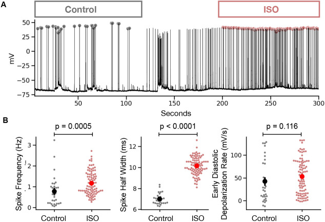
Figure 6. Example analysis of current-clamp recording from a cardiac myocyte.
(A) Time-series plot of a perforated patch current-clamp recording in control and after bath application of ISO. Detected AP peaks are show as a circle with color encoding the condition (gray for control and red for ISO). (B) Summary statistics comparing analysis results in control versus ISO conditions. Statistical p values are for a Mann Whitney U test.

