Abstract
Aim
To investigate the role of neutrophil-to-lymphocyte ratio(NLR) and platelet-to-lymphocyte(PLR) in predicting the risk of in-hospital mortality in elderly acute myocardial infarction(AMI) patients.
Methods
This study was a single-center, retrospective and observational study. From December 2015 to December 2021, a total of 1550 elderly patients (age ≥ 60 years) with AMI with complete clinical history data were enrolled in the Second Hospital of Dalian Medical University. Routine blood tests were performed on admission, and NLR and PLR were calculated based on neutrophil, platelet, and lymphocyte counts. Outcome was defined as all-cause mortality during hospitalization. Cox regression and restricted spline cubic(RCS) models were used to evaluate the association of NLR and in-hospital mortality risk and the association of PLR with in-hospital mortality risk, respectively.
Results
(1) A total of 132 (8.5%) patients died during hospitalization. From the results of blood routine, the white blood cell, neutrophil, NLR and PLR in the death group were higher than those in the non-death group, while the lymphocyte was lower than that in the non-death group, and the difference was statistically significant (P < 0.05). (2) The results of receiver operating characteristic(ROC) curves analysis showed that the predictive ability of NLR (AUC = 0.790) for in-hospital death was better than that of PLR (AUC = 0.637). (3) Multivariate Cox proportional regression hazard models showed that high NLR was associated with the risk of in-hospital mortality in elderly AMI patients (HR = 3.091, 95% CI 2.097–4.557, P < 0.001), while high PLR was not. (4) RCS models showed a nonlinear dose-response relationship between NLR and in-hospital death (P for nonlinear = 0.0007).
Conclusion
High NLR (> 6.69) is associated with the risk of in-hospital mortality in elderly patients with AMI and can be an independent predictor of poor short-term prognosis in elderly patients with AMI.
Keywords: neutrophil to lymphocyte, platelet-to-lymphocyte ratio, acute myocardial infarction, prognosis, restricted cubic splines
Introduction
Acute myocardial infarction(AMI) is considered to be a common critical emergency event in cardiology, and its mortality and morbidity are high. Despite the leapfrog development of percutaneous coronary intervention techniques in recent years, previous studies have shown that mortality within 12 months in patients with AMI is approximately 10%,1,2 and the risk of in-hospital mortality is approximately 4% to 12%.3 The instability and vulnerability of intracoronary plaques are the main causes of death in AMI patients after exposure to certain stimulating factors,4 and the formation and development of plaques are inseparable from the participation of inflammatory responses.
Studies have shown that inflammation is an important mechanism of vulnerable plaque rupture, and vulnerable plaques are pathologically characterized by the presence of thin-cap fibroatheroma (TCFA ≤ 65 μm), large lipid pools, vascular inflammation (macrophage/monocyte infiltration), intimal erosion with plaque rupture bleeding, and platelet aggregation.5,6 Most AMI patients are accompanied by hypertension, hyperlipidemia, and other underlying diseases, resulting in a low-grade inflammatory state overall,7–11 which in turn continues to aggravate their condition and ultimately leads to a poor prognosis. Therefore, new inflammatory markers that can accurately reflect the current inflammatory status of AMI patients are urgently needed to better assess their prognosis. Neutrophil-to-lymphocyte ratio(NLR) and platelet-lymphocyte ratio(PLR) are easy to calculate and can reflect the systemic inflammatory status, which is more stable than conventional blood cell parameters.12,13 Numerous studies have demonstrated the relationship between NLR and PLR and the long-term prognosis of AMI patients. Li and colleagues14 showed that patients with acute coronary syndrome (ACS) had a significantly increased risk of major adverse cardiovascular events (MACEs) at NLR ≥ 2.83, and PLR was significantly associated with no-reflow in STEMI patients as well as long-term survival in AMI patients.15–17 In addition, it has been confirmed that both NLR and PLR can be used as predictors of contrast-induced acute kidney injury (CI-AKI) in STEMI patients after primary percutaneous coronary intervention (pPCI).18,19 Moreover, NLR and PLR are also significantly associated with short-term prognosis in patients with AMI. Previous studies have confirmed that NLR is associated with the risk of in-hospital death in patients with AMI,20 and PLR is associated with the development of MACEs during hospitalization in patients with AMI,21 but these studies have focused less on the association between NLR and PLR and the risk of in-hospital mortality in elderly patients with AMI. It is well-known that age is an important risk factor for poor prognosis in coronary artery disease(CAD), and the risk of death in AMI patients increases with increasing age.22 It is worth mentioning that the systemic immune-inflammation index (SII) is a novel inflammatory indicator that incorporates neutrophil counts (N), platelet counts (P), and lymphocyte counts (L).23 SII is closely related to the risk of MACEs in patients with ACS,14 ACS with chronic kidney disease (CKD),24 and is also associated with long-term mortality in patients with heart failure with reduced ejection fraction,23 which shows that SII also plays an important role in cardiovascular disease, but because SII calculation is cumbersome compared with NLR and PLR, in order to facilitate clinical application, we mainly investigate the relationship between NLR and PLR and short-term prognosis in elderly AMI patients in this article.
The aim of this study was to investigate whether NLR, PLR are associated with the risk of in-hospital mortality in elderly patients with AMI and whether there is a dose-response relationship for this association.
Methods
Study Population
This was a single-center, retrospective and observational study of 1550 elderly patients 60 years of age and older with AMI who were hospitalized at the Second Hospital of Dalian Medical University from December 2015 to December 2021. This study is a observational study, in which patients informed consent can be exempted and ethical requirements in the Declaration of Helsinki have been met, and has been approved by the Ethics Committee of the Second Hospital of Dalian Medical University. Inclusion criteria: 1. Age ≥ 60 years; 2. Diagnosis of acute myocardial infarction (including NSTEMI and STEMI) at admission. Exclusion criteria: 1. Any disease that may affect the neutrophil, lymphocyte, platelet count of patients (such as hematological diseases, malignant tumors, autoimmune diseases, infections and systemic inflammation); 2. History of coronary revascularization (CABG or PCI); 3. Life expectancy less than half a year; 4. Severe CAD, patients requiring CABG.
Data Collection and Processing
The clinical characteristics, medical history, and laboratory test results of the patients at admission and during hospitalization were collected from the electronic medical record system. The laboratory tests performed at admission mainly included blood routine examination, liver and kidney function, blood glucose, blood lipid, serum ion levels, and myocardial enzyme levels. NLR = neutrophil count / lymphocyte count. PLR = platelet count / lymphocyte count. The drug use during hospitalization was also recorded, including lipid-lowering drugs, β-blockers, ACEI/ARBs, and aspirin. The outcome was defined as all-cause mortality during hospitalization.
Statistical Analysis
Data were processed by SPSS 23.0, MedCalc 15.0, and R 4.2.1. For categorical variables, the data were described as the frequency or percentage. For continuous variables, if they conformed to the normal distribution, the data were presented as the mean ± standard deviation; otherwise, data were presented as quartiles [median (quartile 25, 75%)]. If continuous data satisfied normality, comparisons between two groups or among multiple groups were analyzed by the t-test or ANOVA analysis; otherwise, the non-parametric test was used. Fisher´s exact test or the Chi-square test was used for the comparison of categorical variables. Receiver operating characteristic(ROC) curves was employed to explore the performance of NLR and PLR. Univariate and variate Cox regression was performed for determining the hazard ratio (HR) of blood cell variables. In addition, restricted cubic splines(RCS) were used to explore the dose-response relationship between NLR and PLR and in-hospital death. P < 0.05 was considered to indicate a significant difference.
Results
Sample Characteristics
Baseline characteristics of the study cohort are detailed in Table 1. A total of 1550 elderly patients with AMI were included in this retrospective study, and a total of 132 deaths (8.5%) were recorded during hospitalization. Although the mean age and proportion of male patients were more in the death group (71 male, 61 female; mean age 79.55 ± 8.78) than in the non-death group (855 male, 563 female; mean age 72.50 ± 8.30), there was no significant difference between the two groups (P > 0.05). From the results of blood routine, the levels of white blood cells, neutrophils, NLR and PLR in the death group were significantly higher than those in the non-death group, while the levels of lymphocytes and hemoglobin were lower than those in the non-death group, and the differences were statistically significant (P < 0.05).
Table 1.
Basic Characteristics of Enrolling Patients
| Variables | Total Study Population | Without Death | with Death | P value |
|---|---|---|---|---|
| N=1550 | N=1418 | N=132 | ||
| Male, n(%) | 926(59.7) | 855(39.7) | 71(46.2) | 0.145 |
| BMI(kg/m2) | 24.97 ± 3.48 | 25.08 ± 3.51 | 24.07 ± 3.04 | <0.001 |
| Age(years) | 73.10 ± 8.57 | 72.50 ± 8.30 | 79.55 ± 8.78 | 0.818 |
| SBP(mmHg) | 138(120,156) | 139(121,157) | 132(109,141) | 0.001 |
| Diabetes, n(%) | 671(43.3) | 603(42.5) | 68(51.5) | 0.046 |
| Heart failure, n(%) | 1082(69.8) | 959(67.6) | 123(93.2) | <0.001 |
| Type of AMI | 0.048 | |||
| NSTEMI, n(%) | 1102(71.1) | 1018(71.8) | 84(63.6) | |
| STEMI, n(%) | 448(28.9) | 400(28.2) | 48(36.4) | |
| Previous stroke, n(%) | 125(8.1) | 108(7.7) | 16(12.1) | 0.068 |
| Hypertension, n(%) | 1079(69.6) | 994(70.1) | 85(64.3) | 0.173 |
| eGFR class | <0.001 | |||
| >60, n(%) | 1099(70.9) | 1054(74.3) | 45(34.1) | |
| 30~60, n(%) | 228(14.7) | 188(13.3) | 40(30.3) | |
| <30, n(%) | 223(14.4) | 176(12.4) | 47(35.6) | |
| Blood routine examination | ||||
| Neutrophils(109/L) | 5.18(3.93,7.04) | 5.01(3.85,6.63) | 8.96(5.87,12.34) | <0.001 |
| Lymphocytes(109/L) | 1.40(1.05,1.87) | 1.42(1.08,1.89) | 1.07(0.65,1.70) | <0.001 |
| WBC(109/L) | 7.50(0.5.93,9.36) | 7.32(5.86,9.02) | 10.85(8.14,14.39) | <0.001 |
| PLT(109/L) | 205(170,244) | 204.50(170.00,244.00) | 208.50(168.00,252.50) | 0.969 |
| Hb(g/L) | 130(116,142) | 131.00(117.75,142.00) | 117.50(103.25,132.75) | <0.001 |
| NLR | 3.61(2.43,5.83) | 3.43(2.37,5.29) | 7.74(4.71,14.66) | <0.001 |
| PLR | 143.02(107.28,196.92) | 140.07(106.06,189.93) | 185.85(122.81,284.66) | <0.001 |
| Laboratory data | ||||
| Na(mmol/L) | 140(137.89,141.90) | 140.10(138.02,141.92) | 138.90(135.52,141.00) | <0.001 |
| K(mmol/L) | 3.95(3.66,4.26) | 3.93(3.65,4.23) | 4.12(3.71,4.58) | <0.001 |
| ALT(U/L) | 23.07(15.00,37.29) | 22.57(14.96,36.34) | 28.77(16.28,62.95) | <0.001 |
| AST(U/L) | 29.16(19.56,75.73) | 27.83(19.26,66.00) | 75.40(29.04,206.57) | <0.001 |
| Triglycerides(mmol/L) | 1.31(0.96,1.83) | 1.33(0.96,1.85) | 1.17(0.93,1.71) | 0.048 |
| Total cholesterol(mmol/L) | 4.36(3.64,5.20) | 4.38(3.65,5.20) | 4.31(3.45,5.23) | 0.715 |
| LDL-C(mmol/L) | 2.41(1.85,3.04) | 2.42(1.86,3.04) | 2.27(1.73,3.09) | 0.331 |
| HDL-C(mmol/L) | 1.04(0.90,1.21) | 1.03(0.90,1.21) | 1.08(0.84,1.29) | 0.547 |
| CTNI(ug/L) | 1.05(0.55,3.46) | 0.98(0.52,2.58) | 5.26(0.85,20.22) | <0.001 |
| CK-MB(ug/L) | 19.76(14.84,43.05) | 19.59(15.00,40.13) | 22.25(6.10,96.19) | 0.867 |
| Inpatient medication | ||||
| Aspirin, n(%) | 1437(92.7) | 1324(93.4) | 113(85.6) | 0.001 |
| Statins, n(%) | 1512(97.5) | 1391(98.1) | 121(91.7) | <0.001 |
| β-blockers, n(%) | 896(57.8) | 833(58.7) | 63(47.7) | 0.014 |
| ACEI/ARBs, n(%) | 685(44.2) | 653(46.1) | 32(24.2) | <0.001 |
Abbreviations: BMI, body mass index; SBP, systolic blood press; NSTEMI, non-ST segment elevation myocardial infarction; STEMI, ST segment elevation myocardial infarction; eGFR, estimated glomerular filtration rate; WBC, white blood cell; PLT, platelet; Hb, hemoglobin; NLR, neutrophil-lymphocyte ratio; PLR, platelet-lymphocyte ratio; ALT, alanine aminotransferase; AST, aspartate aminotransferase; LDL-C, low-density lipoprotein-cholesterol; HDL-C, high-density lipoprotein-cholesterol; CTNI, cardiac troponin I; CK-MB creatine kinase-MB; ACEIs, angiotensin-converting enzyme inhibitors; ARBs, angiotensin receptor blockers.
Predictive Value of NLR and PLR for the Risk of in-Hospital Mortality in Elderly Patients with AMI
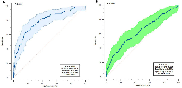
ROC curves were used to evaluate the predictive value of NLR and PLR for in-hospital mortality in elderly patients with AMI. The results showed that the AUC of NLR and PLR were 0.790 (95% CI 0.769–0.810, P < 0.0001) and 0.637 (95% CI 0.613–0.661, P < 0.0001), respectively. The optimal cut-off levels of NLR and PLR were 6.69 (Sensitivity = 62.88%, Specificity = 84.06%) and 187.6 (Sensitivity = 50.00%, Specificity = 74.12%), respectively. NLR was superior to PLR in predicting in-hospital mortality risk in elderly patients with AMI, as detailed in Figure 1.
Figure 1.
The ROC analysis of NLR and PLR predicting in-hospital death in elderly AMI patients. (A) The ROC analysis of NLR predicting in-hospital death in elderly AMI patients; (B) The ROC analysis of PLR predicting in-hospital death in elderly AMI patients.
Abbreviations: ROC curve, receiver operating characteristic curve; NLR, neutrophil-to-Lymphocyte ratio; PLR, platelet-to-lymphocyte ratio; AMI acute myocardial infarction.
Cox Regression Analysis of Risk of in-Hospital Mortality in Elderly Patients with AMI
We transformed NLR and PLR into categorical variables based on their optimal cutoffs. High NLR group was defined as NLR > 6.69 and high PLR was defined as PLR > 187.6. Cox regression analysis was used to analyze the factors affecting in-hospital death in elderly AMI patients. Univariate analysis showed that heart failure, cardiac troponin I (CTNI), creatine kinase-MB (CK-MB), eGFR class, high NLR, and high PLR were associated with the risk of in-hospital death in elderly AMI patients, as detailed in Table 2. We developed three Cox proportional hazards models based on the results of univariate analysis. Model 1 was not adjusted for any confounders, model 2 was adjusted for sex, BMI, presence of diabetes, type of AMI, heart failure and eGFR class. Model 3 was adjusted for CTNI, CK-MB and in-hospital medication (Aspirin, Statins) on the basis of model 2. Cox proportional hazards models showed that elderly AMI patients with high NLR had a 3.091-fold higher risk of in-hospital death events than those with low NLR (95% CI 2.097–4.557, P < 0.001), and high NLR was associated with the risk of in-hospital death in elderly AMI patients; high PLR was also associated with in-hospital death in models 1 and 2, but showed marginally significant (P = 0.088) results in model 3, whcih may require a larger sample sizes for validation, as detailed in Table 3.
Table 2.
Univariate Cox Regression Analysis of NLR and PLR for Predicting in-Hospital Mortality in Elderly AMI Patients
| Variables | HR | 95% CI | P value |
|---|---|---|---|
| Male | 0.794 | 0.563–1.121 | 0.19 |
| BMI | 0.943 | 0.899–0.989 | 0.016 |
| Diabetes | 1.375 | 0.977–1.936 | 0.068 |
| History of stroke | 0.190 | 0.840–2.396 | 1.419 |
| Type of AMI | 1.378 | 0.966–1.966 | 0.077 |
| Heart failure | 4.640 | 2.352–9.151 | <0.001 |
| Hypertension | 0.896 | 0.625–1.823 | 0.547 |
| CTNI | 1.016 | 1.013–1.019 | <0.001 |
| CK-MB | 1.002 | 1.001–1.003 | 0.005 |
| Aspirin | 0.544 | 0.333–0.887 | 0.015 |
| Statins | 0.248 | 0.133–0.460 | <0.001 |
| High NLR | 5.327 | 3.720–7.628 | <0.001 |
| High PLR | 2.064 | 1.462–2.915 | <0.001 |
| eGFR>60(REF) | |||
| 30~60 | 3.465 | 2.256–5.320 | <0.001 |
| <30 | 4.461 | 2.959–6.724 | <0.001 |
Notes: High NLR = NLR > 6.69; High PLR = PLR >187.6; Type of AMI includes NSTEMI and STEMI, Non-ST-segment elevation myocardial infarction was used as a reference.
Abbreviations: NLR, neutrophil-lymphocyte ratio; PLR, platelet-lymphocyte ratio; AMI, acute myocardial infarction; BMI, body mass index; CTNI, cardiac troponin I; CK-MB creatine kinase-MB; eGFR, estimated glomerular filtration rate.
Table 3.
Multivariable Cox Regression of in-Hospital Mortality for Old Patients with AMI
| Variables | High NLR | High PLR | ||||
|---|---|---|---|---|---|---|
| HR | 95% CI | P value | HR | 95% CI | P value | |
| Model 1 | 5.327 | 3.720–7.628 | <0.001 | 2.064 | 1.462–2.915 | <0.001 |
| Model 2 | 3.654 | 2.514–5.312 | <0.001 | 1.430 | 1.006–2.035 | 0.047 |
| Model 3 | 3.091 | 2.097–4.557 | <0.001 | 1.366 | 0.955–1.953 | 0.088 |
Notes: Model 1: we adjusted none; Model 2: we adjusted sex, BMI, diabetes, type of AMI(NSTEMI, STEMI), heart failure and eGFR; Model 3: Model 2 + CTNI, CK-MB, Aspirin use and Statins use. High NLR = NLR > 6.69; High PLR = PLR >187.6.
Abbreviations: AMI, acute myocardial infarction; NLR, neutrophil-lymphocyte ratio; PLR, platelet-lymphocyte ratio; BMI, body mass index; NSTEMI non-ST segment elevation myocardial infarction; STEMI, ST segment elevation myocardial infarction, eGFR, estimated glomerular filtration rate; CTNI, cardiac troponin I; CK-MB creatine kinase-MB.
Dose-Response Relationship of NLR and PLR with in-Hospital Death
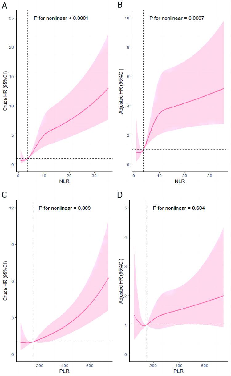
The dose-response relationship between NLR, PLR and in-hospital death in elderly AMI patients is shown in Figure 2. A nonlinear dose-response relationship was found between NLR and in-hospital death (P for crude nonlinear < 0.0001), which persisted after adjusting for multiple confounding factors (P for adjusted nonlinear = 0.0007). There was a linear dose-response relationship between PLR and in-hospital death (P for crude nonlinear = 0.889). All RCS models suggest that the risk of death during hospitalization increases with increasing levels of NLR and PLR.
Figure 2.
Dose-response relationship between NLR, PLR and in-hospital mortality in elderly patients with AMI from RCS analysis. (A) Unadjusted dose-response relationship between NLR and in-hospital mortality in elderly patients with AMI; (B) Adjusted dose-response relationship between NLR and in-hospital mortality in elderly patients with AMI; (C) Unadjusted dose-response relationship between PLR and in-hospital mortality in elderly patients with AMI; (D) Adjusted dose-response relationship between PLR and in-hospital mortality in elderly patients with AMI. Adjusted factors include: sex, BMI, diabetes, type of AMI, heart failure, eGFR, CTNI, CK-MB, Aspirin use and Statins use.
Abbreviations: NLR, neutrophil-to-lymphocyte ratio; PLR, platelet-to-lymphocyte ratio; AMI acute myocardial infarction. RCS, restricted cubic splines; BMI, body mass index; eGFR, estimated glomerular filtration rate; CTNI, cardiac troponin I; CK-MB creatine kinase-MB.
Stratification Analysis of NLR at Cut-off Value in Elderly AMI Patients
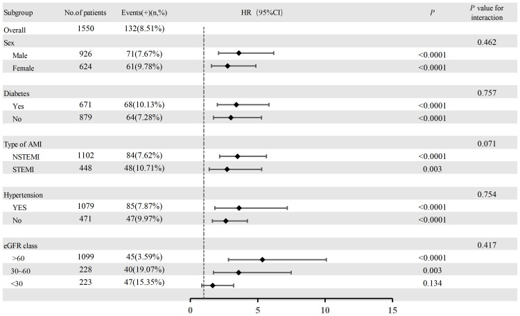
To assess the performance of NLR 6.69 as the optimal threshold for AMI, we perform subgroup stratifying analysis (Figure 3). Stratified by sex (male or female), history of diabetes (yes or no), type of AMI (NSTEMI or STEMI), history of hypertension (yes or no), eGFR class (> 60 mL/min/1.73 m2, 30–60 mL/min/1.73 m2, < 30 mL/min/1.73 m2). When stratified by gender, male patients with NLR > 6.69 (high NLR) had a 3.602-fold higher risk of death compared with patients with NLR ≤ 6.69 (95% CI 2.097–6.188, P < 0.0001), while the HR for female patients was 2.765 (95% CI 1.572–4.863, P < 0.0001). For patients with diabetes, the risk of death in the high NLR group was 3.415-fold higher than that in the low NLR group (95% CI 2.000–5.834, P < 0.0001), while the HR in non-diabetic patients was 3.011 (95% CI 1.717–5.28, P < 0.0001). For patients with NSTEMI, the risk of death in the high NLR group was 3.503-fold higher than that in the low NLR group (95% CI 2.171–5.653, P < 0.0001), while the HR in STEMI patients was 2.726 (95% CI 1.405 to 5.288, P = 0.003). For patients with hypertension, the risk of death in the high NLR group was 3.616-fold higher than that in the low NLR group (95% CI 1.816–7.200, P < 0.0001), while the HR in patients without hypertension was 2.634 (95% CI 1.635–4.243, P < 0.0001). For patients with eGFR class > 60 mL/min/1.73 m2 and 30 to 60 mL/min/1.73 m2, the risk of death in the high NLR group was 5.34 times (95% CI 2.835–10.091, P < 0.0001) and 3.585 times (95% CI 1.718–7.482, P = 0.003) higher than that in the low NLR group, respectively. In patients with eGFR < 30 mL/min/1.73 m2, the HR was 1.659 in the high NLR group, but it was not statistically significant, and we considered that this may be related to the small sample size of this group of patients.
Figure 3.
Hazard ratios of the high NLR for predicting in-hospital death in the subgroup analysis.
Abbreviations: AMI, acute myocardial infarction; NSTEMI, non-ST segment elevation myocardial infarction; STEMI, ST segment elevation myocardial infarction; eGFR, estimated glomerular filtration rate.
Discussion
In this retrospective study of 1550 elderly patients with AMI, we investigated the association between NLR, PLR, and the risk of in-hospital death in elderly patients with AMI. The results of ROC analysis showed that NLR (AUC = 0.790) was superior to PLR (AUC = 0.637) in predicting the risk of in-hospital mortality in elderly patients with AMI. In addition, we divided patients into high NLR group (> 6.69) and high PLR group (> 187.6) according to the optimal cut-off value of NLR and PLR in ROC analysis, and the results showed that high NLR was associated with the risk of in-hospital mortality in elderly AMI (HR = 3.091, 95% CI 2.097–4.557, P < 0.001), and this result was stable in multiple models. Whereas high PLR emerged as marginally significant (HR = 1.366, 95% CI 0.955–1.953, P = 0.088) in the final model, we believe that high PLR may also be independently associated with in-hospital death in elderly AMI patients (at least this trend should exist), but limited by sample size, single-center studies, it showed marginally significant results. Finally, in this study, we investigated for the first time the dose-response relationship between NLR, PLR and the risk of in-hospital death in elderly patients with AMI, and our results showed that NLR had a nonlinear dose-response relationship with the risk of in-hospital death in elderly patients with acute AMI (P for nonlinear = 0.0007), while PLR showed a linear dose-response relationship (P for nonlinear = 0.684), suggesting that with the increasing levels of NLR and PLR, the risk of in-hospital death in elderly patients with AMI also increased.
Age is one of the important risk factors for the occurrence and development of CAD. Mortality increases in AMI patients with increasing age.22 Previous studies have shown that approximately 83% of deaths in patients hospitalized for AMI occur in people over 65 years of age.25 Professor Hayiroğlu26 explored the risk factors for in-hospital mortality in STEMI patients with cardiogenic shock, although the age factor lost statistical significance in multivariate logistic regression analysis in this study, patients in the death group were significantly older than those in the non-death group (71 ± 13 vs 67 ± 12, P = 0.002). Previous studies have shown that Intermountain Risk Score (IMRS) can predict in-hospital mortality risk in STEMI patients27 as well as STEMI patients with cardiogenic shock28 and the patient ‘s age is a major component of the IMRS. The above findings all suggest that age is critical in the short-term prognosis of AMI patients. Therefore, if a convenient and efficient clinical index can be found to predict the risk of death during hospitalization in elderly patients with AMI, and more refined clinical care and treatment options can be adopted according to it, the short-term prognosis of elderly patients with AMI can be significantly improved. NLR and PLR were initially applied in patients with malignant tumors to assess their prognosis and have gradually attracted much attention in the cardiovascular field in recent years.29,30 Inflammation dominates and participates in the whole process of the occurrence and development of CAD, which is the main cause of vulnerable plaque rupture in the coronary artery.5,6 When inflammation is activated in the body, neutrophils produce highly reactive oxygen species to kill pathogens, and the formation of extracellular traps(NETs) by neutrophils also helps destroy pathogens,31 but it also promotes the rupture of vulnerable plaques causing AMI.20 In the early stages of AMI, neutrophils can be recruited and promote platelet activation and deposition before platelets are activated,32 causing further myocardial damage in the infarcted area, and subsequently neutrophils can maintain a chronic inflammatory environment.33 Platelets are one of the basic conditions for coronary thrombosis, and higher platelet counts are associated with worse hospital outcomes in patients with acute STEMI.34 Platelets can be activated to accumulate inflammatory cells recruited to atherosclerotic lesions and release a large number of inflammatory mediators to enrich the inflammatory environment.35 Unlike neutrophils and platelets, lymphocytes are thought to have anti-atherosclerotic effects, but this is dependent on subsets of lymphocytes and the mechanism is complex.36 However, it is clear that low lymphocyte counts are directly associated with worse cardiovascular outcomes,37 which may be due to increased catecholamine and glucocorticoid levels in vivo, promoting lymphocyte apoptosis, inhibiting lymphocyte proliferation and differentiation resulting in decreased peripheral blood lymphocyte counts under this stress state of acute myocardial infarction, while lymphocyte redistribution also affects their numbers.38,39
NLR can systematically and accurately reflect the degree of body inflammation and stress,20 and PLR also reflects platelet activation and prothrombotic state.40 NLR and PLR are more accurate and objective than neutrophils, platelets, and lymphocytes alone in predicting outcomes in patients with AMI because they always combine two indicators that reflect the body ‘s inflammatory status. A cohort study of 2618 hospitalized Chinese patients with AMI showed that a high NLR (> 5.509) was positively associated with the risk of in-hospital mortality in patients with AMI;20 another small study also confirmed that a high NLR (> 6.607) was an important predictor of MACE in patients with AMI.41 Giakoumidakis et al found a relationship between perioperative NLR and increased in-hospital and 30-day mortality and prolonged postoperative hospital stay in their study.42 Although there are differences in the definition of high NLR due to different sample populations, these studies have all shown that high NLR is associated with adverse outcomes in AMI patients. In our study, high NLR was defined as > 6.69. Older AMI patients with high NLR had a 3.091-fold higher risk of in-hospital mortality compared with those with low NLR (95% CI 2.097–4.557, P < 0.001), which is consistent with previous findings. Previous studies have shown that high PLR is associated with the risk of in-hospital death in elderly patients with AMI, but only 45 elderly patients with AMI experienced a death event in this study,40 which is limited by the effective sample size. The study only adjusted for two confounders in multivariate regression: age (≥ 60 years) and presence of hypertension, and the results may not be stable if adjusted for more confounders. Although some studies have confirmed that PLR is also predictive of long-term prognosis in patients with AMI,16,43 a cohort study of 1701 ACS patients treated with PCI has a different voice from previous findings, and this study found that PLR has limited value for long-term prognosis in ACS patients.14 In our study, although a high PLR (> 187.6) was also suggested to be associated with the risk of in-hospital death in elderly AMI patients in the first two models, when we adjusted for in-hospital drug therapy and myocardial enzyme parameters, the results of model 3 showed that high PLR was not significantly associated with the risk of in-hospital death in elderly AMI patients(P = 0.088). We believe this may be related to our sample size, single-center study. But the marginally significant results at least suggest a trend toward a possible association between high PLR and in-hospital death in older AMI patients, and we still need a larger sample size to confirm this. Unlike previous studies, our study is the first to assess the dose-response relationship between NLR, PLR and the risk of in-hospital mortality in elderly AMI patients using RCS. The results showed that there was a nonlinear dose-response relationship between NLR and in-hospital death, while there was a linear dose-response relationship between PLR and in-hospital death. The RCS model suggests that with increasing levels of NLR and PLR, the risk of in-hospital mortality increases in older patients with AMI. When we clinically identify these high-risk patients, we can take the following measures to reduce their risk of death during hospitalization: 1. revascularization as early as possible on the basis of active antithrombotic and anticoagulant therapy;44,45 2. patients should pay close attention to their blood pressure and blood glucose levels after admission, control within the ideal range as far as possible, and pay attention to correcting electrolyte imbalance and acid-base imbalance; 3. pay attention to the prevention and management of AMI complications, such as heart failure, cardiogenic shock, and renal insufficiency; 4. β-blockers (within 24 hours) should be used as soon as possible for AMI patients without contraindications, and angiotensin enzyme inhibitors (ACEIs) or receptor blockers (ARBs) and eplerenone (within 7 days) should be routinely used for AMI patients with impaired left ventricular function;45,46 5. conditional medical institutions may consider telemedicine, especially ECG transmission;47 6. mechanical adjuvant therapy should be performed when necessary; 7. Give more refined clinical care.
This study has the following limitations: First, it is a single-center study and its results may be influenced by patient selection bias. Second, we failed to follow the long-term prognosis of these patients, which limits the predictive value of NLR, PLR for long-term mortality risk in elderly AMI patients. Finally, retrospective studies were unable to identify a causal relationship between NLR, PLR, and risk of in-hospital mortality in older patients with AMI.
Conclusion
Our findings suggest that NLR and PLR are predictive of in-hospital mortality risk in elderly patients with AMI, but NLR has better predictive ability than PLR. In addition, high NLR (> 6.69) is associated with the risk of in-hospital mortality in elderly patients with AMI, and high PLR (> 186.7) may also be associated with in-hospital mortality in elderly patients with AMI. Finally, we found a dose-response relationship between NLR, PLR and the risk of in-hospital mortality in elderly AMI patients. Therefore, clinicians can use NLR, PLR to risk stratify elderly hospitalized patients with AMI to better reduce the incidence of adverse cardiovascular events during hospitalization.
Funding Statement
This study was funded by the Liaoning Provincial “Selecting the Best Candidates by Opening Competition Mechanism” Science and Technology Program (grant number 2022JH1/10400004).
Data Sharing Statement
The raw data supporting the conclusions of this article will be made available by the authors, without undue reservation.
Statement of Ethics
The study protocol has been reviewed and approved by the Ethics Committee of the Second Hospital of Dalian Medical University. The Ethics Committee of the Second Hospital of Dalian Medical University waived the need for informed consent based on the following reasons: (1) the purpose of the study was important; (2) the possible risk to patients was not higher than the minimum one; (3) the waiver of informed consent would not adversely affect the rights and health of patients; (4) the patients’ privacy and personal identity information were well protected. We have desensitized the patient ‘s personal identity to protect patient privacy.
The protocol of the study is compliant with Declaration of Helsinki.
Patient Privacy Protection Statement
We desensitized all the data that can be used to identify patient personal information, such as their names, hospitalization ID and telephone numbers, to protect the privacy of patients.
Author Contributions
All authors made a significant contribution to the work reported, whether that is in the conception, study design, execution, acquisition of data, analysis and interpretation, or in all these areas; took part in drafting, revising or critically reviewing the article; gave final approval of the version to be published; have agreed on the journal to which the article has been submitted; and agree to be accountable for all aspects of the work.
Disclosure
The authors report no conflicts of interest.
References
- 1.Pedersen F, Butrymovich V, Kelbæk H, et al. Short- and long-term cause of death in patients treated with primary PCI for STEMI. J Am Coll Cardiol. 2014;64(20):2101–2108. [DOI] [PubMed] [Google Scholar]
- 2.Fokkema ML, James SK, Albertsson P, et al. Population trends in percutaneous coronary intervention: 20-year results from the SCAAR (Swedish Coronary Angiography and Angioplasty Registry). J Am Coll Cardiol. 2013;61(12):1222–1230. [DOI] [PubMed] [Google Scholar]
- 3.Kristensen SD, Laut KG, Fajadet J, et al. Reperfusion therapy for ST elevation acute myocardial infarction 2010/2011: current status in 37 ESC countries. Eur Heart J. 2014;35(29):1957–1970. [DOI] [PubMed] [Google Scholar]
- 4.Pasterkamp G, den Ruijter HM, Libby P. Temporal shifts in clinical presentation and underlying mechanisms of atherosclerotic disease. Nat Rev Cardiol. 2017;14(1):21–29. [DOI] [PubMed] [Google Scholar]
- 5.Stone GW, Mintz GS, Virmani R. Vulnerable Plaques, Vulnerable Patients, and Intravascular Imaging. J Am Coll Cardiol. 2018;72(17):2022–2026. [DOI] [PubMed] [Google Scholar]
- 6.Arbab-Zadeh A, Fuster V. The myth of the “vulnerable plaque”: transitioning from a focus on individual lesions to atherosclerotic disease burden for coronary artery disease risk assessment. J Am Coll Cardiol. 2015;65(8):846–855. [DOI] [PMC free article] [PubMed] [Google Scholar]
- 7.Ginsberg HN, Packard CJ, Chapman MJ, et al. Triglyceride-rich lipoproteins and their remnants: metabolic insights, role in atherosclerotic cardiovascular disease, and emerging therapeutic strategies-a consensus statement from the European Atherosclerosis Society. Eur Heart J. 2021;42(47):4791–4806. [DOI] [PMC free article] [PubMed] [Google Scholar]
- 8.Musher DM, Abers MS, Corrales-Medina VF. Acute Infection and Myocardial Infarction. N Engl J Med. 2019;380(2):171–176. [DOI] [PubMed] [Google Scholar]
- 9.Webb RJ, Mazidi M, Lip GYH, Kengne AP, Banach M, Davies IG. The role of adiposity, diet and inflammation on the discordance between LDL-C and apolipoprotein B. Nutr Metab Cardiovasc Dis. 2022;32(3):605–615. [DOI] [PubMed] [Google Scholar]
- 10.Mikolajczyk TP, Szczepaniak P, Vidler F, Maffia P, Graham GJ, Guzik TJ. Role of inflammatory chemokines in hypertension. Pharmacol Ther. 2021;223:107799. [DOI] [PubMed] [Google Scholar]
- 11.Harrison DG, Marvar PJ, Titze JM. Vascular inflammatory cells in hypertension. Front Physiol. 2012;3:128. [DOI] [PMC free article] [PubMed] [Google Scholar]
- 12.Sheng J, Liu N, He F, Cheng C, Shen S, Sun Y. Changes in the neutrophil-to-lymphocyte and platelet-to-lymphocyte ratios before and after percutaneous coronary intervention and their impact on the prognosis of patients with acute coronary syndrome. Clinics. 2021;76:e2580. [DOI] [PMC free article] [PubMed] [Google Scholar]
- 13.Kounis NG, Koniari I, Plotas P, et al. Inflammation, Thrombosis, and Platelet-to-Lymphocyte Ratio in Acute Coronary Syndromes. Angiology. 2021;72(1):6–8. [DOI] [PubMed] [Google Scholar]
- 14.Li Q, Ma X, Shao Q, et al. Prognostic Impact of Multiple Lymphocyte-Based Inflammatory Indices in Acute Coronary Syndrome Patients. Front Cardiovasc Med. 2022;9:811790. [DOI] [PMC free article] [PubMed] [Google Scholar]
- 15.Ugur M, Gul M, Bozbay M, et al. The relationship between platelet to lymphocyte ratio and the clinical outcomes in ST elevation myocardial infarction underwent primary coronary intervention. Blood Coagulation Fibrinolysis. 2014;25(8):806–811. [DOI] [PubMed] [Google Scholar]
- 16.Azab B, Shah N, Akerman M, McGinn JT. Value of platelet/lymphocyte ratio as a predictor of all-cause mortality after non-ST-elevation myocardial infarction. J Thromb Thrombolysis. 2012;34(3):326–334. [DOI] [PubMed] [Google Scholar]
- 17.Kurtul A, Yarlioglues M, Murat SN, et al. Usefulness of the platelet-to-lymphocyte ratio in predicting angiographic reflow after primary percutaneous coronary intervention in patients with acute ST-segment elevation myocardial infarction. Am J Cardiol. 2014;114(3):342–347. [DOI] [PubMed] [Google Scholar]
- 18.Kaya A, Kurt M, Tanboğa IH. Neutrophil-to-lymphocyte ratio predicts contrast-induced nephropathy in patients undergoing primary percutaneous coronary intervention. Angiology. 2014;65(1):59. [DOI] [PubMed] [Google Scholar]
- 19.Velibey Y, Oz A, Tanik O, et al. Platelet-to-Lymphocyte Ratio Predicts Contrast-Induced Acute Kidney Injury in Patients With ST-Segment Elevation Myocardial Infarction Undergoing Primary Percutaneous Coronary Intervention. Angiology. 2017;68(5):419–427. [DOI] [PubMed] [Google Scholar]
- 20.Ji Z, Liu G, Guo J, et al. The Neutrophil-to-Lymphocyte Ratio Is an Important Indicator Predicting In-Hospital Death in AMI Patients. Front Cardiovasc Med. 2021;8:706852. [DOI] [PMC free article] [PubMed] [Google Scholar]
- 21.Li XT, Fang H, Li D, et al. Association of platelet to lymphocyte ratio with in-hospital major adverse cardiovascular events and the severity of coronary artery disease assessed by the Gensini score in patients with acute myocardial infarction. Chin Med J. 2020;133(4):415–423. [DOI] [PMC free article] [PubMed] [Google Scholar]
- 22.Xia TL, Huang FY, Li YM, et al. The impact of age on the implementation of evidence-based medications in patients with coronary artery disease and its prognostic significance: a retrospective cohort study. BMC Public Health. 2018;18(1):150. [DOI] [PMC free article] [PubMed] [Google Scholar]
- 23.Hayıroğlu M, Çınar T, Çinier G, et al. Evaluating systemic immune-inflammation index in patients with implantable cardioverter defibrillator for heart failure with reduced ejection fraction. Pacing Clin Electrophysiol. 2022;45(2):188–195. [DOI] [PubMed] [Google Scholar]
- 24.Shi S, Kong S, Ni W, et al. Association of the Systemic Immune-Inflammation Index with Outcomes in Acute Coronary Syndrome Patients with Chronic Kidney Disease. J Inflamm Res. 2023;16:1343–1356. [DOI] [PMC free article] [PubMed] [Google Scholar]
- 25.Alexander KP, Newby LK, Cannon CP, et al. Acute coronary care in the elderly, part I: non-ST-segment-elevation acute coronary syndromes: a scientific statement for healthcare professionals from the American Heart Association Council on Clinical Cardiology: in collaboration with the Society of Geriatric Cardiology. Circulation. 2007;115(19):2549–2569. [DOI] [PubMed] [Google Scholar]
- 26.Hayıroğlu M, Keskin M, Uzun AO, et al. Predictors of In-Hospital Mortality in Patients With ST-Segment Elevation Myocardial Infarction Complicated With Cardiogenic Shock. Heart Lung Circ. 2019;28(2):237–244. [DOI] [PubMed] [Google Scholar]
- 27.Çınar T, Şaylık F, Akbulut T, et al. Evaluation of Intermountain Risk Score for Short- and Long-Term Mortality in ST Elevation Myocardial Infarction Patients. Angiology. 2023;74(4):357–364. [DOI] [PubMed] [Google Scholar]
- 28.Mert Ilker H, Faysal S, Ahmet Çağdaş Y, Murat S, Tufan Ç. Prognostic value of Intermountain Risk Score for short- and long-term mortality in patients with cardiogenic shock. Coron Artery Dis. 2023;34(2):154–159. [DOI] [PubMed] [Google Scholar]
- 29.Templeton AJ, Ace O, McNamara MG, et al. Prognostic role of platelet to lymphocyte ratio in solid tumors: a systematic review and meta-analysis. Cancer Epidemiol Biomarkers Prev. 2014;23(7):1204–1212. [DOI] [PubMed] [Google Scholar]
- 30.Budzianowski J, Pieszko K, Burchardt P, Rzeźniczak J, Hiczkiewicz J. The Role of Hematological Indices in Patients with Acute Coronary Syndrome. Dis Markers. 2017;2017:3041565. [DOI] [PMC free article] [PubMed] [Google Scholar]
- 31.Döring Y, Soehnlein O, Weber C. Neutrophil Extracellular Traps in Atherosclerosis and Atherothrombosis. Circ Res. 2017;120(4):736–743. [DOI] [PubMed] [Google Scholar]
- 32.Darbousset R, Thomas GM, Mezouar S, et al. Tissue factor-positive neutrophils bind to injured endothelial wall and initiate thrombus formation. Blood. 2012;120(10):2133–2143. [DOI] [PubMed] [Google Scholar]
- 33.Drechsler M, Megens RT, van Zandvoort M, Weber C, Soehnlein O. Hyperlipidemia-triggered neutrophilia promotes early atherosclerosis. Circulation. 2010;122(18):1837–1845. [DOI] [PubMed] [Google Scholar]
- 34.Paul GK, Sen B, Bari MA, et al. Correlation of platelet count and acute ST-elevation in myocardial infarction. Mymensingh Med J. 2010;19(3):469–473. [PubMed] [Google Scholar]
- 35.Lievens D, von Hundelshausen P. Platelets in atherosclerosis. Thromb Haemost. 2011;106(5):827–838. [DOI] [PubMed] [Google Scholar]
- 36.Ammirati E, Moroni F, Magnoni M, Camici PG. The role of T and B cells in human atherosclerosis and atherothrombosis. Clin Exp Immunol. 2015;179(2):173–187. [DOI] [PMC free article] [PubMed] [Google Scholar]
- 37.Núñez J, Miñana G, Bodí V, et al. Low lymphocyte count and cardiovascular diseases. Curr Med Chem. 2011;18(21):3226–3233. [DOI] [PubMed] [Google Scholar]
- 38.Bergquist J, Tarkowski A, Ewing A, Ekman R. Catecholaminergic suppression of immunocompetent cells. Immunol Today. 1998;19(12):562–567. [DOI] [PubMed] [Google Scholar]
- 39.Mooren FC, Blöming D, Lechtermann A, Lerch MM, Völker K. Lymphocyte apoptosis after exhaustive and moderate exercise. J Appl Physiol. 2002;93(1):147–153. [DOI] [PubMed] [Google Scholar]
- 40.Li L, Ma Y, Geng XB, et al. Platelet-to-lymphocyte ratio relates to poor prognosis in elderly patients with acute myocardial infarction. Aging Clin Exp Res. 2021;33(3):619–624. [DOI] [PubMed] [Google Scholar]
- 41.Chen B, Yuan L, Chen X, et al. Correlations and Prognostic Roles of the Nutritional Status and Neutrophil-to-lymphocyte Ratio in Elderly Patients with Acute Myocardial Infarction Undergoing Primary Coronary Intervention. Int Heart J. 2020;61(6):1114–1120. [DOI] [PubMed] [Google Scholar]
- 42.Giakoumidakis K, Fotos NV, Patelarou A, et al. Perioperative neutrophil to lymphocyte ratio as a predictor of poor cardiac surgery patient outcomes. Pragmatic Obser Res. 2017;8:9–14. [DOI] [PMC free article] [PubMed] [Google Scholar]
- 43.Ozcan Cetin EH, Cetin MS, Aras D, et al. Platelet to Lymphocyte Ratio as a Prognostic Marker of In-Hospital and Long-Term Major Adverse Cardiovascular Events in ST-Segment Elevation Myocardial Infarction. Angiology. 2016;67(4):336–345. [DOI] [PubMed] [Google Scholar]
- 44.Gheeraert PJ, De Buyzere ML, Taeymans YM, et al. Risk factors for primary ventricular fibrillation during acute myocardial infarction: a systematic review and meta-analysis. Eur Heart J. 2006;27(21):2499–2510. [DOI] [PubMed] [Google Scholar]
- 45.Steg PG, James SK, Atar D, et al. ESC Guidelines for the management of acute myocardial infarction in patients presenting with ST-segment elevation. Eur Heart J. 2012;33(20):2569–2619. [DOI] [PubMed] [Google Scholar]
- 46.Pitt B, White H, Nicolau J, et al. Eplerenone reduces mortality 30 days after randomization following acute myocardial infarction in patients with left ventricular systolic dysfunction and heart failure. J Am Coll Cardiol. 2005;46(3):425–431. [DOI] [PubMed] [Google Scholar]
- 47.Marcolino MS, Maia LM, Oliveira JAQ, et al. Impact of telemedicine interventions on mortality in patients with acute myocardial infarction: a systematic review and meta-analysis. Heart. 2019;105(19):1479–1486. [DOI] [PubMed] [Google Scholar]


