

Abstract
Key Clinical Message
Renal angiomyolipoma is a type of benign hamartoma that may occur sporadically or be associated with tuberous sclerosis complex. Due to their distinct appearance, CT, MRI, or sonography are typically used to diagnose AMLs.
Abstract
The uncommon benign hamartoma known as renal angiomyolipoma (AML) linked with tuberous sclerosis has a poor prognosis and potentially fatal side effects. Due of their distinct appearance, computed tomography (CT), magnetic resonance imaging (MRI), or sonography are typically used to diagnose AMLs.
Keywords: radiology, renal angiomyolipoma, tuberous sclerosis complex
1. CASE DESCRIPTION
A 20‐year‐old female patient with known comorbid epilepsy and mild intellectual disability complained of recent hematuria and one‐year‐long abdominal discomfort. The discomfort began gradually, was limited to the left lumbar area, and was made worse by physical activity and reduced fluid intake. Moreover, she noticed dark urine 2 days back, visible throughout the flow. Intermittent burning sensation during micturition and sporadic pain over the hypogastric region were present.
A contrast‐enhanced computed tomography (CE‐CT) was done (Figure 1). Both kidneys were enlarged. There were multiple varying sizes of mixed‐density lesions in both kidneys, showing areas of fat attenuation, soft tissue component, and enlarged vessels, the largest one in the left kidney measured approximately 8.3 × 7.6 × 14.3 cm in APxTRxCC dimensions. The described imaging features suggested bilateral renal AML with possible rupture in the largest left renal AML (Figure 2).
FIGURE 1.
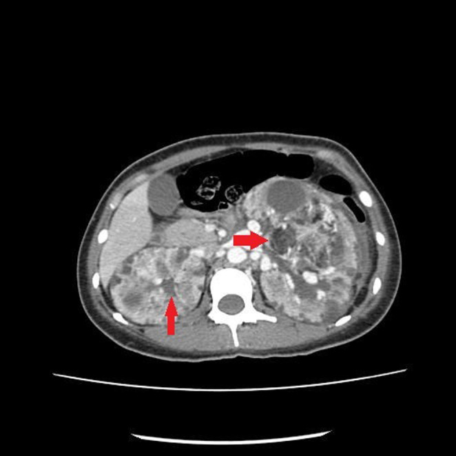
There are multiple varying sizes mixed density lesions in both kidneys, the largest one in the left kidney measures approximately 8.3 × 7.6 × 14.3 cm in APxTRxCC dimensions (Red Arrow).
FIGURE 2.
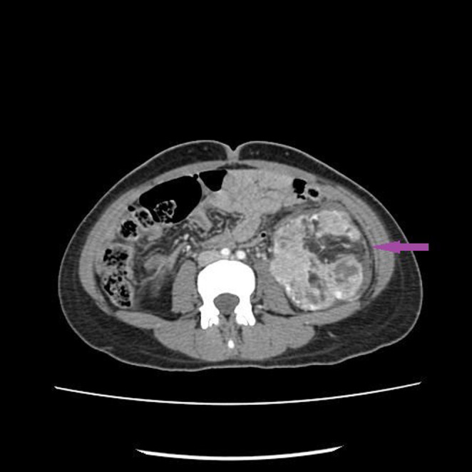
The lesion shows occasional focal breaches in its capsule resulting in the formation of blood clots within the pelvicalyceal system of the left kidney (Purple Arrow).
Additionally, magnetic resonance imaging (MRI) of the brain was performed, revealing subependymal calcification, and cortical tubers.
The definitive diagnosis of tuberous sclerosis complex was reached after reviewing her medical history and radiological findings.
2. DISCUSSION
A benign hamartoma known as renal angiomyolipoma (AML) may develop sporadically or be linked to tuberous sclerosis complex (TSC). 1 , 2 , 3 According to reports, there are four times as many random angiomyolipoma instances as TSC cases. 3 , 4 Adipose tissue, blood arteries, and smooth muscle make up the “triphasic” structure of AML. Signs and symptoms vary by size; small renal AMLs are often asymptomatic, while large renal AMLs have a mass effect on healthy kidney tissue, which, if not treated, can lead to chronic kidney disease (CKD). Using abdominal CT or MRI, one can quickly diagnose AML due to the tumor's histologic fat composition. However, a CT scan is requested in a patient with tuberous sclerosis to search for probable renal AMLs. Sporadic AMLs are typically discovered by coincidence.
Despite the abundance of renal AML, aneurysm formation, or rupture seldom occurs in those less than 4 cm in size. Imaging enables the close monitoring of small lesions. AMLs larger than 4 cm have a higher propensity for acute bleeding; as a result, even while asymptomatic, they can benefit from angiography and embolization as opposed to conventional surgical therapeutic alternatives. Acute arterial bleeding usually necessitates prompt medical attention when symptomatic.
In conclusion, diagnostic imaging is the key to detecting renal AML. AMLs are typically not diagnosed with a biopsy; however, in challenging or complex instances, such as fat‐invisible renal mass lesions, a biopsy may be required to rule out malignancy.
AUTHOR CONTRIBUTIONS
Sajjad Ali: Conceptualization; data curation; formal analysis; project administration; resources; supervision; visualization; writing – original draft; writing – review and editing. Ali Salim Lalani Formal analysis; investigation; methodology; software; writing – original draft. Deblina Mukherjee: Data curation; funding acquisition; validation; visualization; writing – review and editing. Kayhan Nasir Hashmi: Data curation; methodology; resources; validation; visualization.
FUNDING INFORMATION
None.
CONFLICT OF INTEREST STATEMENT
The authors declare that they have no competing interests.
ETHICS STATEMENT
The study was conducted in accordance with the Declaration of Helsinki. The paper is exempt from ethics committee approval as only one case was reported.
CONSENT
The patient gave written consent for their personal or clinical details and any identifying images to be published in this study.
ACKNOWLEDGMENTS
None.
Ali S, Lalani AS, Mukherjee D, Hashmi KN. A rare case of bilateral renal angiomyolipoma: Radiological findings of tuberous sclerosis complex. Clin Case Rep. 2023;11:e7368. doi: 10.1002/ccr3.7368
DATA AVAILABILITY STATEMENT
All data generated or analyzed during this study are included in this published article [and its supplementary information files].
REFERENCES
- 1. Parekh S, Jolapara M, Shah T, Rajpura H. Emergency embolization of actively bleeding renal angiomyolipoma in a patient of tuberous sclerosis. Ren Fail. 2014;36:1114‐1118. doi: 10.3109/0886022X.2014.918784 [DOI] [PubMed] [Google Scholar]
- 2. Azim A, Rajkumar G. Renal angiomyolipomas in tuberous sclerosis‐rare but potentially life‐threatening lesions. Case Reports. 2012;2012. doi: 10.1136/bcr-2012-007720 [DOI] [PMC free article] [PubMed] [Google Scholar]
- 3. Redkar N, Patil MA, Dhakate T, Kolhe P. Tuberous sclerosis complex presenting as bilateral large renal angiomyolipomas. Case Reports. 2012;2012. doi: 10.1136/bcr-2012-006412 [DOI] [PMC free article] [PubMed] [Google Scholar]
- 4. Arora R, Gupta K, Dhupia J, Kushwaha R, Dhawan I. Multifocal renal angiomyolipoma presenting as massive intraabdominal hemorrhage. Indian J Pathol Microbiol. 2010;53:340‐341. doi: 10.4103/0377-4929.64330 [DOI] [PubMed] [Google Scholar]
Associated Data
This section collects any data citations, data availability statements, or supplementary materials included in this article.
Data Availability Statement
All data generated or analyzed during this study are included in this published article [and its supplementary information files].