

Abstract
Degloving is a type of avulsion injury that leads to the separation of the skin from its underlying tissues. It is usually caused by industrial machinery through smashing or traction mechanisms, where the patient typically tries to avoid severe trauma by pulling their hand off, resulting in this particular injury. Although free flaps have now become the standard of treatment in many institutions, the lack of this possibility makes pedicled flaps a good reconstructive option, with advantages such as low donor-site morbidity, low procedure costs, and relatively easy dissection of the flap. Since the description of the pedicled groin flap technique by McGregor and Jackson, this reconstructive option has become a versatile flap for the coverage of wounds on the hand and distal forearm. This axial-patterned cutaneous flap is based on the superficial circumflex arteriovenous system, which can provide soft-tissue coverage for moderate-to-severe injuries, especially those caused by work accidents. This article aims to describe our experience in treating five different cases of traumatic degloving hand injuries using a groin flap for coverage, with excellent aesthetic and functional results. Two of these cases resulted from degloving after a traction accident, one from a firework explosion, one from a gunshot, and finally, one as a result of an electric wound.
Key Words: Degloving, Groin Flap, Skin Coverage, Circumflex Artery, Hand Reconstruction
INTRODUCTION
In the early 1970s, McGregor and Jackson debated the ideas of closed arteriovenous systems in pedicled flaps, concluding that a self-contained vascular territory offered significant advantages for practical purposes1. They postulated that a body area where skin arteries and veins are closely connected at both the source and destination is where the femoral vessels emerge from under the inguinal ligament and give off a series of branches2. This single pedicle flap, lying roughly along the line of the groin and making use of the superficial circumflex iliac artery and vein, was named the “Groin Flap” by these authors, also known as the McGregor flap 3,4. This flap became a workhorse in reconstructive surgery, particularly for traumatic injuries of the hand and forearm, not just because of its vascular configuration, but also due to the simplicity of the surgical technique, low morbidity, and the substantial amount of tissue that can be provided 5,6. Several variations of this technique offer flat tissue coverage of skin and fascia, a “tubular” design that provides 360° coverage for fingers, and a sensitive variant that can deliver some sensitivity to the skin coverage 7.
In Mexico, hand injuries are commonly seen in emergency rooms daily, with a variety of presentations and severity levels, often requiring the attention of plastic surgeons. Many of these cases require a large amount of tissue to achieve proper skin coverage and structure, so local flaps cannot provide that by themselves. Consequently, regional distant flaps play an important role in these cases.
We present a series of five cases that presented to the Emergency Room at the Dr. Manuel Gea Gonzalez General Hospital, Mexico City, with very different injury mechanisms, most of which caused the degloving of structures like tendons and bones, requiring high-quality coverage and friction-resistant tissue. In all of them, we performed a groin flap using the surgical technique further described.
CASE PRESENTATION
This case report has been exempted from ethics approval because it is a retrospective report of anonymized patient data. Written informed consent was obtained from all patients treated in the emergency room for the publication of this case report and any accompanying images. All patient information has been anonymized to protect their confidentiality.
Case 1:
An 11-year-old boy presented to the Emergency Room in 2019 at the Dr. Manuel Gea Gonzalez General Hospital in Mexico City, four hours after suffering from direct firework explosion in his left hand resulting in degloving hand injury. Physical examination revealed a traumatic total avulsion of the fourth and fifth fingers, with preservation of the metacarpal bones and partial avulsion of 80% of the middle phalanx of the third finger, with both vascular pedicles injured. There was a loss of coverage on the palm and ulnar surface, and partial avulsion of the flexor digitorum profundus of the second finger (Fig 1).
Figure 1 .

Total traumatic avulsion of the fourth and fifth fingers, with preservation of the metacarpal bones and partial avulsion of 80% of the middle phalanx of the third finger
In the operating room, the third finger was removed while preserving the metacarpal bone, and the intrinsic muscles and dorsal skin were repaired. The palm and ulnar surface were covered by a groin flap designed to accommodate the size of the defect, and tendon repair was performed to preserve function. After three weeks, during the second surgical procedure, the pedicle of the flap was separated, resulting in good coverage of the skin defect and preservation of thumb opposition, with the option for a future toe transfer (Fig 2).
Figure 2 .

Separation of the pedicled flap, resulting in good coverage of the skin defect and preservation of thumb opposition
Case 2:
A 46-year-old male experienced an accident with an ink injection machine 8 hours before presenting to the Emergency Room, resulting in a smashing injury to the second finger of his left hand. Upon physical examination, he presented with total degloving of the second finger up to the base of the proximal phalanx, and total avulsion of the flexor digitorum profundus within the distal phalanx (Fig 3). However, flexion and extension movements of the middle and proximal phalanges were preserved. The patient was taken to the operating room, and after surgical cleaning, a groin flap was designed to provide tubular coverage for the entire surface of the second finger (Fig 4). After three weeks, the pedicle was separated, resulting in total coverage of the skin and soft tissues while preserving flexion and extension movements of the second finger without any complications.
Figure 3.

Total degloving of the second finger up to the base of the proximal phalanx, and total avulsion of the flexor digitorum profundus within the distal phalanx
Figure 4.
Groin flap tubular design to provide coverage of the entire surface of the second finger
Case 3:
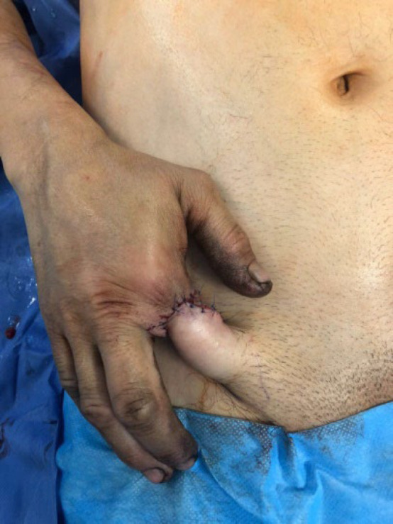
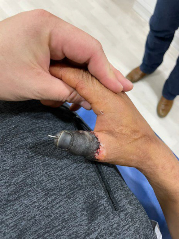
A 71-year-old male sustained an injury to his left hand from a shotgun, causing damage to the thumb 36 hours prior to presenting to the Emergency Room. Upon physical examination, the patient presented with total degloving of the thumb up to the proximal phalanx, with preservation of the insertion and function of all extensor and flexor tendons but lacking coverage of skin and soft tissues (Fig 5). The patient was taken to the operating room, and after surgical cleaning, a groin flap from the ipsilateral flank was performed, providing tubular coverage for the entire circumference of the thumb. Three weeks later, the pedicle of the flap was dissected and separated under local anesthesia, and the patient was referred to rehabilitation. He experienced a recovery of 80% of thumb movements (Fig 6).
Figure 5.

Total degloving injury of the thumb up to the proximal phalanx, with preservation of the insertion and function of all extensor and flexor tendons
Figure 6.

Result of tubular groin flap covering the entire circumference of the thumb after three weeks
Case 4:
A 56-year-old male construction worker who accidentally grabbed a high-voltage cable, suffered an electrical injury that penetrated through his left thumb and exited through his right palm. Both hands had electrical burns, but the left hand, particularly the thumb, sustained more severe damage. The injury presented as a partial degloving on the left palmar surface at the interphalangeal joint. Physical examination revealed partial function of the flexor tendon, preserved abduction and adduction of the thumb, bone and soft tissue loss with bone exposure, and lack of skin and soft tissue coverage (Fig 7). The patient was taken to the operating room for harvesting an ipsilateral groin flap after cleaning and debridement. We preferred to use a tubular design approach to preserve major blood supply and prevent ischemia (Fig 8). The flap was separated three weeks after the surgery under local anesthesia, and thumb function was deemed acceptable with preserved flexor and extensor tendons (Fig 9).
Figure 7.

Partial degloving on the left palmar surface at the first interphalangeal joint resulting from electrical burn
Figure 8.

Tubular design approach to cover affected area, preserve major blood supply, and prevent ischemia
Figure 9.

Result of tubular groin flap covering the entire affected after three weeks
Case 5:
A 16-year-old Mexican “Charro” experienced an avulsion of the right thumb while pulling a horse with a rope, resulting in 3 hours of hot ischemia and partial amputation. Physical examination revealed partial degloving of the proximal phalanx of the thumb, partial avulsion and elongation of both superficial and profundus flexor tendons, as well as partial avulsion of the extensor and long abductor. Both interdigital arteries were injured, and there was minimal attachment of skin and soft tissues on the radial surface. The patient was taken to the operating room for revascularization of at least one artery and two veins, but the injury mechanism only allowed for the revascularization of one artery and one vein. After two weeks, the distal phalanx suffered dry necrosis (Fig 10), so we decided to perform a groin flap for skin coverage of the bone and repaired tendons. An ipsilateral flap was not available due to two previous hernia surgeries the patient had undergone, so we harvested a contralateral flap instead (Fig 11). The pedicle was separated 3 weeks later, and the redundant skin was resected 1 month later. Both the functional and aesthetic results were better than we and the patient expected, considering the injury mechanism and the extent of soft tissue damage (Fig 12).
Figure 10.
Dry necrosis of distal phalanx, two weeks after right thumb avulsion and partial amputation
Figure 1 1 .

Contralateral groin flap covering bone and repaired tendons
Figure 12 .

Result of groin flap covering the affected area after redundant skin resection at one month
Flap design
Tubular Design
DISCUSSION
Preoperative planning for reconstructive hand surgery helps surgeons avoid complications and insufficient tissue coverage. The choice of which flap to use often depends on the surgeon’s experience and available resources. The cases presented in this study were resolved using a relatively simple technique that required minimal surgical time and resulted in a low rate of complications for patients. Initially, we assessed the extent of coverage needed, the quality of the required tissue, and the potential for functional recovery. In some cases, a free flap was considered but ultimately discarded due to the nature of the injury.
Patients were admitted to the emergency room, and although we had access to a microscope for performing free flaps, we opted for the groin flap in cases where recipient vessels were not viable or were limited in size. Surgical debridement was performed during the same surgical event under regional shoulder block anesthesia, which helped immobilize the arm during the initial recovery period and provided pain control.
During surgery, the required tissue amount was determined and transferred to a paper template to define the flap size. Next, the positions of the vessels were drawn on the skin. We then determined the size of the pedicle and pivot point to achieve successful skin coverage, marking the skin with different colors to trace the edges of the vessels and their perforators. After identifying these references, a 15-20 cm length line was drawn along the major axis of the flap, ending at the anterior superior iliac spine as an anatomical boundary, and a 6-8 cm width was marked to allow primary closure and minimize donor site complications. (Figure 13)
Figure 13 .

Landmarks and pivot point in groin flap. The position of the vessels was marked with orange
The pivot point and the end of the undermining dissection were considered to ensure successful coverage, taking into account the major axis of the defect and an additional 2 cm for skin closure. One critical aspect of the technique is the “anatomical positioning” of the flap. As previously mentioned, the arm block prevents involuntary arm movement, and a harmonious flap position reduces the risk of pedicle twisting. Our proximal dissection limit was the inguinal ligament, but we took care not to exclude the deep fascia from the flap structure, ensuring the inclusion of most perforating vessels.
In cases with 360° skin defects, we aimed to create a tubular design flap for complete coverage, folding it and attaching it without tension (Figure 14). After the flap inset and successful skin coverage, the patient was carefully positioned with the arm in the most anatomical relation to the thorax and abdomen, using special bandages to maintain this alignment.
Figure 14 .

Tubular design groin flap
In all our patients, the flap was separated at 21 +/- 2 days postoperatively, using local anesthesia (lidocaine) and sedation for pain control. In all five cases, we successfully achieved skin coverage for the defects, along with satisfactory aesthetic results. Following rehabilitation, patients were able to recover most of their original function. We did not observe dehiscence or infectious complications after surgery or during the waiting period.
CONCLUSION
Skin coverage continues to be a challenge for plastic surgeons, particularly in hand injuries. The scarcity of local flaps that can provide a sufficient amount of tissue is problematic in many cases, especially in degloving injuries, where regional flaps offer a valuable solution for patients. In all the cases presented, the groin flap was the chosen solution. Although technological advancements and the introduction of microsurgery and super-microsurgery have led to a shift from the “reconstructive stairs” concept to the “reconstructive elevator” concept, this new approach should not be considered a universal rule.
Regional flaps, particularly pedicled ones, remain viable options for addressing various skin coverage needs, especially in the hand and forearm regions. This is particularly relevant considering that not every surgeon is an expert in microsurgery, many hospitals lack access to a microscope, and these procedures can be costly. The flap provides enough high-quality tissue and skin coverage, and its versatility allows for various designs to accommodate different types of injuries.
CONFLICT OF INTEREST
The authors declare no conflict of interest.
ACKNOWLEDGMENTS
The financial aspects of this study were primarily supported by the Emergency Department of the Dr. Manuel Gea Gonzalez General Hospital in Mexico City, with funding provided through government assistance. Additionally, patients contributed to covering the costs of some materials used in their treatment. In all cases, a multidisciplinary team, consisting of plastic surgeons, general surgeons, and orthopedic surgeons, collaborated to achieve optimal outcomes for each patient.
References
- 1.Krishnamoorthy R, Karthikeyan G. Degloving injuries of the hand. Indian J Plast Surg. 2011;44(2):227–236. doi: 10.4103/0970-0358.85344. [DOI] [PMC free article] [PubMed] [Google Scholar]
- 2.Rasheed T, Hill C, Riaz M. Innovations in flap design: modified groin flap for closure of multiple finger defects. Burns. 2000;26(2):186–189. doi: 10.1016/s0305-4179(99)00114-x. [DOI] [PubMed] [Google Scholar]
- 3.Thatte R. The history of the groin flap by Ian Jackson. Indian J Plast Surg. 2006;39(2) Available from: https://link.gale.com/apps/doc/A156783339/HRCA?u=googlescholar&sid=bookmark-HRCA&xid=9d08aa2d. [Google Scholar]
- 4.Abdelrahman M, Zelken J, Huang RW, Hsu CC, Lin CH, Lin YT, Lin CH. Suprafascial dissection of the pedicled groin flap: A safe and practical approach to flap harvest. Microsurgery. 2018;38(5):458–65. doi: 10.1002/micr.30238. [DOI] [PubMed] [Google Scholar]
- 5.Chuang DC, Jeng SF, Chen HT, Chen HC, Wei FC. Experience of 73 free groin flaps. Br J Plast Surg. 1992;45(2):81–85. doi: 10.1016/0007-1226(92)90161-p. [DOI] [PubMed] [Google Scholar]
- 6.Arner M, Möller K. Morbidity of the pedicled groin flap A retrospective study of 44 cases. Scand J Plast Reconstr Surg Hand Surg. 1994;28(2):143–146. doi: 10.3109/02844319409071192. [DOI] [PubMed] [Google Scholar]
- 7.Bajantri B, Latheef L, Sabapathy SR. Tips to orient pedicled groin flap for hand defects. Tech Hand Up Extrem Surg. 2013;17(2):68–71. doi: 10.1097/BTH.0b013e31827ddf47. [DOI] [PubMed] [Google Scholar]