Abstract
Background:
Electrophysiological impairments in the magnocellular visual system have been reported among patients with schizophrenia, but previous theories proposed that these deficits may begin in the retina. We therefore sought to evaluate the potential contribution of the retina by comparing retinal and cortical visual electrophysiological impairments between patients with schizophrenia and healthy controls.
Methods:
We recruited patients with schizophrenia and age- and sex-matched healthy controls. We recorded the P100 amplitude and latency using electroencephalography (EEG) while projecting low (0.5 cycles/degree) or high (15 cycles/degree) spatial frequency gratings at a temporal frequency of 0 Hz or 8 Hz. We compared the P100 results with previous results for retinal ganglion cell activity (N95) in these participants. We analyzed data using repeated-measures analysis of variance and correlation analyses.
Results:
We recruited 21 patients with schizophrenia and 29 age- and sex-matched healthy controls. Results showed decreased P100 amplitude and increased P100 latency among patients with schizophrenia compared with healthy controls (p < 0.05). Analyses reported the main effects of spatial and temporal frequency but no interaction effects of spatial or temporal frequency by group. Moreover, correlation analysis indicated a positive association between P100 latency and previous retinal results for N95 latency in the schizophrenia group (p < 0.05).
Discussion:
Alterations in the P100 wave among patients with schizophrenia are consistent with the deficits in early visual cortical processing shown in the literature. These deficits do not seem to correspond to an isolated magnocellular deficit but appear to be associated with previous retinal measurements. Such an association emphasizes the role of the retina in the occurrence of visual cortical abnormalities in schizophrenia. Studies with coupled electroretinography–EEG measurements are now required to further explore these findings.
Clinical trials registration:
Introduction
Sensory distortions are an integral part of the complex experience of schizophrenia.1,2 This point has been reported in relation to vision since it was first characterized by Kraepelin and Bleuler3 and is also commonly described in first-person accounts from patients.4 As knowledge of visual function has developed over the last few decades, visual science studies have reported numerous abnormalities in visual processing among patients with schizophrenia, including colour vision;5 recognition of objects, images or faces;6,7 and contrast sensitivity.8 Electrophysiology, particularly visual evoked potential (VEP) testing, is a key method for highlighting the nature of such visual deficits.
Studies of VEPs have focused on the P100 wave, which provides reliable information about the integrity of early visual cortical processing and, in particular, the primary visual cortex.9 Compared with other VEPs, P100 does not appear to be as sensitive to patient motivational issues or clinical status as more integrative components.10,11 This marker is therefore of considerable importance to highlight deficits related to the early cortical integration of low-level visual stimuli.12,13 Reports in the literature showed decreased P100 amplitude in schizophrenia during various visual tasks, such as contour processing,14 fragmented images15 and perception of simple stimuli.11 These results highlight a deficit in early cortical visual processing in psychiatric illness.16,17 With reference to spatial vision, P100 impairments were found in response to low spatial frequency (LSF) information.18,19 As the magnocellular and parvocellular visual system are preferentially sensitive to the processing of LSF and high spatial frequency (HSF) information,20,21 an LSF deficit could be representative of a preferential magnocellular impairment in schizophrenia.19,22 It should be noted that such visual stimuli cannot specifically activate one pathway over another,23 but SFs remain a useful experimental tool for biasing visual processing toward the magnocellular and the parvocellular visual systems. Studies discussing this potential magnocellular dysfunction in schizophrenia suggest that such a deficit could be driven by early stages of visual processing, such as in the retina or the visual thalamus,22 thus calling into question the origin of P100 alterations.
Furthermore, the literature reports electrophysiological retinal anomalies in schizophrenia.24,25 The most pertinent results indicated both photopic and scotopic alterations in flash electroretinography (ERG) measurements affecting the a-wave and b-wave, thereby highlighting altered cone and rod responses.1,26,27 More recently, using pattern ERG, our team reported a longer N95 latency among patients with schizophrenia compared with controls.1,28 As N95 almost exclusively represents retinal ganglion cell (RGC) activity29,30 and is considered to be the best marker of these cells,31 this provides evidence for a delay in retinal visual transmission caused by RGC dysfunction. This finding of RGC dysfunction was also supported by Moghimi and colleagues24 and Demmin and colleagues32 using the photonegative response in flash ERG. As RGCs are the last retinal layers that make the first link to subsequent brain regions, impaired retinal function may affect both subcortical and cortical visual signals.33
Retinal abnormalities may affect visual cortical processing in ophthalmic diseases34 and neurodegenerative disorders.35,36 For instance, Krasodomska and colleagues37 reported an increased P50 latency, a decrease in N95 and P50 amplitudes, and an increased P100 latency in 30 patients with early-stage Alzheimer disease. Similarly, Heravian and colleagues38 found reductions in the P50 and P100 amplitudes and an increased P100 latency in 40 patients with anisometropic amblyopia. More recently, El-Shazly and colleagues39 reported an increased N95 latency, a decrease in N95 amplitude, a prolonged P100 latency and a decreased P100 amplitude in 60 patients with migraine during aura. Retinal abnormalities may therefore have consequences for visual cortical processing, especially for the P100 wave. With that in mind, in a previous study, our group explored retinal function among patients with schizophrenia and found a delay in N95 latency compared with controls.1 These results suggest an association between retinal dysfunction and subsequent visual processing in schizophrenia. Exploring P100 activity among patients with schizophrenia could provide a way of identifying deficits in early visual cortical processing and could increase understanding of the possible link between retinal and cortical abnormalities in schizophrenia.
The primary goal of this study was to explore early visual cortical processing among patients with schizophrenia compared with healthy controls. Given the theories in the literature supporting a preferential magnocellular impairment in schizophrenia, we sought to measure the amplitude and latency of P100 using electroencephalography (EEG), in response to stimuli biased toward the magnocellular or parvocellular system, such as SF gratings.40,41 Based on previous literature, we hypothesized that we would see P100 alterations, specifically in the magnocellular-biased condition. As a second objective, we sought to compare these cortical results with previous retinal results from the same patients to explore a possible association between both electrophysiological measurements.1
Methods
Clinical assessments and participant ethics statement
This study is part of the CAUSAMAP (Cannabis Use and Magnocellular Processing) project, which aims to examine the neurotoxic effect of cannabis on human vision. We recruited patients with schizophrenia and age- and sex-matched healthy controls. All participants underwent an ERG as well as an EEG on the same day. They provided a detailed psychoactive drug and medical history. All participants had a general psychiatric assessment using the Mini International Neuropsychiatric Interview 5.0.42 Alcohol or cannabis disorder was not an exclusion criterion for recruitment, but we excluded patients with an Alcohol Use Disorders Identification Test (AUDIT) or Cannabis Abuse Screening Test (CAST) score indicating alcohol or cannabis dependence. We chose this criterion because cannabis could have a confounding effect on both retinal and cortical measurements.43,44
Patients with schizophrenia fulfilled the Diagnostic and Statistical Manual of Mental Disorders Fourth Edition, Text Revision (DSM-IV-TR) Axis I Disorders criteria for schizophrenia. They were clinically stable on antipsychotic medication and had no history of neurologic disease. Their urine toxicology test for illicit drug or opiate substitution treatment use was negative. Healthy controls had no actual or past psychiatric disorder, no family or personal history of schizophrenia or bipolar disorder, no history of neurologic disease, no alcohol or cannabis dependance and no history of ophthalmic disease or visual symptoms.
Participants received €100 in vouchers. They signed informed consent detailing all aspects of the research in compliance with the Helsinki declaration.45 All experiments were performed in compliance with the Ethics Committee of Nancy Regional University Hospital Center (2013-A00097–38 CPP 13.02.02). The study was first registered on Aug. 12, 2016, and is available on clinicaltrials.gov (ID NCT02864680).
Experimental procedure, recording and EEG data processing
We used the method described previously by our team.43 Stimuli were generated using the Visual Stimulus Generator system (Cambridge Research System). They consisted of black and white, sinusoidal, vertical Gabor gratings at 6° of visual angle.41 We chose LSF and HSF gratings (0.5 and 15 cycles per degree, respectively) to preferentially stimulate the magnocellular and parvocellular pathways.46,47 The gratings had a light and dark contrast of 80% and were presented against an isoluminant grey background. Both types of gratings were presented under 2 different temporal frequency (TF) conditions. In the dynamic condition, which preferentially stimulates activity in the magnocellular pathway,21 black and white stripes alternated sinusoidally at a frequency of 8 Hz. In the static condition, the stripes did not alternate (0 Hz). Overall, 20% of the stimuli were invisible control stimuli at a contrast of 0%. We used 5 stimuli, namely LSF–static, LSF–dynamic, HSF–static, HSF–dynamic and control stimuli.
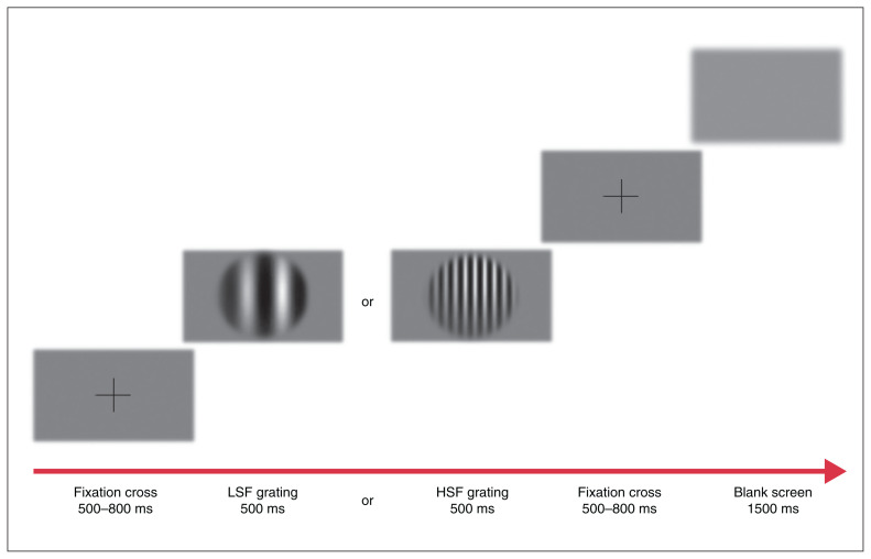
Figure 1 shows the experimental procedure. A total of 300 stimuli were projected onto a cathode-ray tube screen with a sampling rate of 120 Hz in an electrically shielded room with no surrounding light. The participants sat on a chair at a distance of 57 cm from the screen. In each trial, a central fixation cross was displayed for 500–800 ms, allowing participants to maintain their attention on the central zone of the screen. A randomized grating was then presented centrally for 500 ms. A blank screen followed, lasting 1500 ms, during which time participants had to indicate via a response button if they had seen a grating. This task was designed to maintain participants’ attention. Each trial was separated by a supplementary blank screen of 1500 ms. The entire procedure consisted of 300 trials (60 trials per stimulus condition) and was divided into 2 blocks of 150 trials.
Figure 1.
Representation of the experimental procedure, with both low (LSF) and high (HSF) spatial frequency gratings.
For EEGs, recording was performed with Ag/AgCl electrodes using a 64-electrode Micromed headset (10–10 system, QuickCap; Compumedics Neuroscan), with both ear lobe electrodes as reference. The signal was recorded at a sampling rate of 512 Hz (SD64 Headbox, Micromed) with a bandwidth from 0.15 Hz to 200 Hz. We kept electrode impedances below 10 kΩ. We used vertical and horizontal ocular electrodes for eye-artifact rejection. Each epoch included 1000 ms prestimulus and lasted to 1000 ms poststimulus for each SF modality. We processed data with Brain Vision Analyzer 2.0 software (Brain Products GmbH). We used a band-pass filter on the raw EEG signal (0.5–40 Hz). We performed artifact rejection for noise, eyes blinking, muscular activity and nonbiological components using independent component analysis.48 We conducted a manual artifact rejection based on visual inspection to exclude the last remaining artifacts. We excluded the participant from the study if more than 10% of the trials had to be removed. To limit bias, the person who decided to exclude the participants was blinded to the group, and this action was performed before the VEP averaging. Thereafter, data collection focused on 3 pairs of electrodes in the left (O1, PO3, PO7) and right (O2, PO4, PO8) hemispheres. An overall average across all conditions determined the global aspect of peak P100 amplitude. To determine a temporal time window for the extraction of the P100 amplitude, we calculated the root mean square by squaring the peak amplitude of the sine wave, dividing it by 2 and taking the square root of that value. Consequently, we extracted the P100 peak with a 28 ms interval around the maximum peak. We extracted the P100 latency based on its mean appearance on the overall average. As a result, P100 latency was determined at 116 ms for patients with schizophrenia and 113 ms for healthy controls.
Experimental procedure, recording and ERG data processing
Detailed information about the stimulation and recording processes for the ERG has been provided previously.49 Briefly, we used the MonPackOne system (Metrovision) for stimulation, recording and analysis. We explored RGC function using pattern reversal checkerboard stimuli, according to standards of the International Society for Clinical Electrophysiology of Vision.31 Pattern ERG markers were the N95 and P50 waves.
Statistical analysis
We analyzed data using STATISTICA (version 10.0). We conducted both descriptive and comparative analyses according to the nature and the distribution of the variables, assessing normality using the Shapiro–Wilk test. We used frequencies and percentages for categorical variables, and means and standard deviations (SDs) for continuous variables. Since sociodemographic and clinical characteristics followed a normal distribution, given the nonsignificant Shapiro–Wilk test, we analyzed the differences between groups using an independent sample t test. Given that behavioural and EEG data followed a normal distribution, as indicated by a nonsignificant Shapiro–Wilk test, and their variances did not differ according to a nonsignificant Levene test, we used parametric tests. We analyzed behavioural data using 2 × 2 analysis of variance (ANOVA), whereby the 2 factors were TF (dynamic or static) and SF (LSF or HSF). We analyzed P100 amplitude and latency using 2 × 3 × 2 × 2 ANOVA (hemisphere [left or right] × electrodes [O1/2, PO3/4, PO7/8] × TF [dynamic or static] × SF [LSF or HSF]). We used Pearson r coefficients to assess correlations between experimental variables. More detail on statistical analysis of pattern ERGs was described by Bernardin and colleagues.1 For all tests, we used an α value of 0.05 to determine statistical significance.
Results
We recruited 26 patients with schizophrenia and 30 age- and sex-matched healthy controls. All participants underwent an ERG as well as an EEG on the same day. However, not all electrophysiological plots were usable owing to artifacts in the recording. Our final sample consisted of 21 patients with schizophrenia (mean age 29.00 yr, standard deviation [SD] 8.15 yr) and 29 healthy controls (mean age 25.89 [SD 5.49] yr). All participants were aged 19–46 years. Fundoscopic examination was normal and visual acuity was normal or corrected to normal, as verified using the Monoyer chart. Among the 21 patients with schizophrenia, 2 were cannabis users without dependence and 12 were alcohol users without dependence. Sociodemographic and clinical characteristics of the participants are summarized in Table 1.
Table 1.
Participant sociodemographic and clinical characteristics
| Characteristic | Mean ± SD* | p value | |
|---|---|---|---|
|
| |||
| Healthy controls n = 29 |
Patients with schizophrenia n = 21 |
||
| Sex | 0.25 | ||
| No. (%) female | 8 (28) | 3 (14) | |
| No. (%) male | 21 (72) | 18 (86) | |
| Age, yr, mean | 25.89 ± 5.49 | 29.00 ± 8.15 | 0.12 |
| Education, yr | 15.03 ± 1.57 | 12.05 ± 1.40 | < 0.01 |
| AUDIT score | 3.35 ± 2.69 | 2.43 ± 3.41 | 0.47 |
| Disease duration, mo | NA | 94.67 ± 93.21 | – |
| Fagerström score | NA | 2.19 ± 2.77 | – |
| CAST score | NA | 0.43 ± 1.36 | – |
| PANSS Global | NA | 65.24 ± 13.40 | – |
| PANSS Positive | NA | 14.48 ± 4.31 | – |
| PANSS Negative | NA | 18.29 ± 5.57 | – |
| PANSS General | NA | 32.48 ± 6.76 | – |
| Chlorpromazine equivalent, mg/d | NA | 544.55 ± 241.29 | – |
| Diazepam equivalent, mg/d | NA | 1.56 ± 9.64 | – |
AUDIT = Alcohol Use Disorders Identification Test; CAST = Cannabis Abuse Screening Test; NA = not applicable; PANSS = Positive and Negative Syndrome Scale.
Unless indicated otherwise.
Behavioural data
Patients with schizophrenia and healthy controls had mean reaction times of 595.15 (SD 46.60) ms and 393.01 (SD 14.87) ms, respectively. The ANOVA analysis showed a significant main effect of group (F1,47 = 22.98, p < 0.01), indicating a higher mean reaction time among patients with schizophrenia, compared with healthy controls, regardless of the type of stimuli.
EEG results
P100 amplitude
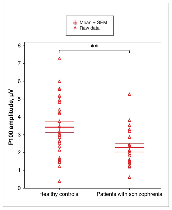
The mean P100 amplitude was 2.25 (SD 0.52) μV among patients with schizophrenia and 3.42 (SD 0.62) μV among healthy controls. The ANOVA analysis showed a main effect of group (F1,48 = 7.88, p < 0.01), indicating a lower P100 amplitude among patients with schizophreniathan among healthy controls (Figure 2).
Figure 2.
Main group effect on P100 amplitude between patients with schizophrenia and healthy controls. Data were obtained from the average activity of 3 pairs of electrodes (O1/O2, PO3/PO4, PO7/PO8). Means are displayed with their standard error (SEM). **p < 0.01.
A main SF effect (F1,48 = 51.34, p < 0.001) highlighted a larger P100 amplitude for LSF gratings (mean 3.80 [SD 1.94] μV) compared with HSF gratings (mean 1.89 [SD 1.49] μV). The interaction of SF by group was not significant (F1,48 = 0.04, p = 0.85). Analysis indicated a main effect of TF (F1,48 = 8.79, p < 0.01) explained by a greater P100 amplitude for the dynamic condition (mean 3.02 [SD 1.51] μV) compared with the static condition (mean 2.67 [SD 1.49] μV). The interaction of TF by group was not significant (F1,48 = 1.17, p = 0.29). No main effect was found for other variables.
P100 latency
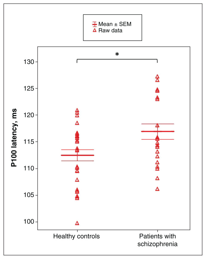
The mean P100 latency was 117.04 (SD 2.65) ms among patients with schizophrenia and 112.46 (SD 2.31) ms among healthy controls. The ANOVA analysis showed a main effect of group (F1,48 = 6.45, p < 0.05) indicating a longer P100 latency among patients with schizophrenia than healthy controls (Figure 3).
Figure 3.
Main group effect on P100 latency between patients with schizophrenia and healthy controls. Data were obtained from the average activity of 3 pairs of electrodes (O1/O2, PO3/PO4, PO7/PO8). Means are displayed with their standard error (SEM). *p < 0.05.
Analysis exhibited a main SF effect (F1,48 = 17.89, p < 0.001) explained by a shorter P100 latency for LSF gratings (mean 112.72 [SD 7.04] ms) than HSF gratings (mean 116.72 [SD 6.85] ms). The interaction of SF by group was not significant (F1,48 = 0.34, p = 0.56). No main effect was found for other variables.
P100 correlation analysis
Among both patients with schizophrenia and healthy controls, we did not observe correlations between P100 amplitude and latency on the one hand (n = 50, r = −0.01, p = 0.92), and between P100 amplitude or latency and CAST score, AUDIT score, number of joints per week and medication use on the other hand (p > 0.05).
Pattern ERG results
Pattern ERG results were previously described by Bernardin and colleagues.1 The main result was a significant effect of group for N95 latency (F1,54 = 18.0, p < 0.001) indicating a longer N95 latency among patients with schizophrenia (mean 95.7 [SD 7.5] ms) than among healthy controls (mean 88.4 [SD 5.4] ms).
Association between EEG and pattern ERG results
We found a strong and a positive correlation between the N95 latency and the P100 latency among patients with schizophrenia (n = 21, r = 0.85, p < 0.05). Thus, the longer the N95 latency, the longer the P100 latency among patients with schizophrenia. No other significant correlations were found (p > 0.05).
Discussion
The first aim of this study was to explore visual cortical processing among patients with schizophrenia compared with healthy controls, with stimuli biased toward the magnocellular or the parvocellular system. We found decreased P100 amplitude and increased P100 latency among patients with schizophrenia compared with healthy controls. These P100 alterations in the schizophrenia group are consistent with the findings of other studies, which reported impairments in the very first step of early visual cortical processing and, in particular, the primary visual cortex.12,50 Analysis also indicated greater P100 amplitude and shorter P100 latency for LSF stimuli and the dynamic condition, consistent with the sensitivity of the magnocellular system to LSF information and movements.21 Despite this, our P100 results failed to find any interactions in SF or TF by group. Such results undermine the case for alterations in the magnocellular visual system in schizophrenia, although this is well described in the literature.
A study by Butler and colleagues,22 conducted in a very similar setting, reported a decrease in P100 in response to magnocellular-biased stimuli. In our study, patients were younger (mean 29.0 [SD 8.2] yr v. mean 35.9 [SD 2.2] yr), with half as long a duration of illness (mean 94.67 [SD 93.21] mo v. mean 16.7 [SD 1.8] yr) and used an average dose of anti-psychotic medications that was nearly 3 times smaller (mean 544.55 [SD 241.29] mg/d v. 1365.4 [SD 157.7] mg/d). In this regard, the literature indicates that visual abnormalities in schizophrenia may be more pronounced with age, duration, stage and severity of the illness.51 Similarly, the use of anti-psychotic medication may impair visual performance, including contrast perception, to a greater extent than without the use of antipsychotic medication.5,52 For instance, a recent study by Almeida and colleagues53 found greater deficits in contrast sensitivity among patients with longer duration of illness. These deficits were also related to the combined effect of duration of illness and the use of atypical antipsychotic medications. In short, magnocellular alteration could be a delayed process in schizophrenia, influenced by treatment and disease progression. Similar to previous studies, we hypothesize that these late visual deficits are preceded by early abnormalities in visual processing that begin in the retina or thalamus because these are the only subcortical visual relays.22
A second aim of this study was to compare the present cortical results with previous retinal results obtained from the same patients to see if an association existed between both visual stages.1 Correlation analysis showed a positive association between N95 latency in the retina and P100 latency in the cortex among patients with schizophrenia, which was not found in the control group. This result also supports previous studies that hypothesized that retinal abnormalities could have cortical repercussions in ophthalmic and neurologic disorders such as anisometropic amblyopia,38 Alzheimer disease37 and migraine,39 as evidenced by multiple N95 and P100 wave abnormalities. This potential association between N95 latency and P100 latency in the present study may support the existence of a link between retinal and cortical visual electrophysiological impairments in psychiatric disorders. Therefore, visual impairments may begin in the retina and contribute to deficits further along the visual pathways.
Our findings raise potential arguments for a relationship between retinal and cortical visual abnormalities in psychosis. More specifically, our findings describe an association between N95 latency on pattern ERG and P100 latency on EEG and, thus, makes the case for retinal contributions to P100 abnormalities. This potential link would suggest that retinal abnormalities could have repercussions for cortical visual processing in schizophrenia. Electrophysiology provides good objectivity, reliability and reproducibility in results, especially in low-level visual measures with poor sensitivity to attentional factors. As visual deficits are associated with severe psychopathology, a poor prognosis and a high risk of death in schizophrenia, electrophysiology could improve the therapeutic management of patients.54,55
Limitations
Although we found no interaction effects of magnocellular- or parvocellular-biased stimuli, these results should be tempered by the fact that the gratings are only an experimental model that cannot completely activate 1 pathway over another and can therefore lack specificity.23 Moreover, since RGCs are separated into M-type and P-type cells, which lead to the cortex via magnocellular and parvocellular pathways, respectively,56,57 a deficit of M-type cells may occur as early as the retina. Samani and colleagues58 found a decrease in retinal contrast sensitivity to LSF among patients with schizophrenia compared with controls and hypothesized that these alterations may signify a disease-related loss of magnocellular ganglion cells already visible in the retina. Studies also report neurotransmission abnormalities in RGCs, particularly with respect to N-methyl-D-aspartate receptors, thus implying a magnocellular dysfunction in schizophrenia.24 On a further point, the fact that N95 retinal amplitude was normal among patients with schizophrenia, whereas P100 cortical amplitude is abnormal, raises the possibility that retinal abnormalities do not fully explain the P100 amplitude deficit. Indeed, other mechanisms could be involved in the retina and beyond. For example, the activity of the central retina, in particular the photoreceptors and rods, could contribute to the delay in N95 latency or P100 latency. Although our previous results showed alterations in cone and rod responses,1 we found no significant correlations between the a-wave and b-wave, or the N95 or P100 waves. Other research is also raising interest in components linked to the retinotectal pathway in the beginning of psychotic disorders.59
Although we did not observe any differences between groups in terms of clinical characteristics, substance use must be considered. Indeed, Schwitzer and colleagues44 found an increase in N95 latency among people who used cannabis regularly compared with healthy controls. At the cortical level, we recently found a P100 impairment among people who used cannabis regularly in response to the same stimuli as described in this study.43 Similarly, smoking nicotine would appear to affect P100 latency in studies using visual modality tasks,60 and alcohol may induce a P100 latency delay, even among healthy individuals.61 Nevertheless, we found no correlations between the amount of cannabis, cigarettes or alcohol consumed daily and the P100 results. Moreover, only 2 patients were cannabis users and did not show dependence. Additional studies involving patients with schizophrenia without substance use are necessary. Although we investigated the same patients as in our previous retina study,1 stimuli were different for both electrophysiological methods, measures were decoupled and we did not investigate all ERG examinations, such as those related to cone or rod response. However, the stimuli used assessed low-level visual processing for both techniques, allowing us to compare the N95 and P100 waves with each other. Overall, ERG examinations and simultaneous ERG-EEG recordings are required to further clarify these findings.
Conclusion
Electrophysiological deficits that affect early visual processing among patients with schizophrenia have become increasingly well established in recent years, particularly regarding the retina and the visual cortex. We previously reported alterations in N95 latency in the retina among patients with schizophrenia. Looking at the cortex, the present results showed an alteration affecting P100 amplitude and latency in the same patients. These alterations were not specific to visual stimuli that were strongly biased toward the magnocellular system, a hypothesis that is supported in the psychiatric illness. Moreover, we showed specific associations between N95 retinal latency and P100 cortical latency in patients, involving a link between the alterations of both visual stages. Based on our findings, we suggest that deficits in cortical processing are partially driven by RGC dysfunction. Our results reinforce the role of the retina in the occurrence of cortical visual deficits in psychosis and suggest that cortical abnormalities could potentially be caused by retinal abnormalities in schizophrenia. Further studies with simultaneous and comparable electrophysiological methods are now necessary to confirm the association between both visual stages. The use of reliable, objective and reproducible electrophysiological measures to routine clinical assessments, such as ERG and EEG, would considerably improve diagnosis and patient follow-up in health services, and would increase the prevention and early detection of mental illness.
Acknowledgement
The authors thank all members of the CAUSAMAP study group.
Footnotes
Competing interests: Vincent Laprévote reports travel support from Boehringer Ingelheim France and Janssen-Cilag. No other competing interests were declared.
Contributors: Julian Krieg, Louis Maillard, Raymond Schwan, Thomas Schwitzer and Vincent Laprévote contributed to the conception and design of the work. All of the authors contributed to the the acquisition, analysis and interpretation of data. Irving Remy, Florent Bernardin and Vincent Laprévote drafted the manuscript. All of the authors revised it critically for important intellectual content, gave final approval of the version to be published and agreed to be accountable for all aspects of the work.
Data sharing: The data are not publicly available because the information could compromise the privacy of research participants. Nevertheless, raw electroencephalography data and processed data are fully available from the corresponding author, upon reasonable request, for the verification of research results supporting the findings of this present study.
Funding: This study was supported by grant ANR-12-SAMA-0016-01 from the French National Research Agency and by the French Mission Interministérielle contre les Drogues et les Conduites Addictives. The funding sources have no role in the design and conduct of the study; collection, management, analysis and interpretation of the data; preparation, review or approval of the manuscript; and decision to submit the manuscript for publication.
References
- 1.Bernardin F, Schwitzer T, Angioi-Duprez K, et al. Retinal ganglion cells dysfunctions in schizophrenia patients with or without visual hallucinations. Schizophr Res 2020;219:47–55. [DOI] [PubMed] [Google Scholar]
- 2.Javitt DC, Freedman R. Sensory processing dysfunction in the personal experience and neuronal machinery of schizophrenia. Am J Psychiatry 2015;172:17–31. [DOI] [PMC free article] [PubMed] [Google Scholar]
- 3.Javitt DC. When doors of perception close: bottom-up models of disrupted cognition in schizophrenia. Annu Rev Clin Psychol 2009;5:249–75. [DOI] [PMC free article] [PubMed] [Google Scholar]
- 4.Chapman J. Schizophrenia from the inside. Ment Health (Lond) 1966;25:6–8. [PMC free article] [PubMed] [Google Scholar]
- 5.Fernandes TMP, Silverstein SM, Butler PD, et al. Color vision impairments in schizophrenia and the role of antipsychotic medication type. Schizophr Res 2019;204:162–70. [DOI] [PubMed] [Google Scholar]
- 6.Schneider F, Gur RC, Koch K, et al. Impairment in the specificity of emotion processing in schizophrenia. Am J Psychiatry 2006;163:442–7. [DOI] [PubMed] [Google Scholar]
- 7.Oker A, Del Goleto S, Vignes A, et al. Schizophrenia patients are impaired in recognition task but more for intentionality than physical causality. Conscious Cogn 2019;67:98–107. [DOI] [PubMed] [Google Scholar]
- 8.Kéri S, Antal A, Szekeres G, et al. Spatiotemporal visual processing in schizophrenia. J Neuropsychiatry Clin Neurosci 2002;14:190–6. [DOI] [PubMed] [Google Scholar]
- 9.Liu J, Harris A, Kanwisher N. Stages of processing in face perception: an MEG study. Nat Neurosci 2002;5:910–6. [DOI] [PubMed] [Google Scholar]
- 10.Johnson SC, Lowery N, Kohler C, et al. Global-local visual processing in schizophrenia: evidence for an early visual processing deficit. Biol Psychiatry 2005;58:937–46. [DOI] [PubMed] [Google Scholar]
- 11.Yeap S, Kelly SP, Sehatpour P, et al. Early visual sensory deficits as endophenotypes for schizophrenia: high-density electrical mapping in clinically unaffected first-degree relatives. Arch Gen Psychiatry 2006;63:1180–8. [DOI] [PubMed] [Google Scholar]
- 12.Earls HA, Curran T, Mittal V. Deficits in early stages of face processing in schizophrenia: a systematic review of the P100 component. Schizophr Bull 2016;42:519–27. [DOI] [PMC free article] [PubMed] [Google Scholar]
- 13.Jeantet C, Laprevote V, Schwan R, et al. Time course of spatial frequency integration in face perception: an ERP study. Int J Psychophysiol 2019;143:105–15. [DOI] [PubMed] [Google Scholar]
- 14.Foxe JJ, Murray MM, Javitt DC. Filling-in in schizophrenia: a high-density electrical mapping and source-analysis investigation of illusory contour processing. Cereb Cortex 2005;15:1914–27. [DOI] [PubMed] [Google Scholar]
- 15.Doniger GM, Foxe JJ, Murray MM, et al. Impaired visual object recognition and dorsal/ventral stream interaction in schizophrenia. Arch Gen Psychiatry 2002;59:1011–20. [DOI] [PubMed] [Google Scholar]
- 16.Butler PD, Javitt DC. Early-stage visual processing deficits in schizophrenia. Curr Opin Psychiatry 2005;18:151–7. [DOI] [PMC free article] [PubMed] [Google Scholar]
- 17.Foxe JJ, Doniger GM, Javitt DC. Early visual processing deficits in schizophrenia: impaired P1 generation revealed by high-density electrical mapping. Neuroreport 2001;12:3815–20. [DOI] [PubMed] [Google Scholar]
- 18.Lee JS, Park G, Song MJ, et al. Early visual processing for low spatial frequency fearful face is correlated with cortical volume in patients with schizophrenia. Neuropsychiatr Dis Treat 2015;12:1–14. [DOI] [PMC free article] [PubMed] [Google Scholar]
- 19.Martínez A, Hillyard SA, Bickel S, et al. Consequences of magno-cellular dysfunction on processing attended information in schizophrenia. Cereb Cortex 2012;22:1282–93. [DOI] [PMC free article] [PubMed] [Google Scholar]
- 20.Derrington AM, Lennie P. Spatial and temporal contrast sensitivities of neurones in lateral geniculate nucleus of macaque. J Physiol 1984;357:219–40. [DOI] [PMC free article] [PubMed] [Google Scholar]
- 21.Milner AD, Goodale MA. Two visual systems re-viewed. Neuropsychologia 2008;46:774–85. [DOI] [PubMed] [Google Scholar]
- 22.Butler PD, Martinez A, Foxe JJ, et al. Subcortical visual dysfunction in schizophrenia drives secondary cortical impairments. Brain 2007;130:417–30. [DOI] [PMC free article] [PubMed] [Google Scholar]
- 23.Skottun BC, Skoyles JR. Contrast sensitivity and magnocellular functioning in schizophrenia. Vision Res 2007;47:2923–33. [DOI] [PubMed] [Google Scholar]
- 24.Moghimi P, Torres Jimenez N, McLoon LK, et al. Electoretinographic evidence of retinal ganglion cell-dependent function in schizophrenia. Schizophr Res 2020;219:34–46. [DOI] [PMC free article] [PubMed] [Google Scholar]
- 25.Demmin DL, Davis Q, Roché M, et al. Electroretinographic anomalies in schizophrenia. J Abnorm Psychol 2018;127:417–28. [DOI] [PubMed] [Google Scholar]
- 26.Hébert M, Mérette C, Paccalet T, et al. Light evoked potentials measured by electroretinogram may tap into the neurodevelopmental roots of schizophrenia. Schizophr Res 2015;162:294–5. [DOI] [PubMed] [Google Scholar]
- 27.Warner R, Laugharne J, Peet M, et al. Retinal function as a marker for cell membrane omega-3 fatty acid depletion in schizophrenia: a pilot study. Biol Psychiatry 1999;45:1138–42. [DOI] [PubMed] [Google Scholar]
- 28.McCulloch DL, Marmor MF, Brigell MG, et al. ISCEV standard for full-field clinical electroretinography (2015 update). Doc Ophthalmol 2015;130:1–12. [DOI] [PubMed] [Google Scholar]
- 29.Froehlich J, Kaufman DI. The pattern electroretinogram: N95 amplitudes in normal subjects and optic neuritis patients. Electroencephalogr Clin Neurophysiol 1993;88:83–91. [DOI] [PubMed] [Google Scholar]
- 30.Hull BM, Thompson DA. A review of the clinical applications of the pattern electroretinogram. Ophthalmic Physiol Opt 1989;9: 143–52. [DOI] [PubMed] [Google Scholar]
- 31.Bach M, Brigell MG, Hawlina M, et al. ISCEV standard for clinical pattern electroretinography (PERG): 2012 update. Doc Ophthalmol 2013;126:1–7. [DOI] [PubMed] [Google Scholar]
- 32.Demmin DL, Netser R, Roché MW, et al. People with current major depression resemble healthy controls on flash Electroretinogram indices associated with impairment in people with stabilized schizophrenia. Schizophr Res 2020;219:69–76. [DOI] [PubMed] [Google Scholar]
- 33.Silverstein SM, Fradkin SI, Demmin DL. Schizophrenia and the retina: towards a 2020 perspective. Schizophr Res 2020;219:84–94. [DOI] [PMC free article] [PubMed] [Google Scholar]
- 34.Atilla H, Tekeli O, Ornek K, et al. Pattern electroretinography and visual evoked potentials in optic nerve diseases. J Clin Neurosci 2006;13:55–9. [DOI] [PubMed] [Google Scholar]
- 35.Nightingale S, Mitchell KW, Howe JW. Visual evoked cortical potentials and pattern electroretinograms in Parkinson’s disease and control subjects. J Neurol Neurosurg Psychiatry 1986;49:1280–7. [DOI] [PMC free article] [PubMed] [Google Scholar]
- 36.Calzetti S, Franchi A, Taratufolo G, et al. Simultaneous VEP and PERG investigations in early Parkinson’s disease. J Neurol Neurosurg Psychiatry 1990;53:114–7. [DOI] [PMC free article] [PubMed] [Google Scholar]
- 37.Krasodomska K, Lubinski W, Potemkowski A, et al. Pattern electroretinogram (PERG) and pattern visual evoked potential (PVEP) in the early stages of Alzheimer’s disease. Doc Ophthalmol 2010;121:111–21. [DOI] [PMC free article] [PubMed] [Google Scholar]
- 38.Heravian J, Daneshvar R, Dashti F, et al. Simultaneous pattern visual evoked potential and pattern electroretinogram in strabismic and anisometropic amblyopia. Iran Red Crescent Med J 2011;13:21–6. [PMC free article] [PubMed] [Google Scholar]
- 39.El-Shazly AAE-F, Farweez YA, Hamdi MM, et al. Pattern visual evoked potential, pattern electroretinogram, and retinal nerve fiber layer thickness in patients with migraine during and after aura. Curr Eye Res 2017;42:1327–32. [DOI] [PubMed] [Google Scholar]
- 40.Bullier J. Integrated model of visual processing. Brain Res Rev 2001;36:96–107. [DOI] [PubMed] [Google Scholar]
- 41.DeValois RL, Burke P, DeValois KK. Spatial vision. Oxford University Press on Demand; 1990 [Google Scholar]
- 42.Sheehan DV, Lecrubier Y, Sheehan KH, et al. The Mini-International Neuropsychiatric Interview (M.I.N.I): The development and validation of a structured diagnostic psychiatric interview for DSM-IV and ICD-10. J Clin Psychiatry 1998;59:22–33. [PubMed] [Google Scholar]
- 43.Remy I, Schwitzer T, Albuisson É, et al. Impaired P100 among regular cannabis users in response to magnocellular biased visual stimuli. Prog Neuropsychopharmacol Biol Psychiatry 2022;113:110437. [DOI] [PubMed] [Google Scholar]
- 44.Schwitzer T, Schwan R, Albuisson E, et al. Association between regular cannabis use and ganglion cell dysfunction. JAMA Ophthalmol 2017;135:54–60. [DOI] [PubMed] [Google Scholar]
- 45.World Medical Association. World Medical Association Declaration of Helsinki: ethical principles for medical research involving human subjects. JAMA 2013;310:2191–4. [DOI] [PubMed] [Google Scholar]
- 46.Atkinson J. Early visual development: differential functioning of parvocellular and magnocellular pathways. Eye (Lond) 1992;6:129–35. [DOI] [PubMed] [Google Scholar]
- 47.Butler PD, Schechter I, Zemon V, et al. Dysfunction of early-stage visual processing in schizophrenia. Am J Psychiatry 2001;158:1126–33. [DOI] [PubMed] [Google Scholar]
- 48.Jung T-P, Makeig S, Westerfield M, et al. Removal of eye activity artifacts from visual event-related potentials in normal and clinical subjects. Clin Neurophysiol 2000;111:1745–58. [DOI] [PubMed] [Google Scholar]
- 49.Schwitzer T, Schwan R, Angioi-Duprez K, et al. Delayed bipolar and ganglion cells neuroretinal processing in regular cannabis users: the retina as a relevant site to investigate brain synaptic transmission dysfunctions. J Psychiatr Res 2018;103:75–82. [DOI] [PubMed] [Google Scholar]
- 50.Tanaka S, Maezawa Y, Kirino E. Classification of schizophrenia patients and healthy controls using p100 event-related potentials for visual processing. Neuropsychobiology 2013;68:71–8. [DOI] [PubMed] [Google Scholar]
- 51.Joseph J, Bae G, Silverstein SM. Sex, symptom, and premorbid social functioning associated with perceptual organization dysfunction in schizophrenia. Front Psychol 2013;4:547. [DOI] [PMC free article] [PubMed] [Google Scholar]
- 52.Chen Y, Levy DL, Sheremata S, et al. Effects of typical, atypical, and no antipsychotic drugs on visual contrast detection in schizophrenia. Am J Psychiatry 2003;160:1795–801. [DOI] [PubMed] [Google Scholar]
- 53.Almeida NL, Fernandes TP, Lima EH, et al. Combined influence of illness duration and medication type on visual sensitivity in schizophrenia. Braz J Psychiatry 2020;42:27–32. [DOI] [PMC free article] [PubMed] [Google Scholar]
- 54.Clark ML, Waters F, Vatskalis TM, et al. On the interconnectedness and prognostic value of visual and auditory hallucinations in first-episode psychosis. Eur Psychiatry 2017;41:122–8. [DOI] [PubMed] [Google Scholar]
- 55.Chouinard V-A, Shinn AK, Valeri L, et al. Visual hallucinations associated with multimodal hallucinations, suicide attempts and morbidity of illness in psychotic disorders. Schizophr Res 2019;208:196–201. [DOI] [PMC free article] [PubMed] [Google Scholar]
- 56.Kolb H, Fernandez E, Nelson R, editors. Webvision: the organization of the retina and visual system. Salt Lake City (UT): University of Utah Health Sciences Center; 1995. Available: http://www.ncbi.nlm.nih.gov/books/NBK11530/ (accessed 2020 Apr. 2). [PubMed] [Google Scholar]
- 57.Hoon M, Okawa H, Della Santina L, et al. Functional architecture of the retina: development and disease. Prog Retin Eye Res 2014;42:44–84. [DOI] [PMC free article] [PubMed] [Google Scholar]
- 58.Samani NN, Proudlock FA, Siram V, et al. Retinal layer abnormalities as biomarkers of schizophrenia. Schizophr Bull 2018;44:876–85. [DOI] [PMC free article] [PubMed] [Google Scholar]
- 59.Koropouli E, Melanitis N, Dimitriou VI, et al. New-onset psychosis associated with a lesion localized in the rostral tectum: insights into pathway-specific connectivity disrupted in psychosis. Schizophr Bull 2020;46:1296–305. [DOI] [PMC free article] [PubMed] [Google Scholar]
- 60.Pritchard W, Sokhadze E, Houlihan M. Effects of nicotine and smoking on event-related potentials. Nicotine Tob Res 2004;6:961–84. [DOI] [PubMed] [Google Scholar]
- 61.Kim JT, Yun CM, Kim S-W, et al. The effects of alcohol on visual evoked potential and multifocal electroretinography. J Korean Med Sci 2016;31:783–9. [DOI] [PMC free article] [PubMed] [Google Scholar]


