

Abstract
Introduction:
Sexual dimorphism practically involves differences in size and appearance of the same species which does not involve the difference in sexual organs. A significant variation also occurs with the tooth size, shape, etc., which plays a critical role in sex determination. Forensic investigations are used in defining the number of missing people whose skeletal remains are unknown. Depending on the available bones and their state, a variety of methods with varying degrees of reliability are available for identifying unknown remains.
Materials and Methods:
Fifty male and 50 female patients in the age group 20–30 years were randomly selected after taking a detailed history. All maxillary impressions were made with alginate and poured into a dental stone. These casts were measured for intercanine width, interpremolar width, and intermolar width using a digital vernier caliper, and findings were correlated with sexual dimorphism.
Results:
Intercanine width between the tips of right and left maxillary canine was measured in males and females at 36.08 ± 2.04 mm (Range: 30.05–41.64 mm) and in females, the mean intercanine width was 34.31 ± 1.75 mm (Range: 28.35–39.01 mm), respectively. Interpremolar width between the distal pits of right and left first premolars was measured in males and females at 38.97 ± 2.10 mm (Range: 33.94–45.21 mm) and in females, the mean interpremolar width was 36.92 ± 1.87 mm (Range: 31.34 mm), respectively. Intermolar width between the central fossae of right and left first molars was measured in males and females at 50.43 ± 2.25 mm (Range: 44.16–56.84 mm) and in females, the mean intermolar width was 47.90 ± 2.06 mm (Range: 42.66–54.63 mm), respectively.
Conclusion:
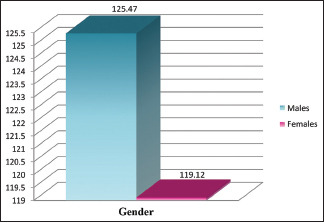
In males, the mean value of the combination of intercanine, interpremolar, and intermolar widths was 125.47 ± 5.61 mm (Range: 108.15–141.86 mm) and in females, it was 119.12 ± 5.05 mm (Range: 103.25–134.36 mm). Mean values of all combinations were larger in males than in females. Thereby, maxillary arch widths contribute to accuracy in determining the gender of the individual.
Keywords: Arch width, intercanine width, intermolar width, interpremolar width, sexual dimorphism
INTRODUCTION
Dentition in itself is a unique feature for an individual. Just like other identification features such as fingerprints and DNA; teeth (considering number, shape, size, color, texture, and consistency) also have a substantial role to play in one's life. Being one of the most calcified structures presents in the human body, it has a pivotal role to play as it is the hardest and chemically the most stable tissue in the body.[1,2]
Variations in dental traits according to geographic location, ethnic groups, and racial differences are observed globally which was proposed by Charles Darwin in 1871.[3,4] The above-mentioned reason is why we chose our study population of the central Indian population (Maharashtra). In addition, regarding dimensions of teeth with respect to gender, the males exhibit larger tooth dimensions than females.[5] Furthermore, distinct teeth such as canines, upper laterals, and lower centrals also help contribute to a greater percentage of sex difference. This statement was supported by Aluko IA et al.,[6] where the author mentioned the significant sexual dimorphism in maxillary arch widths and intermolar width of mandibular arch in both early and late dentitions.
The study aims to evaluate the maxillary intercanine, interpremolar, and intermolar widths in the study population thereby studying the sexual dimorphism in the maxillary arch in Maharashtrian young adults.
MATERIALS AND METHODS
The study was conducted in the department of Oral & Maxillofacial Pathology and Oral Microbiology, with the approval of institutional ethical committee and D. Y. Patil Deemed to be University, School of Dentistry, Navi Mumbai. The subjects selected for the study included 100 individuals (50 males and 50 females) with the following inclusion and exclusion criteria: participants whose age ranging from 20 to 30 years and having a healthy state of periodontium were included in the study. Exclusion criteria include carious maxillary canine, first premolar and first molar teeth, presence of crowding, spacing, missing teeth, and habits like thumb sucking, mouth breathing, previous orthodontic treatment, and any localized or systemic hard or soft tissue pathology related to teeth and jaws. Later, relevant detailed case history of the subjects was recorded along with informed consent.
Armamentarium used [Figures 1 and 2]
Figure 1.

Shows the armamentarium (alginate, spatula, mixing bowls, metal trays, dental stone, etc.) used in the study
Figure 2.

Shows the measuring device Vernier caliper which was used to measure the intercanine, interpremolar, and intermolar width of the participants
Sterile disposable gloves
Mouth mirror and probe
Perforated metal trays
Rubber bowl (Large and Small)
Alginate spatula
Alginate impression material
Cotton
Plaster spatula
Dental stone
Digital Vernier caliper (Forbes Gokak Ltd. Model no. 111-322).
After taking a detail case history and informed consent, the alginate hydrocolloid impression of the maxillary arch was made using perforated metal trays. For making impressions, first, the perforated metal tray was selected. Then, the patient was asked to sit upright in the dental chair with their head tilted slightly downward. Alginate impression material was mixed with water in a large rubber bowl to obtain a smooth workable mix. Then, the tray was loaded with sufficient impression material and impression of maxillary arch was made. Impressions were poured immediately into the dental stone to avoid any dimensional change in the impression [Figure 3a and b].
Figure 3.

Shows alginate impression of maxillary arch (a) and cast prepared (b)
To study morphometric data of maxillary arch, the following measurements were taken from casts of every individual selected for study:
Intercanine width—measured between the tip of right and left canines [Figure 4].
Figure 4.

Shows measurement of intercanine width
Interpremolar width—measured between the distal pit of right and left first premolars [Figure 5].
Figure 5.

Shows measurement of interpremolar width
Intermolar width—measured between the central fossa of right and left first molars [Figure 6].
Figure 6.
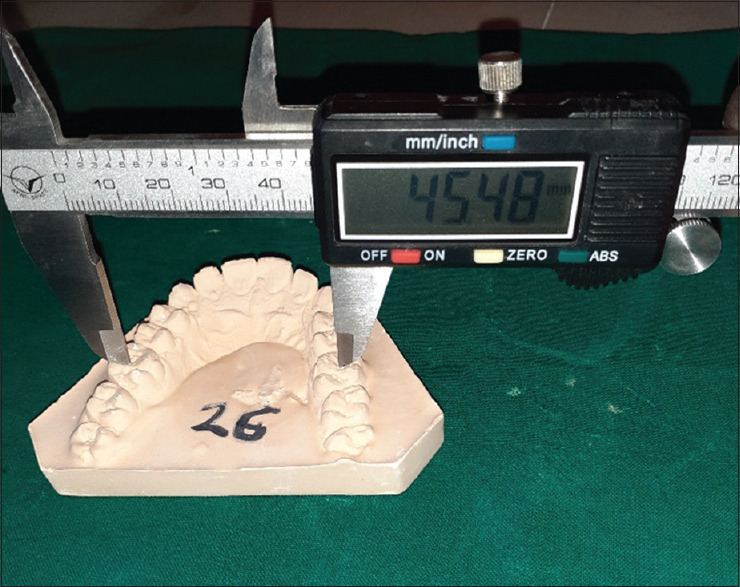
Shows measurement of intermolar width
All the measurements were taken in millimeters (mm) using a digital Vernier caliper. The Vernier caliper has measurements from 0 to 150 mm (0–6 inches) with a resolution of 0.01 mm and accuracy of ±0.02 mm.
All measurements were undertaken by a single observer. To minimize inter-observer bias, three readings were taken at separate times for each variable measured.
Statistical analysis: (SPSS software version 17)
A comparison of these parameters between males and females was done using an unpaired “t” test. Difference was considered statistically significant when P value was <0.05 (confidence interval = 95%).
The diagnostic performance (accuracy to discriminate males and females) of each parameter was quantified by plotting the “receiver operating characteristic” (ROC) curve. To make the ROC graph, the X-axis was 1 minus the specificity (the false positive rate), and the Y-axis was the sensitivity (the true positive rate). [In brief to make ROC graph, there is a need to calculate the sensitivity and specificity of the diagnostic parameter for each possible cut point value. The area under the ROC curve (AUC) is widely recognized as the measure of a diagnostic test's discriminatory power. The maximum value for the AUC is 1.0, thereby indicating a (theoretically) perfect test (i.e., 100% sensitive and 100% specific). An AUC value of 0.5 indicates no discriminative value (i.e., 50% sensitive and 50% specific) and is represented by a straight, diagonal line extending from the lower left corner to the upper right corner.].
Differences in the diagnostic performance of all parameters were assessed by merely comparing AUC values. An optimal cut-off point of each parameter was also determined using the respective ROC curve. [Ideally, the best cut-off value provides both the highest sensitivity and the highest specificity, easily located on the ROC curve by finding the highest point on the vertical axis and the furthest to the left on the horizontal axis (upper left corner)].
RESULTS
The present study was conducted on 100 individuals. The subjects included in the study were without any hard and soft tissue pathology related to the teeth. The following parameters were assessed in these subjects:
Intercanine width of maxillary arch,
Interpremolar width of maxillary arch,
Intermolar width of maxillary arch,
Combination of intercanine and interpremolar widths,
Combination of interpremolar and intermolar widths,
Combination of intercanine and intermolar widths,
Combination of intercanine, interpremolar and intermolar widths,
The results obtained in the present study annexure were analyzed and interpreted as follows:
Intercanine width of maxillary arch
Intercanine width between the tips of right and left maxillary canine was measured in males and females. In males, the mean intercanine width was 36.08 ± 2.04 mm (Range: 30.05–41.64 mm) and in females, the mean intercanine width was 34.31 ± 1.75 mm (Range: 28.35–39.01 mm). The mean intercanine width in males was larger than in females. Table 1 shows intercanine width in males as well as in females.
Table 1.
Mean value of intercanine width of maxillary arch in males and females
| Intercanine±width | ||
|---|---|---|
|
| ||
| Range (mm) | Mean±SD (mm) | |
| Males (n=50) | 30.05-41.64 | 36.08±2.04 |
| Females (n=50) | 28.35-39.01 | 34.31±1.75 |
Interpremolar width of maxillary arch
Interpremolar width between the distal pits of right and left first premolars was measured in males and females. In males, the mean interpremolar width was 38.97 ± 2.10 mm (Range: 33.94–45.21 mm) and in females, the mean interpremolar width was 36.92 ± 1.87 mm (Range: 31.34 mm). The mean interpremolar width in males was larger than the females. Table 2 shows interpremolar width in males as well as females.
Table 2.
Mean value of interpremolar width of maxillary arch in males and females
| Interpremolar width | ||
|---|---|---|
|
| ||
| Range (mm) | Mean±SD (mm) | |
| Males (n=50) | 33.94-45.21 | 38.97±2.10 |
| Females (n=50) | 31.34-42.83 | 36.92±1.87 |
Intermolar width of maxillary arch
Intermolar width between the central fossae of right and left first molars was measured in males and females. In males, the mean intermolar width was 50.43 ± 2.25 mm (Range: 44.16–56.84 mm) and in females, the mean intermolar width was 47.90 ± 2.06 mm (Range: 42.66–54.63 mm). The mean intermolar width in males was larger than the females. Table 3 shows intermolar width in males as well as females.
Table 3.
Mean value of intermolar width of maxillary arch in males and females
| Intermolar width | ||
|---|---|---|
|
| ||
| Range (mm) | Mean±SD (mm) | |
| Males (n=50) | 44.16-56.84 | 50.43±2.25 |
| Females (n=50) | 42.66-54.63 | 47.90±2.06 |
Combinations of intercanine, interpremolar, and intermolar widths
In males, the mean value of the combination of intercanine and interpremolar widths was 75.04 ± 3.88 mm (Range: 63.99–86.18 mm) and in females, it was 71.22 ± 3.37 mm (Range: 59.73–80.59 mm). The mean value of the combination of interpremolar and intermolar widths was 89.40 ± 4.03 mm (Range: 78.10–100.40 mm) in males and 84.82 ± 3.68 mm (Range: 74.86–97.42 mm) in females. The mean value of the combination of intercanine and intermolar widths was 86.50 ± 3.72 mm (Range: 74.21–97.19 mm) in males and 82.21 ± 3.38 mm (Range: 71.87–92.35 mm) in females. In males, the mean value of the combination of intercanine, interpremolar, and intermolar widths was 125.47 ± 5.61 mm (Range: 108.15–141.86 mm) and in females, it was 119.12 ± 5.05 mm (Range: 103.25–134.36 mm). The mean values of all combinations were larger in males than in females.
Table 4 shows the combination of arch widths in males as well as in females.
Table 4.
Mean values of combinations of arch widths in males and females
| Combinations | Sex | Range (mm) | Mean±SD (mm) |
|---|---|---|---|
| Intercanine + interpremolar | Males (n=50) | 63.99-86.18 | 75.04±3.88 |
| Females (n=50) | 59.73-80.59 | 71.22±3.37 | |
| Interpremolar + intermolar | Males (n=50) | 78.10-100.40 | 89.40±4.03 |
| Females (n=50) | 74.86-97.42 | 84.82±3.68 | |
| Intercanine + intermolar | Males (n=50) | 74.21-97.19 | 86.50±3.72 |
| Females (n=50) | 71.87-92.35 | 82.21±3.38 | |
| Intercanine + interpremolar + intermolar | Males (n=50) | 108.15-141.86 | 125.47±5.61 |
| Females (n=50) | 103.25-134.36 | 119.12±5.05 |
Comparison of the intercanine width of maxillary arch between males and females [Table 5 and Graph 1]
Table 5.
Comparison of the intercanine width of maxillary arch between males and females
| Groups | Intercanine Width | P (Significance) | ||
|---|---|---|---|---|
|
| ||||
| n | Range | Mean±SD | ||
| Males | 50 | 30.05-41.64 | 36.08±2.04 | <0.001 |
| Females | 50 | 28.35-39.01 | 34.31±1.75 | HS |
HS=Highly significant
Graph 1.

Comparison of mean values of intercanine width of maxillary arch in males and females
The comparison of the intercanine width of maxillary arch between males and females was done using the unpaired “t” test. There was a statistically significant difference in the intercanine width between males and females (P < 0.001). The intercanine width in males was significantly greater than that in females.
Comparison of the interpremolar width of maxillary arch between males and females [Table 6 and Graph 2]
Table 6.
Comparison of the interpremolar width of maxillary arch between males and females
| Groups | Interpremolar Width | P (Significance) | ||
|---|---|---|---|---|
|
| ||||
| n | Range | Mean±SD | ||
| Males | 50 | 33.94-45.21 | 38.97±2.10 | <0.001 |
| Females | 50 | 31.34-42.83 | 36.92±1.87 | HS |
HS=Highly significant
Graph 2.

Comparison of mean values of interpremolar width of maxillary arch in males and females
The comparison of the interpremolar width of maxillary arch between males and females was performed using unpaired “t” test. There was a statistically significant difference in the interpremolar width between males and females (P < 0.001). Males had significantly larger interpremolar width than females.
Comparison of the intermolar width of maxillary arch between males and females [Table 7 and Graph 3]
Table 7.
Comparison of the intermolar width of maxillary arch between males and females
| Groups | Intermolar Width | P (Significance) | ||
|---|---|---|---|---|
|
| ||||
| n | Range | Mean±SD | ||
| Males | 50 | 44.16-56.84 | 50.43±2.25 | <0.001 |
| Females | 50 | 42.66-54.63 | 47.90±2.06 | HS |
HS=Highly significant
Graph 3.

Comparison of mean values of intermolar width of maxillary arch in males and females
The intermolar width of maxillary arch was compared between males and females using unpaired “t” test. A statistically significant difference was found between males and females for intermolar width (P < 0.001). The intermolar width was significantly higher in males than in females.
Comparison of combination of the intercanine and interpremolar widths of maxillary arch between males and females [Table 8 and Graph 4]
Table 8.
Comparison of combination of the intercanine and interpremolar widths of maxillary arch between males and females
| Groups | Intercanine + Interpremolar Widths | P (Significance) | ||
|---|---|---|---|---|
|
| ||||
| n | Range | Mean±SD | ||
| Males | 50 | 63.99-86.18 | 75.04±3.88 | <0.001 |
| Females | 50 | 59.73-80.59 | 71.22±3.37 | HS |
HS=Highly significant
Graph 4.

Comparison of mean values of combination of the intercanine and interpremolar widths in males and females
The comparison of the combination of the intercanine and interpremolar widths between males and females was done using unpaired “t” test. There was a statistically significant difference between males and females in the combination of intercanine and interpremolar widths (P < 0.001). Males had significantly higher values for this combination as compared to females.
Comparison of combination of the interpremolar and intermolar widths of maxillary arch between males and females [Table 9 Graph 5]
Table 9.
Comparison of combination of the interpremolar and intermolar widths of maxillary arch between males and females
| Groups | Interpremolar + Intermolar Widths | P (Significance) | ||
|---|---|---|---|---|
|
| ||||
| n | Range | Mean±SD | ||
| Males | 50 | 78.10-100.40 | 89.40±4.03 | <0.001 |
| Females | 50 | 74.86-97.42 | 84.82±3.68 | HS |
HS=Highly significant
Graph 5.

Comparison of mean values of combination of the interpremolar and intermolar widths in males and females
The combination of the interpremolar and intermolar widths was compared between males and females using unpaired “t” test. A statistically significant difference was found between males and females for the combination of the interpremolar and intermolar widths (P < 0.001). The value of this combination was significantly greater in males than in females.
Comparison of combination of the intercanine and intermolar widths of maxillary arch between males and females [Table 10 and Graph 6]
Table 10.
Comparison of combination of the intercanine and intermolar widths of maxillary arch between males and females
| Groups | Intercanine + Intermolar Widths | P (Significance) | ||
|---|---|---|---|---|
|
| ||||
| n | Range | Mean±SD | ||
| Males | 50 | 74.21- | 86.50±3.72 | <0.001 |
| Females | 50 | 71.87- | 82.21±3.38 | HS |
HS=Highly significant
Graph 6.

Comparison of mean values of combination of the intercanine and intermolar widths in males and females
The comparison of the combination of the intercanine and intermolar widths between males and females was performed using unpaired “t” test. There was a statistically significant difference between males and females in the combination of the intercanine and intermolar widths (P < 0.001). The value of this combination in males was significantly larger than that in females.
Comparison of combination of the intercanine, interpremolar, and intermolar widths of maxillary arch between males and females [Table 11 and Graph 7]
Table 11.
Comparison of combination of the intercanine, interpremolar and intermolar widths of maxillary arch between males and females
| Groups | Intercanine + Interpremolar + Intermolar Widths | P (Significance) | ||
|---|---|---|---|---|
|
| ||||
| n | Range | Mean±SD | ||
| Males | 50 | 108.15- | 125.47±5.61 | <0.001 |
| Females | 50 | 103.25- | 119.12±5.05 | HS |
HS=Highly significant
Graph 7.
Comparison of mean values of combination of the intercanine, interpremolar, and intermolar widths in males and females
The combination of the intercanine, interpremolar, and intermolar widths of maxillary arch was compared between males and females with the help of unpaired “t” test. A statistically significant difference was noticed between males and females for this combination. Males had significantly higher values of this combination than that of females.
Diagnostic performance (accuracy to discriminate males and females) of all arch widths and combinations of arch widths [Table 12 and Graph 8]
Table 12.
Area under receiver operating characteristic curve for all arch widths and combinations of arch widths
| Parameters | AUC | Standard error | P | 95% confidence interval | |
|---|---|---|---|---|---|
|
| |||||
| Lower bound | Upper bound | ||||
| Intercanine width (ICW) | 0.742 | 0.022 | 0.000 | 0.699 | 0.785 |
| Interpremolar width (IPW) | 0.764 | 0.021 | 0.000 | 0.723 | 0.806 |
| Intermolar width (IMW) | 0.801 | 0.020 | 0.000 | 0.762 | 0.839 |
| ICW + IPW | 0.771 | 0.021 | 0.000 | 0.730 | 0.812 |
| IPW + IMW | 0.809 | 0.019 | 0.000 | 0.771 | 0.846 |
| ICW + IMW | 0.801 | 0.019 | 0.000 | 0.763 | 0.839 |
| ICW + IPW + IMW | 0.804 | 0.019 | 0.000 | 0.766 | 0.842 |
Graph 8.

Comparison of diagnostic performances of all arch widths and combinations of arch widths
Diagnostic performances of all parameters studied were determined by measuring the area under the respective ROC curves. Intercanine width had area under ROC curve (AUC) of 0.742 (lower bound = 0.699, upper bound = 0.785). AUC for interpremolar width was 0.764 (lower bound = 0.723, upper bound = 0.806). AUC for intermolar width was 0.801 (lower bound = 0.762, upper bound = 0.839). Combination of intercanine and interpremolar widths had AUC of 0.771 (lower bound = 0.730, upper bound = 0.812). AUC for combination of interpremolar and intermolar widths was 0.809 (lower bound = 0.771, upper bound = 0.846). AUC for combination of intercanine and intermolar widths was 0.801 (lower bound = 0.763, upper bound = 0.839). Combination of intercanine, interpremolar, and intermolar widths had AUC of 0.804 (lower bound = 0.766, upper bound = 0.842).
The area under the ROC curve was highest for the combination of interpremolar and intermolar widths, followed by the combination of intercanine, interpremolar, and intermolar widths, the combination of intercanine and intermolar widths, intermolar width, combination of intercanine and interpremolar widths, and interpremolar width and intercanine width.
Determination of optimal “cut-off” points [Table 13]
Table 13.
Optimal cut-off points for all arch widths and combinations of arch widths with percentage accuracy of sex identification
| Parameters | Cut-off point (mm) | % accuracy in male | % accuracy in female |
|---|---|---|---|
| Intercanine width (ICW) | 35.04 | 70.8 | 68.0 |
| Interpremolar width (IPW) | 37.80 | 68.4 | 70.8 |
| Intermolar width (IMW) | 48.91 | 75.6 | 72.4 |
| ICW + IPW | 72.83 | 69.6 | 68.0 |
| IPW + IMW | 86.94 | 71.2 | 72.4 |
| ICW + IMW | 84.12 | 74.4 | 74.0 |
| ICW + IPW + IMW | 121.89 | 72.4 | 71.2 |
Optimal cut-off points of all parameters were determined by assessing points that had provided the highest sensitivity and highest specificity. Optimal cut-off points for various parameters were as follows:
Intercanine width—35.04 mm with the 70.8% and 68% accuracy of sex identification in male and female, respectively.
Interpremolar width—37.8 mm with the 68.4% and 70.8% accuracy of sex identification in male and female, respectively.
Intermolar width—48.91 mm with the 75.6% and 72.4% accuracy of sex identification in male and female, respectively.
Combination of intercanine and interpremolar widths—72.83 mm with the 69.6% and 68% accuracy of sex identification in male and female, respectively.
Combination of interpremolar and intermolar widths—86.94 mm with the 71.2% and 72.4% accuracy of sex identification in male and female, respectively.
Combination of intercanine and intermolar widths—84.12 mm with the 74.4% and 74% accuracy of sex identification in male and female, respectively.
Combination of intercanine, interpremolar and intermolar widths—121.89 mm with the 72.4% and 71.2% accuracy of sex identification in male and female, respectively.
Thus, in short, there was a statistically significant difference (P < 0.001) between males and females in all arch widths and a combination of arch widths. The highest diagnostic performance to discriminate male and female was noticed for the combination of interpremolar and intermolar widths (AUC = 0.809).
DISCUSSION
The present study comprised 50 males and 50 females. The subjects were between the age group of 20–30 years as attrition is an age-dependent process, which means that increase in this process is directly proportional to the increased functional activities and the cumulative effect of etiologic agents. Also, the growth in width of dental arches tends to be completed before the adolescent growth spurt and is affected minimally by adolescent growth changes.[7,8]
As mobility can affect proper measurements of arch widths, subjects with healthy periodontium were only included. Subjects having parafunctional habits like thumb sucking, tongue thrusting, mouth breathing, etc., were excluded since these habits lead to constriction of maxillary arch which can adversely affect measurements of arch widths. Subjects those having significant crowding, spacing, and carious maxillary canine, first premolar, or first molar were also excluded. Measurements were taken from the casts of the subjects using a digital Vernier caliper with a resolution of 0.01 mm. Combinations were made by simply adding the arch widths. Statistical analyses were performed on www.spss.com using Statistical Package for Social Sciences SPSS software version 17 for windows.
Intercanine width of the maxillary arch was measured between the tips of right and left maxillary canine. The mean value of intercanine width was 36.08 ± 2.04 mm in males and 34.31 ± 1.75 mm in females. The mean value of intercanine width of maxillary arch in the present study is consistent with the findings of Younes SAES et al.[9] In their study, mean value of intercanine width of maxillary arch in Saudi males was 35.43 ± 1.59 mm and 33.69 ± 1.01 mm in Saudi females. In the same study, they have shown the intercanine width of the maxillary arch in Egyptian males 36.79 ± 1.77 mm, and 33.99 ± 0.81 mm in Egyptian females, respectively. They have concluded that Saudi maxillary arch dimension was greater in males than in females, and similar results were found for the Egyptian group by sex. The Egyptian maxillary arch dimension was greater than that of the Saudi subjects.
The present study was planned to assess sexual dimorphism in maxillary arch widths. Intercanine, interpremolar, and intermolar arch widths were selected for comparison purpose. The reason behind canine teeth selection was that they are the least frequently extracted teeth and being less affected by periodontal disease. Canine teeth have also been reported to survive in air and hurricane disasters.[7] First premolar and first molar were chosen since these are the least frequently missing or malformed teeth. According to Butler, mammalian dentition can be divided into several developmental fields like incisors, premolars, and molars fields which correlated with our findings.[10] Among field, dental variability manifests itself more strongly in the terminal or most posterior tooth than anterior tooth.[8] Forster et al. (2008)[11] noticed sexual dimorphism in maxillary intercanine, interpremolar, and intermolar arch widths with higher significance level (P < 0.001) as compared to mandibular arch widths (ranging from P < 0.05 to P < 0.001).
Aluko et al. (2009)[6] also found significant sexual dimorphism in all maxillary arch widths and intermolar width of mandibular arch in both early and late permanent dentitions. Intercanine and interpremolar widths of the mandibular arch were not significantly different between males and females in early and late permanent dentition, respectively.
Dilpreet Singh Grewal et al. (2017)[12] concluded that linear odontometric parameters intercanine width, interpremolar width, along with arch length, and combined width shows significant sexual dimorphism. Hence, odontometric parameters offer simple, reliable, and cost-effective way of determining sex in any forensic investigation. Only a few studies intended to assess sexual dimorphism in maxillary arch dimensions.
This shows that observations of these studies are also consistent with the finding of the present study. Aluko et al. (2009) studied the Nigerian population were the values measured was way too higher than the present study, though they measured interpremolar width between buccal cusp tips (difference is about 4–7 mm) because Nigerian people have bigger size jaw and interpremolar width mean value for males 45.91 and females 42.62. Rajbir (2015)[13] studied in Modinagar, Uttar Pradesh India which shows maxillary interpremolar width mean value in males 44.38 mm and 42.99 mm in females. Dilpreet Singh Grewal et al. (2017) studied in Punjab population found interpremolar width mean values for male 45.60 mm and females 43.17 mm and is clearly higher than that of the present study.
CONCLUSION
The highest percentage accuracy (75.6%) of sex identification in males was shown by the cut-off point for intermolar width. The cut-off point for the combination of intercanine and intermolar width showed the highest percentage accuracy (74%) of sex identification in females. The present study emphasizes the contribution of maxillary arch widths in forensic sciences. Thus, these arch widths can be used as a supplementary method along with other methods to increase the accuracy of sex identification.
Declaration of patient consent
The authors certify that they have obtained all appropriate patient consent forms. In the form, the patient(s) has/have given his/her/their consent for his/her/their images and other clinical information to be reported in the journal. The patients understand that their names and initials will not be published and due efforts will be made to conceal their identity, but anonymity cannot be guaranteed.
Financial support and sponsorship
Nil.
Conflicts of interest
There are no conflicts of interest.
Acknowledgement
The authors would like to sincerely thank D.Y.Patil Dental College for providing the opportunity to write a case study on the above topic, Dr. Avinash Tamgadge for guiding in every stage of this research paper. Without his support, it would have been extremely difficult to prepare the paper so meaningful and interesting, and Dr. Treville Pereira who has helped during this course of research paper in several ways.
REFERENCES
- 1.Frayer DW, Wolpoff MH. Sexual Dimorphism. Annu Rev Anthropol. 1985;14:429–73. [Google Scholar]
- 2.Vodanović M, Demo Ž, Njemirovskij V, Keros J, Brkić H. Odontometrics: A useful method for sex determination in an archaeological skeletal population? J Archaeol Sci. 2007;34:905–13. [Google Scholar]
- 3.Gustafsson A, Lindenfors P. Human size evolution: No evolutionary allometric relationship between male and female stature. J Hum Evol. 2004;47:253–66. doi: 10.1016/j.jhevol.2004.07.004. [DOI] [PubMed] [Google Scholar]
- 4.Alvesalo L. Sex chromosomes and human growth. Hum Genet. 1997;101:1–5. doi: 10.1007/s004390050575. [DOI] [PubMed] [Google Scholar]
- 5.Keiser-Nielsen S. Bristol: John Wright & Sons Ltd; 1980. Person identification by means of the teeth. [Google Scholar]
- 6.Aluko IA, DaCosta OO, Isiekwe M. Dental arch widths in the early and late permanent dentitions of a Nigerian population. Niger Dent J. 2010:17. doi: 10.4314/ndj.v17i1.54342. [Google Scholar]
- 7.Al-Rifaiy MQ, Abdullah MA, Ashraf I, Khan N. Dimorphism of mandibular and maxillary canine teeth in establishing sex identity. Saudi Dent J. 1997;9:17–20. [Google Scholar]
- 8.Vastardis H. The genetics of human tooth agenesis: New discoveries for understanding dental anomalies. Am J Orthod Dentofacial Orthop. 2000;117:650–6. [PubMed] [Google Scholar]
- 9.Abd-el Samad Younes S. Maxillary arch dimensions in Saudi and Egyptian population sample. Am J Orthod. 1984;85:83–8. doi: 10.1016/0002-9416(84)90126-x. [DOI] [PubMed] [Google Scholar]
- 10.Butler PM. Studies of the mammalian dentition.–Differentiation of the post-canine dentition. Proc Zool Soc Lond. 1939;B109:1–36. [Google Scholar]
- 11.Forster CM, Sunga E, Chung CH. Relationship between dental arch width and vertical facial morphology in untreated adults. European Journal Orthodontics. 2008;30:288–94. doi: 10.1093/ejo/cjm113. [DOI] [PubMed] [Google Scholar]
- 12.Grewal DS, Khangura RK, Sircar K, Tyagi KK, Kaur G, David S. Morphometric analysis of odontometric parameters for gender determination. J Clin Diagn Res. 2017;11:ZC09–13. doi: 10.7860/JCDR/2017/26680.10341. [DOI] [PMC free article] [PubMed] [Google Scholar]
- 13.Khangura RK, Sircar K, Grewal DS. Four odontometric parameters as a forensic tool in stature estimation. J Forensic Dent Sci. 2015;7:132–6. doi: 10.4103/0975-1475.146367. [DOI] [PMC free article] [PubMed] [Google Scholar]