Abstract
Background
American foulbrood (AFB) disease caused by Paenibacillus larvae is dangerous, and threatens beekeeping. The eco-friendly treatment method using probiotics is expected to be the prospective method for controlling this pathogen in honey bees. Therefore, this study investigated the bacterial species that have antimicrobial activity against P. larvae.
Results
Overall, 67 strains of the gut microbiome were isolated and identified in three phyla; the isolates had the following prevalence rates: Firmicutes 41/67 (61.19%), Actinobacteria 24/67 (35.82%), and Proteobacteria 2/67 (2.99%). Antimicrobial properties against P. larvae on agar plates were seen in 20 isolates of the genus Lactobacillus, Firmicutes phylum. Six representative strains from each species (L. apis HSY8_B25, L. panisapium PKH2_L3, L. melliventris HSY3_B5, L. kimbladii AHS3_B36, L. kullabergensis OMG2_B25, and L. mellis OMG2_B33) with the largest inhibition zones on agar plates were selected for in vitro larvae rearing challenges. The results showed that three isolates (L. apis HSY8_B25, L. panisapium PKH2_L3, and L. melliventris HSY3_B5) had the potential to be probiotic candidates with the properties of safety to larvae, inhibition against P. larvae in infected larvae, and high adhesion ability.
Conclusions
Overall, 20 strains of the genus Lactobacillus with antimicrobial properties against P. larvae were identified in this study. Three representative strains from different species (L. apis HSY8_B25, L. panisapium PKH2_L3, and L. melliventris HSY3_B5) were evaluated to be potential probiotic candidates and were selected for probiotic development for the prevention of AFB. Importantly, the species L. panisapium isolated from larvae was identified with antimicrobial activity for the first time in this study.
Supplementary Information
The online version contains supplementary material available at 10.1186/s12866-023-02902-0.
Keywords: Paenibacillus larvae, American foulbrood, Probiotics, Gut microbiome, Apis mellifera, Lactobacillus
Background
Honey bees provide important products to humans, such as honey, pollen, royal jelly, and propolis, and play a vital role in pollinating wild plants and crops [1, 2]. It is estimated that 87.5% of all flowering plants and 50% of global crops are pollinated by honey bees (Apis mellifera) [3–5]. However, honey bees are facing great challenges of colony losses of up to 30% in some countries [6, 7]. Pests and diseases are considered the major factors that result in honey bee loss [6]. Therefore, investigating efficient methods for disease control to mitigate colony losses is one of the major concerns in beekeeping.
American foulbrood (AFB) is one of the most destructive diseases of honey bees caused by Paenibacillus larvae, a spore-forming, gram-positive bacterium [8]. The spores germinate in the gut of infected larvae 12 h after ingestion and rapidly proliferate to kill the larvae [9]. Spores from dead larvae are spread to the hive by worker bees during the removal of the dead larvae, resulting in the collapse of the infected colony [10]. The traditional method of controlling P. larvae is using antibiotics such as oxytetracycline and tylosin tartrate [10]. However, there are great concerns about the use of antibiotics because of the adverse effects on honey bees by disturbing the gut microbiome [11, 12], and the P. larvae develop resistance to the antibiotics [13–15]. Therefore, it is necessary to develop an alternative eco-friendly method to control and treat AFB.
The digestive tract of honey bees contains a complex of microbial communities [16, 17]. The microbial composition in the gut varies depending on the food sources, season, geographical region, living stage, and different strains of honey bees [18–21]. The bacterial brood diseases caused by Melissococcus plutonius and P. larvae could result in changes in the gut bacterial community of honey bees, affecting honey bee health [22, 23]. The gut microbiome, specifically the lactic acid bacteria (LAB), was demonstrated to have a beneficial function on honey bees’ health [24]. In addition, the antimicrobial activity of LAB for biological control of Melissococcus plutonius and P. larvae, the causative agents of European and American foulbrood, was shown [25, 26]. The LAB belonging to the genus Lactobacillus and Bifidobacterium were isolated from different sources including the gut of adult bees, brood, brood comb, and honey, and were demonstrated to be the potential probiotic candidates for the inhibition of P. larvae [27–30]. Further investigations are necessary to identify bacterial species that inhibit P. larvae, are safe to honey bee larvae, and meet the requirements for probiotic bacteria.
Accordingly, this study was conducted to isolate the gut bacteria from honey bees Apis mellifera and investigate the potential bacterial strains for P. larvae inhibition. The evaluation of potential probiotic candidates for AFB disease treatment was performed using the in vitro rearing larvae method.
Results
Isolation of gut microbiome
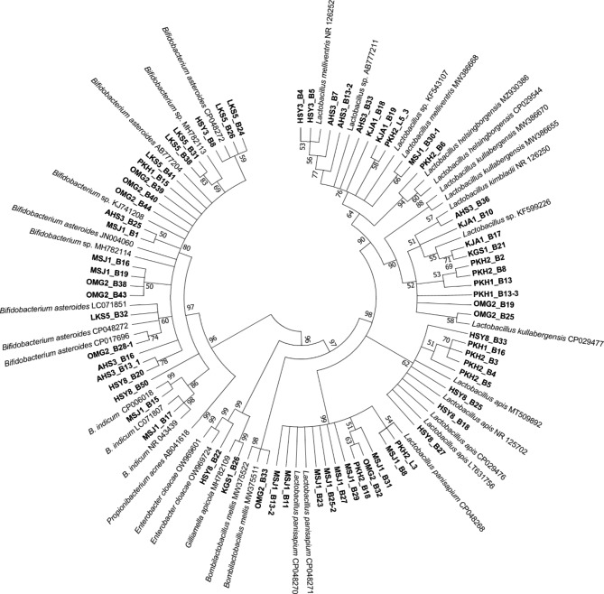
Overall, 67 strains of honey bee gut bacteria were isolated and identified to belong to three phyla, with the prevalence of the isolates in descending order as follows: Firmicutes 41/67 (61.19%), Actinobacteria 24/67 (35.82%), and Proteobacteria 2/67 (2.99%). The Firmicutes phylum consisted of only one genus, Lactobacillus, with six identified species, including L. panisapium (9 isolates), L. apis (4), L. mellis (3), L. melliventris (3), L. kimbladii (1), and L. kullabergensis (1), and other 20 unidentified strains (Fig. 1; Supplementary Table S1). Actinobacteria phylum had one genus, Bifidobacterium, with two species (B. asteroids and B. indicum) and 17 unclassified strains. Meanwhile, the Proteobacteria phylum contained two genera, Gilliamella (G. apicola) and Enterobacter (Fig. 1; Supplementary Table S1).
Fig. 1.
Phylogenetic tree of isolated bacterial strains. A maximum likelihood tree was created using the 16 S rRNA gene of 67 isolates with bootstrapping 500. The names of the isolated strains are written in bold, and the reference species name with NCBI accession number is given. Only the percentage of bootstrap ≥ 50% is shown
Antimicrobial activity of isolated lactic acid bacteria
The antimicrobial effect of the isolated strains was evaluated based on the inhibition zone observed surrounding the isolated strains against P. larvae, and 20 of 67 isolates of only one genus, Lactobacillus, showed antimicrobial activity (Fig. 2; Supplementary Figure S2; Supplementary Table S1). Six representative strains of the six identified species with the largest inhibition zone were selected to evaluate probiotic candidates (Fig. 2). The six strains consisted of OMG2_B25 (L. kullabergensis), HSY8_B25 (L. apis), PKH2_L3 (L. panisapium), OMG2_B33 (L. mellis), HSY3_B5 (L. melliventris), and AHS3_B36 (L. kimbladii). Two strains, HSY3_B5 (L. melliventris) and AHS3_B36 (L. kimbladii), had the largest size inhibition zone (20.0 ± 0.0 mm), followed by HSY8_B25 (L. apis; 18.0 ± 2.0 mm), OMG2_B33 (L. mellis; 12.7 ± 1.2 mm), OMG2_B25 (L. kullabergensis; 12.7 ± 2.3 mm), and PKH2_L3 (L. panisapium; 11.3 ± 1.2 mm) (Fig. 2). Other two strains of Lactobacillus, KJA1_B10 and AHS3_B13-2, also showed a large inhibition zone with 18.7 and 20 mm, respectively (Fig. 2; Supplementary Table S1). However, the species names of the two strains were not identified (Fig. 1). Therefore, these two unidentified strains were not selected for further evaluation. Of the six selected probiotic candidates, only one strain, PKH2_L3 (L. panisapium), was isolated from larvae, and the other five strains were from adult honey bees (Supplementary Table S1).
Fig. 2.
Antimicrobial activity of isolated bacteria against P. larvae identified by inhibition zone on an agar plate
Twenty of the 67 isolates showed antimicrobial activity with different sizes of inhibition zone, and six of the 20 isolates (indicated by arrows) belonging to different species were selected as probiotic candidates; the selected strains were OMG2_B25 (L. kullabergensis; Lk), HSY8_B25 (L. apis; La), PKH2_L3 (L. panisapium; Lp), OMG2_B33 (L. mellis; Lm), HSY3_B5 (L. melliventris; Lmv), and AHS3_B36 (L. kimbladii; Lkb)
Safety examination of isolated LAB on honey bee larvae
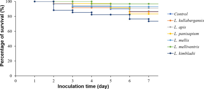
The safety challenge of the six selected LAB to honey bee larvae showed that the survival rates of larval groups that received different strains were not significantly different (p = 0.285) during the evaluation period. The larvae received three strains, including L. apis HSY8_B25, L. mellis OMG2_B33, and L. melliventris HSY3_B5, which showed higher survival rates, of 91%, 93%, and 97%, respectively than that of the control group without LAB administration (87%) (Fig. 3). The survival rates of two groups that received L. kullabergensis OMG2_B25 and L. panisapium PKH2_L3 (86% and 83%, respectively) were comparable with that of the control group. Meanwhile, L. kimbladii AHS3_B36 reduced the survival rate of larvae to 74% compared with 87% in the control group. The results demonstrated that the administration of five strains (L. apis HSY8_B25, L. mellis OMG2_B33, L. melliventris HSY3_B5, L. kullabergensis OMG2_B25, and L. panisapium PKH2_L3) was safe for the honey bee larvae, with the exception of L. kimbladii AHS3_B36.
Fig. 3.
The survival rate of honey bee larvae that received six isolated LAB strains. Six species of isolated LAB were supplied to six groups of larvae, the living number of larvae was examined, and the survival rate was calculated daily till day 7. The control group was fed with only a feeding solution
Biological control of probiotic candidates against P. larvae in infected larvae
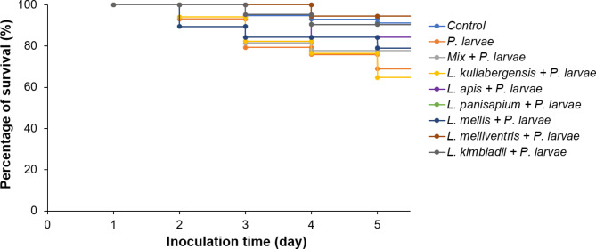
The inhibitory effect of the LAB against P. larvae in the infected larvae was evaluated for 5 days after infection. The results showed that the survival rate of larvae fed with L. kullabergensis OMG2_B25 (64.71%) was lower than that of the only P. larvae infected group (68.97%). Meanwhile, the other five strains and the mixture of the six strains protected larvae by increasing the survival rate. The survival rate after 5 days of infection was 77.78%, 78.95%, 84.21%, 90.48%, 90.48%, and 94.44% for the mixture, L. mellis OMG2_B33, L. apis HSY8_B25, L. panisapium PKH2_L3, L. kimbladii AHS3_B36, and L. melliventris HSY3_B5, respectively (Fig. 4). Three species, L. panisapium PKH2_L3, L. kimbladii AHS3_B36, and L. melliventris HSY3_B5, helped increase the survival rate to the same level as that of the control group without P. larvae infection and no LAB administration (91.23%; p = 0.865).
Fig. 4.
The survival rate of P. larvae infected larvae fed with isolated LAB. The six isolated strains were singly supplied to each group of larvae, and one group was provided with a mixture of six strains. On the first day, after the administration of LAB, the larvae were infected with P. larvae. The number of living larvae was examined daily in each group to calculate the survival rate until the fifth day. The control group was neither infected with P. larvae nor fed with the LAB. Another group was infected with P. larvae without the administration of LAB.
DNA copy number of P. larvae in different groups was calculated based on the Ct value of P. larvae detection in real-time polymerase chain reaction (PCR) (Table 1). The results demonstrated that the mixture of six LAB strains had the highest inhibition to P. larvae with the lowest number of P. larvae DNA copies (8.37 × 10⁵ copies), followed by the group fed with L. panisapium PKH2_L3 (1.42 × 10⁶ copies), L. apis HSY8_B25 (3.64 × 10⁶ copies), L. kullabergensis OMG2_B25 (4.56 × 10⁶ copies), L. mellis OMG2_B33 (5.49 × 10⁶ copies), L. melliventris HSY3_B5 (6.93 × 10⁶ copies), and L. kimbladii AHS3_B36 (1.07 × 10⁶ copies). Meanwhile, the P. larvae infected group without LAB feeding showed the highest number of P. larvae DNA copies, 1.32 × 10⁷ copies. The control group, with no P. larvae infection and no LAB administration, showed negative results in P. larvae detection (Table 1).
Table 1.
Quantification of P. larvae DNA in larval groups that received different strains of LAB
| Group | Only P. larvae | Mixture of LAB + P. larvae |
L. kullabergensis + P. larvae | L. apis + P. larvae | L. panisapium + P. larvae | L. mellis + P. larvae | L. melliventris + P. larvae | L. kimbladii + P. larvae | No P. larvae, no LAB |
|---|---|---|---|---|---|---|---|---|---|
| Ct value | 18.46 | 22.49 | 20.01 | 20.34 | 21.72 | 19.74 | 19.4 | 18.74 | Negative |
| P. larvae DNA copy | 1.32 × 10⁷ | 8.37 × 10⁵ | 4.56 × 10⁶ | 3.64 × 10⁶ | 1.42 × 10⁶ | 5.49 × 10⁶ | 6.93 × 10⁶ | 1.09 × 10⁷ | 0 |
Hydrophobicity and auto-aggregation
The Bacterial Adherence to Hydrocarbons (BATH) assay results showed that the adhesion ability of the six LAB strains was different. L. kullabergensis OMG2_B25 and L. mellis OMG2_B33 demonstrated low hydrophobicity with adhesion to toluene (22.10% and 14.83%) and xylene (19.27% and 9.16%), respectively. Meanwhile, the other four strains showed moderate to high hydrophobicity, with the adhesion to toluene ranging from 56.16% (L. melliventris HSY3_B5) to 71.03% (L. kimbladii AHS3_B36) and the adhesion to xylene ranging from 50.55% (L. melliventris HSY3_B5) to 70.09% (L. panisapium PKH2_L3; Table 2).
Table 2.
Adhesion to toluene and xylene of the six isolated lactic acid bacteria
| Hydrophobicity (%) Toluene | ||||||
|---|---|---|---|---|---|---|
| Time (min) | L. kullabergensis (OMG2_B25) | L. apis (HSY8_B25) | L. panisapium (PKH2_L3) | L. mellis (OMG2_B33) | L. melliventris (HSY3_B5) | L. kimbladii (AHS3_B36) |
| 60 | 22.10 ± 1.48 | 68.70 ± 0.39 | 58.19 ± 1.20 | 14.83 ± 2.09 | 56.16 ± 0.84 | 71.03 ± 0.85 |
| Hydrophobicity (%) Xylene | ||||||
| 60 | 19.27 ± 2.00 | 55.45 ± 1.10 | 70.09 ± 0.45 | 9.16 ± 1.77 | 50.55 ± 1.70 | 56.70 ± 1.15 |
The auto-aggregation results showed that there was no significant difference (p = 0.978) in auto-aggregation percentage (AA%) among the six isolated LAB. All strains showed progressive aggregation over time. The AA% after 24 h was all above 50%, with the lowest value of 82.80% (L. panisapium PKH2_L3) and the highest value of 90.21% (L. melliventris HSY3_B5) (Table 3). The L. melliventris HSY3_B5 showed the fastest auto-aggregation ability with 22.90% at 1 h and reached 87.49% after 5 h, followed by L. mellis OMG2_B33 (69.46%), and L. kimbladii AHS3_B36 (53.02%) (Table 3). However, the other three strains showed a lower auto-aggregation ability with AA% at 5 h of 45.79%, 43.42%, and 40.50% for L. apis HSY8_B25, L. kullabergensis OMG2_B25, and L. panisapium PKH2_L3, respectively (Table 3).
Table 3.
Auto-aggregation test of the six isolated lactic acid bacteria
| Auto-Aggregation (%) | ||||||
|---|---|---|---|---|---|---|
| Time (h) | L. kullabergensis (OMG2_B25) | L. apis (HSY8_B25) | L. panisapium (PKH2_L3) | L. mellis (OMG2_B33) | L. melliventris (HSY3_B5) | L. kimbladii (AHS3_B36) |
| 1 | 9.86 ± 0.31 | 2.77 ± 1.38 | 14.45 ± 2.04 | 18.65 ± 1.08 | 22.90 ± 1.22 | 7.61 ± 0.23 |
| 5 | 45.79 ± 1.13 | 43.42 ± 0.29 | 40.50 ± 1.45 | 69.46 ± 1.08 | 87.49 ± 0.19 | 53.02 ± 0.20 |
| 18 | 76.45 ± 1.05 | 77.00 ± 0.81 | 63.06 ± 0.72 | 82.06 ± 0.91 | 89.97 ± 1.56 | 78.30 ± 0.98 |
| 24 | 86.18 ± 0.29 | 85.63 ± 0.21 | 82.80 ± 0.57 | 89.08 ± 0.47 | 90.21 ± 0.84 | 85.03 ± 0.65 |
Discussion
The gut microbiome of honey bees was isolated and identified as belonging to the genera Bifidobacterium, Lactobacillus, Gilliamella, and Enterobacter, with Lactobacillus spp. being the dominant species. These are the major gut microbiome of honey bees [16, 21], of which Bifidobacterium and Lactobacillus are known to be the lactic acid producers [31] and Gilliamella is identified to have a vital role in improving the dietary tolerances of honey bees [32]. The structure of the honey bee gut microbiome varies depending on the geographical region, specific environmental landscape, humidity, temperature, and seasonality [33–35]. The gut bacterial community can be perturbed by pathogen infections as well as miticide and pesticide exposure [36–39]. Dysbiosis of the gut microbiota could lead to an increased susceptibility of honeybees to pathogens [40, 41]. Therefore, it is crucial to understand the microbial structure of the honey bee gut microbiome and select probiotic candidates that can help maintain a healthy balance of the gut microbiota to enhance honey bee immunity.
Honey bee gut-originated species of Bifidobacterium and Lactobacillus were demonstrated to have inhibitory effects on P. larvae [42]. These bacteria produce organic acids that increase the acidity of the digestive tract and inhibit the growth of pathogenic microorganisms [43–45]. In addition, the Bifidobacterium and Lactobacillus bacteria enhance the honey bee immune system by upregulating antimicrobial peptides in honey bees [46]. However, in this study, only Lactobacillus sp. showed inhibitory properties against P. larvae growth on agar plates (Table S1; Figure S2). The Bifidobacterium strains showed no inhibition against P. larvae (strain ATCC9545) ERIC genotype I [47] used in this study. The result was consistent with that of Forsgren et al. [42] who showed that the strains of Bifidobacterium had an antimicrobial effect against only the ERIC genotype III and IV of P. larvae and no inhibition against ERIC I.
The LAB isolated from the honey bee gut has been demonstrated to be helpful for the inhibition of P. larvae [48]. The LAB species with an inhibitory effect against P. larvae were previously isolated from the gut of A. mellifera L. and identified as strains of 11 species in the genus Lactobacillus (L. kunkeei, L. plantatarum, L. apinorum, L. mellis, L. kimbladii, L. kullabergensis, L. mellifer, L. apis, L. helsingborgensis, L. brevis, and L. melliventris) [25, 29, 30, 42, 48, 49], and strains of two species in genus Bifidobacterium (B. asteroides and B. coryneforme) [42, 48]. Five of the six Lactobacillus species with inhibitory effects against P. larvae, which were identified in this study, were similar to those reported in previous studies. Notably, another species was newly determined, L. panisapium. This species was isolated from both larva and adult bees. Therefore, the species could be safe to apply in both living stages of honey bees.
The differences in the structure of the digestive tract and food sources between the adult and larval stages in honey bees could result in the difference in gut microbiome between the two stages [21, 50, 51]. Therefore, it could be harmful when the larvae receive a large amount of LAB isolated from adult bees. It was demonstrated that the brood size was reduced when the LAB mixture was supplementally administered to the colonies for P. larvae treatment [52]. Consequently, LAB originating from larvae may be the optimal probiotic candidate for safe application in the colony. The L. panisapium PKH2_L3 strain isolated from larvae in this study with antimicrobial effect against P. larvae and no harm to larvae could be selected for further evaluation as a potential probiotic candidate.
The combination of all isolated strains of different LAB was expected to increase the diversity of metabolites and antimicrobial peptides that help inhibit P. larvae [42, 48]. However, the influence of each LAB strain and the mixture on the healthy larvae were not previously evaluated. Feeding larvae with the combination of six strains in different species isolated in this study also showed high efficiency of P. larvae inhibition compared to feeding larvae with individual strains. However, the survival rate of larvae fed with the combination of six LAB was lower than that of the group that was singly fed with only L. apis HSY8_B25, L. panisapium PKH2_L3, L. mellis OMG2_B33, L. melliventris HSY3_B5, or L. kimbladii AHS3_B36 (Fig. 4). In addition, administration of the six LAB strains to the healthy larvae (no P. larvae infection) demonstrated that some species help enhance the survival rate of the larvae, such as L. apis HSY8_B25, L. mellis OMG2_B33, and L. melliventris HSY3_B5. However, L. kimbladii AHS3_B36 is harmful to the larvae (Fig. 3). Therefore, the combination of all LAB strains could result in an increase in larval mortality, and safety screening is necessary to select the potential LAB strain for AFB treatment.
Adhesion ability is an essential property of the desirable probiotic bacteria to demonstrate effective antimicrobial activity [53]. The bacteria with high adhesion ability can prevent elimination by peristalsis and develop in the host intestinal tract to protect against the colonization of other harmful microorganisms [25, 28, 54]. The potentiality of the bacteria to adhere to the intestinal tract of honey bees can be tested by its adhesion to hydrocarbon (BATH), and the hydrophobicity and auto-aggregation ability are helpful in understanding the adherence ability of the probiotic bacteria [55, 56]. Among the six strains selected in this study as probiotic candidates, two species (L. kullabergensis OMG2_B25 and L. mellis OMG2_B33) demonstrated low adhesion to toluene and xylene, and the other four species had moderate to high adhesion ability in comparison with other probiotic species [54, 57]. The auto-aggregation of the six species was higher than those of other probiotic candidates for honey bees analyzed in previous studies [25]. Therefore, the four strains (L. apis HSY8_B25, L. panisapium PKH2_L3, L. melliventris HSY3_B5, and L. kimbladii AHS3_B36) with high adhesion ability could be selected for further analysis.
Conclusions
Isolation of gut bacteria from honey bees was conducted to find bacteria with antimicrobial effects against P. larvae; 20 of the 67 isolated strains belonging to only the Lactobacillus genus showed inhibitory properties to P. larvae on agar plates. Six representative strains from six different species (L. apis HSY8_B25, L. panisapium PKH2_L3, L. melliventris HSY3_B5, L. kimbladii AHS3_B36, L. kullabergensis OMG2_B25, and L. mellis OMG2_B33) were selected for evaluation of the probiotic properties, of which three species (L. apis HSY8_B25, L. panisapium PKH2_L3, and L. melliventris HSY3_B5) passed the tests for probiotic properties, including safety to larvae, inhibition of P. larvae growth in infected larvae, and adhesion ability. These strains could be potential candidates for probiotics to control AFB disease.
Methods
Isolation of the gut microbiome from honey bees
Live honey bees (Apis mellifera L.) in the combs were collected from 18 different apiaries in South Korea and carried to the laboratory. Subsequently, they were washed twice using autoclaved distilled water. Overall, 90 gut samples (larvae [n = 36] and adult bees [n = 54]) collected from 18 different apiaries were isolated (Supplementary Figure S1). The three guts of adult bees or two guts of larvae collected from each apiary were placed into a tissue grinding tube with steel beads (SNC, Hanam, Korea). Next, 500 µL phosphate-buffered saline (PBS) solution was added, and the samples were vortexed for 10 s. The homogenate was briefly centrifuged at 200 ×g for 30 s; subsequently, 100 µL of the supernatant was spread on different agar plates as follows: brain heart infusion (BHI), and the De Man, Rogosa and Sharpe (MRS) [58]. The plates were incubated at 37 ℃ in an anaerobic condition until white round colonies were seen (24–72 h). The anaerobic jar system Anoxomat AN2CTS (MART Microbiology B.V., 9207 JB Drachten, Netherlands) was used to provide an anaerobic condition. Finally, the colonies were selected from agar plates and transferred to a broth medium for cultivation in the same condition for further analysis.
Species identification of isolated microbiome
The colonies of bacteria BHI or MRS agar were singly selected and inoculated in broth medium for 24 h at 37 ℃ in anaerobic conditions. Stock bacteria were made from the cultivated bacteria by adding glycerol to the final 20% and stored at -80℃, and the remaining solution of each cultivation was used for species identification. Bacteria from the medium were collected by centrifuging at 13,000 ×g for 5 min and discarding the supernatant. Deoxyribonucleic acid (DNA) was extracted using the FastDNA Spin Kit for Soil (MP Biochemicals GmbH, Eschwege, Germany) following the manufacturer’s instructions. The 16S rRNA gene of bacteria was amplified and sequenced using primer pair 518F: 5’-CCA GCA GCC GCG GTA ATA CG-3’/800R: 5’-TAC CAG GGT ATC TAA TCC-3’ (Macrogen, Inc., Seoul, South Korea). The sequences were analyzed by comparing them with the National Center for Biotechnology Information (NCBI) database using the Basic Local Alignment Search Tool (BLAST-NCBI). Notably, sequences on NCBI with the highest similarity to that of isolated strains were selected for alignment using Clustal X version 2.0 [59]. A maximum likelihood phylogenetic tree was constructed using the Kimura 2-parameter model [60], gamma distribution, and bootstrapping 500 times with the software MEGA version 7 [61].
Screening of antimicrobial activity
The inhibition assay was conducted according to a previous method [62] with some modifications. The vegetative form of P. larvae ATCC 9545 strain, enterobacterial repetitive intergenic consensus (ERIC) genotype I, was cultivated at 35 ℃ for 48 h in a BHI medium. The concentration adjustment of OD600 = 0.7 was made, and the cultivated medium of P. larvae was spread on a BHI agar plate using a sterile cotton swab, and three replicate plates were used. The isolated strains of LAB were cultured on MRS agar at 37 ℃ for 3 days. Afterwards, a single colony was selected and cultured in MRS broth for 18 h at 37 ℃ in an anaerobic condition. The LAB in cultured MRS broth were collected by centrifugation at 13,000 ×g for 5 min, and the supernatant was discarded. The pellet was washed twice by suspending it in PBS solution and centrifuging under the same condition. Finally, the pellet was suspended by PBS and adjusted to OD600 = 0.7; subsequently, 10 µL of the suspended bacterial solution was dispensed onto a sterile filter paper (6 mm, Whatman, USA) and placed on a BHI agar plate, where the P. larvae were spread. The inhibition zone was observed after 48 h incubation at 37 ℃, microaerobic condition, 5% CO2.
Safety examination of the isolated LAB on honey bee larvae
The representative strain of each species with the largest inhibition zone was selected for the safety examination of in vitro rearing larvae. LAB strains were cultured on MRS agar at 37 ℃ in an anaerobic condition for 3 days; a single colony was selected and cultured in MRS broth for 18 h at 37 ℃ in an anaerobic condition. The cultivated bacteria were collected by centrifugation at 13,000 ×g for 5 min, and the bacterial pellet was suspended by feeding diet to a concentration of 1 × 10⁴ cells/µL [63]. The feeding diet was prepared with 6% glucose, 6% fructose, 1% yeast extract, and 50% royal jelly [64]. Larvae (1 to 2 instar) of A. mellifera L. were transferred to a 6-well plate with 10 larvae/well. Each LAB strain was evaluated with larvae in three wells (n = 30). Feeding solution of 200 µL containing LAB (1 × 10⁴ cells/µL) was supplied to the larvae in one dose on day 1; subsequently, the larvae were fed daily with feeding solution without LAB until day 7. The control groups were fed with feeding solution without LAB. Finally, the number of dead larvae was recorded daily. Notably, dead larvae were identified by lack of body elasticity or color change to brown.
Inhibition of Paenibacillus larvae in artificially infected larvae
The antibacterial effect of the isolated LAB was evaluated in an in vivo larval model. Honey bee larvae (A. mellifera L.) 1 to 2 instar were transferred to a 48-well plate, one larva/well. Larvae in each group, including three plates (n = 144), were used for each LAB strain. One group was fed with a mixture of six selected LAB strains, another group was infected with only P. larvae with no LAB administration, and the control group was fed with only feeding solution without P. larvae and LAB. The feeding diet containing LAB was prepared as described above. In preparing P. larvae spores for infection, colonies of P. larvae were suspended in BHI broth, then spread on Columbia sheep blood agar (BD, Franklin Lakes, NJ, USA) and incubated for 10 days at 37 ℃. A medium containing spores was collected, and the spores were suspended in a feeding solution with 1,000 spores/µL. The larvae were supplied by a feeding diet containing LAB in one dose (50 µL/larva) on day 2, and a feeding diet containing P. larvae spores (50 µL/larva) was supplied to each larva on day 3. Then, 50 µL of feeding diet was supplied daily to each larva until day 5. The larvae were inspected daily, and the number of dead larvae in each group was recorded. At the end of the period, three living larvae of each group were randomly selected for real-time PCR detection of P. larvae using primer pairs AFB-F: AAA TCA TCA TGC CCC TTA TG/ AFB-R: CGA TTA CTA GCA ATT CCG ACT, and the probe: FAM-CGT ACT ACA ATG GCC GGT ACA ACG–BHQ-1 [65]. The digestive tract of the larvae was separated after washing five times with autoclaved distilled water and used for DNA extraction employing the FastDNA Spin Kit for Soil (MP Biochemicals GmbH, Eschwege, Germany) following the manufacturer’s instructions. Finally, 50 µL of DNA solution was acquired from each group. The PCR reaction mix (20 µL) was composed of 1 µL (10 pmol) of each primer, 1 µL (5 pmol) of the probe, 2 µL of the extracted DNA solution, 5 µL of ddH2O, and 10 µL of Iq™ supermix as PCR premix (Bio-Rad, Hercules, CA, USA). PCR was performed at 95 ℃ for 5 min, followed by 40 PCR cycles for 10 s at 95 ℃ and 30 s at 60 ℃. DNA copy of P. larvae was calculated based on the cycle threshold (Ct) of P. larvae detection from each group using a standard linear regression. Standard curves (Supplementary Figure S3) representing the relationship between Ct value and initial DNA copy were established from the amplification using P. larvae recombinant DNA 108-101 copies (10-fold dilution).
Hydrophobicity and auto-aggregation
Adhesion ability is an essential property of desired probiotics by which the bacteria can adhere to intestinal epithelial cells [54]. The ability of cells to adhere to the intestine can be affected by the composition and structure of the cell surface, and the hydrophobicity of the cell surface is known to be a major factor [66]. Therefore, the auto-aggregation and hydrophobicity ability of the LAB were measured. Auto-aggregation capacity was evaluated according to a previously reported method [54]. The LAB strains were cultured at 37 ℃ for 18 h under anaerobic conditions. The LAB bacteria were collected by centrifugation at 8,000 ×g, at 4 ℃, for 10 min, and were washed twice in PBS. The bacteria were suspended in PBS and adjusted to a concentration of OD580 = 0.5. The bacteria suspensions were incubated at 37 ℃, and the OD580 was measured at 0, 1, 5, 18, and 24 h, respectively. The auto-aggregation percentage was calculated using the formula: Auto-aggregation % = [1 – (ODfinal/OD0)] × 100, in which OD0 was measured at the time 0 h, and OD final was measured at 1, 5, 18, and 24 h [67], respectively. On the other hand, the hydrophobicity ability of LAB was evaluated by its ability to adhere to the hydrocarbons xylene and toluene [25]. Preparation of LAB was done as described above. After measuring the OD580 = 0.5 ± 0.05, xylene or toluene was added to each washed bacterial strain in a ratio of 1:1 (v/v). The mixtures were vortexed for 2 min and incubated for 60 min at room temperature, and the aqueous phase was removed for measurement of OD580. Hydrophobicity was calculated using the formula:
Hydrophobicity % = [1 – ODfinal/OD0] × 100, where ODfinal and OD0 represent the absorbance values after 60 min incubation and before adding xylene or toluene, respectively. The level of hydrophobicity was classified as low (0–35%), moderate (36–70%), or high (71–100%) [57].
Statistical analysis
Survival rates of larvae were analyzed using the Kaplan–Meier method [68, 69]. To compare the results among the six strains in the auto-aggregation test, the survival rate of larvae in the safety examination, and the P. larvae DNA levels in the infected groups that received different strains of LAB, one-way analysis of variance (ANOVA) was performed. The PAST version 4.03 software was used [70]. The statistical level of significance was set at p < 0.05.
Tables.
Electronic supplementary material
Below is the link to the electronic supplementary material.
Acknowledgements
The authors would like to thank Bo-Ran Yun for her assistance in the isolation and characterization of the strain.
Abbreviations
- AFB
American foulbrood
- LAB
lactic acid bacteria
- BATH
Bacterial Adherence to Hydrocarbons
- AA
auto-aggregation
- CT
cycle threshold
- PCR
polymerase chain reaction
- OD
optical density
- ANOVA
analysis of variance
- DNA
Deoxyribonucleic acid
- ERIC
Enterobacterial repetitive intergenic consensus
Author contributions
YSC and MSY provided conceptualization and methodology. JEK did experiment performance and data acquisition. TTN and SYY performed formal analysis of result. ATT performed data analysis and wrote the main manuscript text. SSY and YSC reviewed and edited the manuscript. All authors read and approvaled the final manuscript.
Funding
This research was supported by the Animal and Plant Quarantine Service (Grant No. M-1543081-2022-24-01).
Data Availability
All data generated or analyzed during this study are included in this article and its supplementary information files.
Declarations
Competing interests
The authors declare no competing interests.
Supplementary Information
Additional file 1: Table S1. Antimicrobial activity of isolated lactic acid bacteria against P. larvae.
Additional file 2: Figure S1. Extraction of the digestive tract from adult bees and larvae; Figure S2. Antimicrobial activity of the isolated bacteria against P. larvae; Figure S3. Standard curves of P. larvae DNA amplification.
Ethics approval and consent to participate
not applicable.
Consent for publication
not applicable.
Footnotes
Publisher’s Note
Springer Nature remains neutral with regard to jurisdictional claims in published maps and institutional affiliations.
References
- 1.Al-Kahtani SN, Taha EK, Khan KA, Ansari MJ, Farag SA, Shawer DMB, et al. Effect of harvest season on the nutritional value of bee pollen protein. PLoS ONE. 2020;15(12):e0241393. doi: 10.1371/journal.pone.0241393. [DOI] [PMC free article] [PubMed] [Google Scholar] [Retracted]
- 2.Pasupuleti VR, Sammugam L, Ramesh N, Gan SH. Honey, propolis, and royal jelly: a comprehensive review of their biological actions and health benefits. Oxid Med Cell Longev. 2017;2017:1259510. doi: 10.1155/2017/1259510. [DOI] [PMC free article] [PubMed] [Google Scholar]
- 3.Kleijn D, Winfree R, Bartomeus I, Carvalheiro LG, Henry M, Isaacs R, et al. Delivery of crop pollination services is an insufficient argument for wild pollinator conservation. Nat Commun. 2015;6:7414. doi: 10.1038/ncomms8414. [DOI] [PMC free article] [PubMed] [Google Scholar]
- 4.Ollerton J, Winfree R, Tarrant S. How many flowering plants are pollinated by animals? Oikos. 2011;120:321–6. doi: 10.1111/j.1600-0706.2010.18644.x. [DOI] [Google Scholar]
- 5.Stanley DA, Msweli SM, Johnson SD. Native honeybees as flower visitors and pollinators in wild plant communities in a biodiversity hotspot. Ecosphere. 2020;11(2). 10.1002/ecs2.2957.
- 6.Chauzat MP, Cauquil L, Roy L, Franco S, Hendrikx P, Ribière-Chabert M. Demographics of the european apicultural industry. PLoS ONE. 2013;8(11):e79018. doi: 10.1371/journal.pone.0079018. [DOI] [PMC free article] [PubMed] [Google Scholar]
- 7.Hristov P, Shumkova R, Palova N, Neov B. Factors associated with honey bee colony losses: a mini-review. Vet Sci. 2020;7(4):166. doi: 10.3390/vetsci7040166. [DOI] [PMC free article] [PubMed] [Google Scholar]
- 8.de Graaf DC, Alippi AM, Antúnez K, Aronstein KA, Budge G, De Koker DD, et al. Standard methods for american foulbrood research. J Apic Res. 2013;52(1):1–28. doi: 10.3896/IBRA.1.52.1.11. [DOI] [PMC free article] [PubMed] [Google Scholar]
- 9.Genersch E. American Foulbrood in honeybees and its causative agent, Paenibacillus larvae. J Invertebr Pathol. 2010;103(Suppl 1):10–S9. doi: 10.1016/j.jip.2009.06.015. [DOI] [PubMed] [Google Scholar]
- 10.Tsourkas PK. Paenibacillus larvae bacteriophages: obscure past, promising future. Microb Genom. 2020;6(2):e000329. doi: 10.1099/mgen.0.000329. [DOI] [PMC free article] [PubMed] [Google Scholar]
- 11.Bulson L, Becher MA, McKinley TJ, Wilfert L. Long-term effects of antibiotic treatments on honeybee colony fitness: a modelling approach. J Appl Ecol. 2021;58(1):70–9. doi: 10.1111/1365-2664.13786. [DOI] [PMC free article] [PubMed] [Google Scholar]
- 12.Raymann K, Shaffer Z, Moran NA. Antibiotic exposure perturbs the gut microbiota and elevates mortality in honeybees. PLOS Biol. 2017;15(3):e2001861. doi: 10.1371/journal.pbio.2001861. [DOI] [PMC free article] [PubMed] [Google Scholar]
- 13.Evans JD. Diverse origins of tetracycline resistance in the honey bee bacterial pathogen Paenibacillus larvae. J Invertebr Pathol. 2003;83:46–50. doi: 10.1016/S0022-2011(03)00039-9. [DOI] [PubMed] [Google Scholar]
- 14.Miyagi T, Peng CYS, Chuang RY, Mussen EC, Spivak MS, Doi RH. Verification of oxytetracycline-resistant american foulbrood pathogen Paenibacillus larvae in the United States. J Invertebr Pathol. 2000;75:95–6. doi: 10.1006/jipa.1999.4888. [DOI] [PubMed] [Google Scholar]
- 15.Tian B, Fadhil NH, Powell JE, Kwong WK, Moran NA. Long-term exposure to antibiotics has caused accumulation of resistance determinants in the gut microbiota of honeybees. mBio. 2012;3:e00377–12. doi: 10.1128/mBio.00377-12. [DOI] [PMC free article] [PubMed] [Google Scholar]
- 16.Kwong WK, Moran NA. Gut microbial communities of social bees. Nat Rev Microbiol. 2016;14(6):374–84. doi: 10.1038/nrmicro.2016.43. [DOI] [PMC free article] [PubMed] [Google Scholar]
- 17.Moran NA, Hansen AK, Powell JE, Sabree ZL. Distinctive gut microbiota of honey bees assessed using deep sampling from individual worker bees. PLoS ONE. 2012;7(4):e36393. doi: 10.1371/journal.pone.0036393. [DOI] [PMC free article] [PubMed] [Google Scholar]
- 18.Ellegaard KM, Engel P. Genomic diversity landscape of the honey bee gut microbiota. Nat Commun. 2019;10(1):446. doi: 10.1038/s41467-019-08303-0. [DOI] [PMC free article] [PubMed] [Google Scholar]
- 19.Martinson VG, Moy J, Moran NA. Establishment of characteristic gut bacteria during development of the honeybee worker. Appl Environ Microbiol. 2012;78:2830–40. doi: 10.1128/AEM.07810-11. [DOI] [PMC free article] [PubMed] [Google Scholar]
- 20.Mohr KI, Tebbe CC. Diversity and phylotype consistency of bacteria in the guts of three bee species (Apoidea) at an oilseed rape field. Environ Microbiol. 2006;8(2):258–72. doi: 10.1111/j.1462-2920.2005.00893.x. [DOI] [PubMed] [Google Scholar]
- 21.Yun BR, Truong AT, Choi YS, Lee MY, Kim BY, Seo M, et al. Comparison of the gut microbiome of sacbrood virus-resistant and -susceptible Apis cerana from South Korea. Sci Rep. 2022;12(1):10010. doi: 10.1038/s41598-022-13535-0. [DOI] [PMC free article] [PubMed] [Google Scholar]
- 22.Erban T, Ledvinka O, Kamler M, Hortova B, Nesvorna M, Tyl J, et al. Bacterial community associated with worker honeybees (Apis mellifera) affected by european foulbrood. PeerJ. 2017;5:e3816. doi: 10.7717/peerj.3816. [DOI] [PMC free article] [PubMed] [Google Scholar]
- 23.Erban T, Ledvinka O, Kamler M, Nesvorna M, Hortova B, Tyl J, et al. Honeybee (Apis mellifera)-associated bacterial community affected by american foulbrood: detection of Paenibacillus larvae via microbiome analysis. Sci Rep. 2017;7(1):5084. doi: 10.1038/s41598-017-05076-8. [DOI] [PMC free article] [PubMed] [Google Scholar]
- 24.Audisio MC, Benítez-Ahrendts MR. Lactobacillus johnsonii CRL1647, isolated from Apis mellifera L. bee-gut, exhibited a beneficial effect on honey bee colonies. Benef Microbes. 2011;2:29–34. doi: 10.3920/BM2010.0024. [DOI] [PubMed] [Google Scholar]
- 25.Iorizzo M, Ganassi S, Albanese G, Letizia F, Testa B, Tedino C, et al. Antimicrobial activity from putative probiotic lactic acid bacteria for the biological control of american and european foulbrood diseases. Vet Sci. 2022;9(5):236. doi: 10.3390/vetsci9050236. [DOI] [PMC free article] [PubMed] [Google Scholar]
- 26.Royan M. Mechanisms of probiotic action in the honey bee. Crit Rev Eukaryot Gene Expr. 2019;29(2):95–103. doi: 10.1615/CritRevEukaryotGeneExpr.2019025358. [DOI] [PubMed] [Google Scholar]
- 27.Alippi AM, Reynaldi FJ. Inhibition of the growth of Paenibacillus larvae, the causal agent of american foulbrood of honeybees, by selected strains of aerobic spore-forming bacteria isolated from apiarian sources. J Invertebr Pathol. 2006;91(3):141–6. doi: 10.1016/j.jip.2005.12.002. [DOI] [PubMed] [Google Scholar]
- 28.Iorizzo M, Testa B, Lombardi SJ, Ganassi S, Ianiro M, Letizia F, et al. Antimicrobial activity against Paenibacillus larvae and functional properties of lactiplantibacillus plantarum strains: potential benefits for honey bee health. Antibiot (Basel) 2020;9(8):442. doi: 10.3390/antibiotics9080442. [DOI] [PMC free article] [PubMed] [Google Scholar]
- 29.Mudroňová D, Toporčák J, Nemcová R, Gancarčíková S, Hajdučková V, Rumanovská K. Lactobacillus sp. as a potential probiotic for the prevention of Paenibacillus larvae infection in honey bees. J Apic Res. 2011;50(4):323–4. doi: 10.3896/IBRA.1.50.4.11. [DOI] [Google Scholar]
- 30.Pietropaoli M, Carpana E, Milito M, Palazzetti M, Guarducci M, Croppi S, et al. Use of Lactobacillus plantarum in preventing clinical cases of american and european foulbrood in central Italy. Appl Sci. 2022;12:1388. doi: 10.3390/app12031388. [DOI] [Google Scholar]
- 31.Saez-Lara MJ, Gomez-Llorente C, Plaza-Diaz J, Gil A. The role of probiotic lactic acid bacteria and bifidobacteria in the prevention and treatment of inflammatory bowel disease and other related diseases: a systematic review of randomized human clinical trials. BioMed Res Int. 2015;2015:505878. doi: 10.1155/2015/505878. [DOI] [PMC free article] [PubMed] [Google Scholar]
- 32.Zheng H, Nishida A, Kwong WK, Koch H, Engel P, Steele MI, et al. Metabolism of toxic sugars by strains of the bee gut symbiont Gilliamella apicola. mBio. 2016;7(6):e01326–16. doi: 10.1128/mBio.01326-16. [DOI] [PMC free article] [PubMed] [Google Scholar]
- 33.Jabal-Uriel C, Alba C, Higes M, Rodríguez JM, Martín-Hernández R. Effect of Nosema ceranae infection and season on the gut bacteriome composition of the european honeybee (Apis mellifera) Sci Rep. 2022;12(1):9326. doi: 10.1038/s41598-022-13337-4. [DOI] [PMC free article] [PubMed] [Google Scholar]
- 34.Jones JC, Fruciano C, Hildebrand F, Al Toufalilia H, Balfour NJ, Bork P, et al. Gut microbiota composition is associated with environmental landscape in honey bees. Ecol Evol. 2017;8(1):441–51. doi: 10.1002/ece3.3597. [DOI] [PMC free article] [PubMed] [Google Scholar]
- 35.Tola YH, Waweru JW, Hurst GDD, Slippers B, Paredes JC. Characterization of the kenyan honey bee (Apis mellifera) gut microbiota: a first look at tropical and sub-saharan african bee associated microbiomes. Microorganisms. 2020;8(11):1721. doi: 10.3390/microorganisms8111721. [DOI] [PMC free article] [PubMed] [Google Scholar]
- 36.Huang Q, Evans JD. Targeting the honey bee gut parasite Nosema ceranae with siRNA positively affects gut bacteria. BMC Microbiol. 2020;20:258. doi: 10.1186/s12866-020-01939-9. [DOI] [PMC free article] [PubMed] [Google Scholar]
- 37.Paris L, Peghaire E, Moné A, Diogon M, Debroas D, Delbac F, et al. Honeybee gut microbiota dysbiosis in pesticide/parasite co-exposures is mainly induced by Nosema ceranae. J Invertebr Pathol. 2020;172:107348. doi: 10.1016/j.jip.2020.107348. [DOI] [PubMed] [Google Scholar]
- 38.Rouzé R, Moné A, Delbac F, Belzunces L, Blot N. The honey bee gut microbiota is altered after chronic exposure to different families of insecticides and infection by Nosema ceranae. Microbes Environ. 2019;34(3):226–33. doi: 10.1264/jsme2.ME18169. [DOI] [PMC free article] [PubMed] [Google Scholar]
- 39.Yu L, Yang H, Cheng F, Wu Z, Huang Q, He X, et al. Honey bee Apis mellifera larvae gut microbial and immune, detoxication responses towards flumethrin stress. Environ Pollut. 2021;290:118107. doi: 10.1016/j.envpol.2021.118107. [DOI] [PubMed] [Google Scholar]
- 40.Dosch C, Manigk A, Streicher T, Tehel A, Paxton RJ, Tragust S. The gut microbiota can provide viral tolerance in the honey bee. Microorganisms. 2021;9(4):871. doi: 10.3390/microorganisms9040871. [DOI] [PMC free article] [PubMed] [Google Scholar]
- 41.Schwarz RS, Moran NA, Evans JD. Early gut colonizers shape parasite susceptibility and microbiota composition in honey bee workers. Proc Natl Acad Sci U S A. 2016;113(33):9345–50. doi: 10.1073/pnas.1606631113. [DOI] [PMC free article] [PubMed] [Google Scholar]
- 42.Forsgren E, Olofsson TC, Vásquez A, Fries I. Novel lactic acid bacteria inhibiting Paenibacillus larvae in honey bee larvae. Apidologie. 2010;41(1):99–108. doi: 10.1051/apido/2009065. [DOI] [Google Scholar]
- 43.Fuselli SR, de la García SB, Eguaras MJ, Fritz R. Chemical composition and antimicrobial activity of Citrus essences on honeybee bacterial pathogen Paenibacillus larvae, the causal agent of american foulbrood. World J Microbiol Biotechnol. 2008;24:2067–72. doi: 10.1007/s11274-008-9711-9. [DOI] [Google Scholar]
- 44.Olofsson TC, Vásquez A. Detection and identification of a novel lactic acid bacterial flora within the honey stomach of the honey bee Apis mellifera. Curr Microbiol. 2008;57(4):356–63. doi: 10.1007/s00284-008-9202-0. [DOI] [PubMed] [Google Scholar]
- 45.Van der Meulen R, Adriany T, Verbrugghe K, De Vuyst L. Kinetic analysis of bifidobacterial metabolism reveals a minor role for succinic acid in the regeneration of NAD + through its growth-associated production. Appl Environ Microbiol. 2006;72(8):5204–10. doi: 10.1128/AEM.00146-06. [DOI] [PMC free article] [PubMed] [Google Scholar]
- 46.Evans JD, Lopez DL. Bacterial probiotics induce an immune response in the honeybee (Hymenoptera: Apidae) J Econ Entomol. 2004;97:752–6. doi: 10.1093/jee/97.3.752. [DOI] [PubMed] [Google Scholar]
- 47.Amšiejute P, Jurgelevičius V, Mačiulskis P, Butrimaite-Ambrozevičiene C, Pilevičiene S, Janeliunas Z, et al. Molecular diversity of Paenibacillus larvae strains isolated from lithuanian apiaries. Front Vet Sci. 2022;9:959636. doi: 10.3389/fvets.2022.959636. [DOI] [PMC free article] [PubMed] [Google Scholar]
- 48.Lamei S, Stephan JG, Riesbeck K, Vasquez A, Olofsson T, Nilson B, et al. The secretome of honey bee-specific lactic acid bacteria inhibits Paenibacillus larvae growth. J Apic Res. 2019;58(3):405–12. doi: 10.1080/00218839.2019.1572096. [DOI] [Google Scholar]
- 49.Daisley BA, Pitek AP, Chmiel JA, Al KF, Chernyshova AM, Faragalla KM, et al. Novel probiotic approach to counter Paenibacillus larvae infection in honey bees. ISME J. 2020;14(2):476–91. doi: 10.1038/s41396-019-0541-6. [DOI] [PMC free article] [PubMed] [Google Scholar]
- 50.Kowallik V, Mikheyev AS. Honey bee larval and adult microbiome life stages are effectively decoupled with vertical transmission overcoming early life perturbations. mBio. 2021;12(6):e0296621. doi: 10.1128/mBio.02966-21. [DOI] [PMC free article] [PubMed] [Google Scholar]
- 51.Lanh PT, Duong BTT, Thu HT, Hoa NT, Yoo MS, Cho YS, et al. The gut microbiota at different developmental stages of Apis cerana reveals potential probiotic bacteria for improving honey bee health. Microorganisms. 2022;10:1938. doi: 10.3390/microorganisms10101938. [DOI] [PMC free article] [PubMed] [Google Scholar]
- 52.Stephan JG, Lamei S, Pettis JS, Riesbeck K, de Miranda JR, Forsgren E. Honeybee-specific lactic acid bacterium supplements have no effect on american foulbrood-infected honeybee colonies. Appl Environ Microbiol. 2019;85(13):e00606–19. doi: 10.1128/AEM.00606-19. [DOI] [PMC free article] [PubMed] [Google Scholar]
- 53.Santarmaki V, Kourkoutas Y, Zoumpopoulou G, Mavrogonatou E, Kiourtzidis M, Chorianopoulos N, et al. Survival, intestinal mucosa adhesion, and immunomodulatory potential of Lactobacillus plantarum strains. Curr Microbiol. 2017;74:1061–7. doi: 10.1007/s00284-017-1285-z. [DOI] [PubMed] [Google Scholar]
- 54.Cozzolino A, Vergalito F, Tremonte P, Iorizzo M, Lombardi SJ, Sorrentino E, et al. Preliminary evaluation of the safety and probiotic potential of Akkermansia muciniphila DSM 22959 in comparison with Lactobacillus rhamnosus GG. Microorganisms. 2020;8(2):189. doi: 10.3390/microorganisms8020189. [DOI] [PMC free article] [PubMed] [Google Scholar]
- 55.Duary RK, Rajput YS, Batish VK, Grover S. Assessing the adhesion of putative indigenous probiotic lactobacilli to human colonic epithelial cells. Indian J Med Res. 2011;134:664–71. doi: 10.4103/0971-5916.90992. [DOI] [PMC free article] [PubMed] [Google Scholar]
- 56.Botes M, Loos B, van Reenen CA, Dicks LM. Adhesion of the probiotic strains Enterococcus mundtii ST4SA and Lactobacillus plantarum 423 to Caco-2 cells under conditions simulating the intestinal tract, and in the presence of antibiotics and anti-inflammatory medicaments. Arch Microbiol. 2008;190:573–84. doi: 10.1007/s00203-008-0408-0. [DOI] [PubMed] [Google Scholar]
- 57.Ekmekci H, Aslim B, Ozturk S. Characterization of vaginal lactobacilli coaggregation ability with Escherichia coli. Microbiol Immunol. 2009;53(2):59–65. doi: 10.1111/j.1348-0421.2009.00115.x. [DOI] [PubMed] [Google Scholar]
- 58.Carina Audisio M, Torres MJ, Sabaté DC, Ibarguren C, Apella MC. Properties of different lactic acid bacteria isolated from Apis mellifera L. bee-gut. Microbiol Res. 2011;166(1):1–13. doi: 10.1016/j.micres.2010.01.003. [DOI] [PubMed] [Google Scholar]
- 59.Larkin MA, Blackshields G, Brown NP, Chenna R, McGettigan PA, McWilliam H, et al. Clustal W and Clustal X version 2.0. Bioinformatics. 2007;23(21):2947–8. doi: 10.1093/bioinformatics/btm404. [DOI] [PubMed] [Google Scholar]
- 60.Kimura M. A simple method for estimating evolutionary rates of base substitutions through comparative studies of nucleotide sequences. J Mol Evol. 1980;16:111–20. doi: 10.1007/BF01731581. [DOI] [PubMed] [Google Scholar]
- 61.Kumar S, Stecher G, Tamura K. MEGA7: molecular evolutionary genetics analysis version 7.0 for bigger datasets. Mol Biol Evol. 2016;33:1870–4. doi: 10.1093/molbev/msw054. [DOI] [PMC free article] [PubMed] [Google Scholar]
- 62.Yoshiyama M, Wu M, Sugimura Y, Takaya N, Kimoto-Nira H, Suzuki C. Inhibition of Paenibacillus larvae by lactic acid bacteria isolated from fermented materials. J Invertebr Pathol. 2013;112(1):62–7. doi: 10.1016/j.jip.2012.09.002. [DOI] [PubMed] [Google Scholar]
- 63.Arredondo D, Castelli L, Porrini MP, Garrido PM, Eguaras MJ, Zunino P, et al. Lactobacillus kunkeei strains decreased the infection by honey bee pathogens Paenibacillus larvae and Nosema ceranae. Benef Microbes. 2018;9(2):279–90. doi: 10.3920/BM2017.0075. [DOI] [PubMed] [Google Scholar]
- 64.Crailsheim K, Brodschneider R, Aupinel P, Behrens D, Genersch E, Vollmann J, et al. Standard methods for artificial rearing of Apis mellifera larvae. J Apic Res. 2013;52(1):1–16. doi: 10.3896/IBRA.1.52.1.05. [DOI] [Google Scholar]
- 65.Truong A-T, Yoo MS, Yun BR, Kang JE, Noh J, Hwang TJ et al. Prevalence and pathogen detection of Varroa and Tropilaelaps mites in Apis mellifera (Hymenoptera, Apidae) apiaries in South Korea. J Apic Res. 2022:1–9; doi: 10.1080/00218839.2021.2013425.
- 66.Salas-Jara MJ, Ilabaca A, Vega M, García A. Biofilm forming Lactobacillus: new challenges for the development of probiotics. Microorganisms. 2016;4:35. doi: 10.3390/microorganisms4030035. [DOI] [PMC free article] [PubMed] [Google Scholar]
- 67.Del Re B, Sgorbati B, Miglioli M, Palenzona D. Adhesion, autoaggregation and hydrophobicity of 13 strains of Bifidobacterium longum. Lett Appl Microbiol. 2000;31:438–42. doi: 10.1046/j.1365-2672.2000.00845.x. [DOI] [PubMed] [Google Scholar]
- 68.Bland JM, Altman DG. Survival probabilities (the Kaplan-Meier method) BMJ. 1998;317:1572. doi: 10.1136/bmj.317.7172.1572. [DOI] [PMC free article] [PubMed] [Google Scholar]
- 69.Kaplan EL, Meier P. Nonparametric estimation from incomplete observations. J Am Stat Assoc. 1958;53:457–81. doi: 10.1080/01621459.1958.10501452. [DOI] [Google Scholar]
- 70.Hammer Ø, Harper DAT, Ryan PD. Past: paleontological statistics software package for education and data analysis. Palaeontol Electron. 2001;4(1):9. [Google Scholar]
Associated Data
This section collects any data citations, data availability statements, or supplementary materials included in this article.
Supplementary Materials
Data Availability Statement
All data generated or analyzed during this study are included in this article and its supplementary information files.


