Abstract
Systemic lupus erythematosus (SLE) is a complex autoimmune disorder of unknown etiology. Multifactorial interaction among various susceptible factors such as environmental, hormonal, and genetic factors makes it more heterogeneous and complex. Genetic and epigenetic modifications have been realized to regulate the immunobiology of lupus through environmental modifications such as diet and nutrition. Although these interactions may vary from population to population, the understanding of these risk factors can enhance the perception of the mechanistic basis of lupus etiology. To recognize the recent advances in lupus, an electronic search was conducted among search engines such as Google Scholar and PubMed, where we found about 30.4% publications of total studies related to genetics and epigenetics, 33.5% publications related to immunobiology and 34% related to environmental factors. These outcomes suggested that management of diet and lifestyle have a direct relationship with the severity of lupus that influence via modulating the complex interaction among genetics and immunobiology. The present review emphasizes the knowledge about the multifactorial interactions between various susceptible factors based on recent advances that will further update the understanding of mechanisms involved in disease pathoetiology. Knowledge of these mechanisms will further assist in the creation of novel diagnostic and therapeutic options.
Keywords: Lupus, Multifactorial interactions, Epigenetics, Immunobiology, Environmental factors, Etiology
Introduction
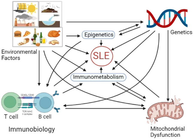
Systemic lupus erythematosus (SLE), a rare multifactorial disease, has shown an upward trend in prevalence. A potentially severe autoimmune systemic disease characterized by the production of autoantibodies to components of the cell nucleus results in a diverse group of clinical manifestations. While the exact pathology of SLE is still unknown, patients depict an inflammatory milieu, deposition of immune complexes (ICs) in various organs, and vasculopathy. Clinical heterogeneity of SLE suggested that various susceptible factors including genetic, epigenetic, environmental, infections, and hormones modulate the disease pathology [1]. Multiple genes, the interaction of sex hormones along with defective immune regulatory mechanisms [2], including impaired clearance of apoptotic cell debris and immune complex deposition, are the important contributors to the development of SLE [3]. Various epigenetic modifications such as methylation, acetylation, and small RNA have also been found to modulate the disease pathology; however, these modifications can vary individually, and thus personalized approach is required to elaborate the role of these mechanisms among lupus patients [4]. Altered immunometabolism observed in various immune cells had a positive correlation with cellular differentiation and lupus severity. Aberrant cell signaling mechanisms have been reported to trigger abnormalities in cell differentiation and over-activation of immune cells thereby enhancing the autoantibody generation [5]. Environmental triggers such as chemical/physical factors, dietary factors, and infectious agents probably contribute to the initiation of SLE disease. The interplay of these factors (as shown in Fig. 1) is associated with disease heterogeneity and complexity; therefore, the understanding of these pathological factors can assist in understanding this dreadful disease. In this review, the focus is on the recent advances in information relevant to factors that can contribute to SLE susceptibility.
Fig. 1.

The complex interactions between the various susceptible factors implicated in lupus etiology. Implications of genetics known to directly regulate the disease manifestations or indirectly through epigenetic modifications induced by environmental factors. These indirect modifications can also regulate the immunobiology and mitochondrial health which combines to affect the immunometabolism, an important aspect in lupus pathology. Diet is equally important as it can regulate the disease severity and hence assist in disease management
Search strategy
An electronic search was conducted among various databases such as google scholar/PubMed and articles published from January 2019 to September 2022 were retrieved and shortlisted based on the search words; “Susceptible or pathological factor” and “SLE or lupus etiology”. A further search was conducted based on the keywords such as genetics, epigenetics, immunobiology, and environmental factors in lupus pathology. Studies that included general clinical manifestations in lupus were excluded. Out of 128 articles analyzed, 70.4% were research articles and clinical reports, while 29.5% were review articles and 47% were published in 2021–2022.
SLE immunobiology
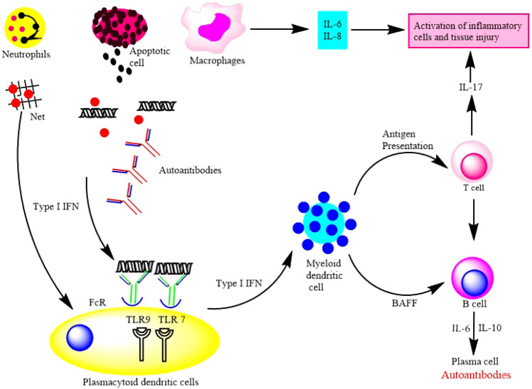
The pathophysiology of SLE is complex involving the interplay between factors and cells of innate/adaptive immunity in the microenvironment and triggers an immune response (Fig. 2). A very important innate trigger is neutrophil, which is deregulated in SLE due to high reactive oxygen species (ROS) levels and get triggered by enhanced IL-18 receptors [6]. Neutrophil extracellular traps (NETs) are enhanced and are known to be associated with enhanced IgG2 production from B cells in lupus [7]. Neutrophil degranulation is known to increase the secretion of pro-inflammatory cytokines (IFN-ϒ) that can fan the flames of lupus etiology [8]. Innate immune receptors such as the Toll-like receptors (TLRs) have the unique capacity to recognize pathogen-associated molecular patterns (PAMPs) while inducing immune system activation by linking the innate and adaptive immunity responses. The TLRs 1, 2, 4, 5, and 6 for bacterial or fungal PAMPs are located on the cell surface and TLRs 3, 7, 8, and 9 for nucleic acid (single-stranded/double-stranded RNA or DNA) are located on the endosomal membrane [9]. TLRs 7 and 9 have been strongly implicated in SLE pathogenesis since they mediate IFNα production by plasmacytoid dendritic cells (pDCs) upon their activation by circulating immune complexes containing self-nucleic acid components [10]. TLR-7 can regulate the extra-follicular B cells response in the germinal center which may enhance autoantibody generation, while TLR-9 can limit the TLR-7 stimulatory activity, demonstrating its protective function in lupus etiology [11]. Platelets have functional TLR-7, which is responsible for their activation, leading to platelet–leukocyte aggregates possibly involved in priming the immune system upon viral attack [12]. Other TLRs such as TLR-2 and 4 were also found to be highly expressed in the saliva of SLE patients, however, their low expression was demonstrated with the presence of chronic periodontitis in lupus patients [13].
Fig. 2.
Immunobiology of systemic lupus erythematosus. Interaction between dysregulated innate and adaptive immune system leads to production of inflammatory cytokines and autoantibody. Over-activation of innate system further interacts with the adaptive immune system leads to over-activation of various immune cells. Self-antigen presentation by dendritic cells leads to the activation of T cells that further activates the autoreactive B cells and secretes the autoantibodies. FcR Immunoglobulin Fc receptors, TLR Toll-like receptor, BAFF B cell activating factors, IL Interleukin
The dendritic cells play an important role in linking the innate and adaptive immune systems and serve as both a “break” and “engine” for lupus pathology [14]. It has been shown that monocytes from healthy individuals may be differentiated into myeloid dendritic cells (mDCs) in the presence of serum from a lupus patient [15]. These mDCs can also have the ability to phagocytize nuclear material and present antigens to naive CD4+ T cells, which can later activate the cytotoxic activity of CD8+ T cells and thus support the proliferation and differentiation of B cells [16]. Autoreactive IgE, such as anti-dsDNA IgE, was found to be elevated in SLE patients and can also elicit an increase in IFNα production by binding the Fc epsilon RI (FcεRI) of plasmacytoid dendritic cells (pDCs) [17]. The binding of IgE to pDCs also enhances the follicular T cell expansion and reduces the Treg population, which increased the inflammatory condition in lupus patients [18]. IFN-α can also activate the IL-1 receptor-associated kinase that further induces apoptosis in Treg cells from SLE patients [19]. Tregs have potent anti-inflammatory activity, therefore their apoptosis will further enhance the immune response in lupus patients. In a recent study, P-Selectins was also demonstrated as a suppressor of Treg cell’s function in SLE pathogenesis [20]. The suppressive function of PD-1+ Treg cells was found to be impaired, which leads to the over-activation of T and B cells in lupus patients [21]. Over-activation of T cells has led to its exhaustion, which can be further correlated with tolerance mechanisms such as prolonged remission in lupus patients [22]. The type-I interferon has been demonstrated to affect the metabolic fitness of CD8+ T cells, which may increase their death and lupus severity [23]. The microenvironment in systemic lupus patients favors the generation of double-negative T cells (CD4−, CD8−, TCRαβ+), which facilitates the secretion of IL-17 and increases disease severity [24]. IL-17 can further expand the proliferation of Th17 and autoreactive T and B cells which can worsen the lupus symptoms. The autoreactive T cells such as nuclear antigen-specific CD4+ T cells are elevated in lupus patients and were further correlated with kidney manifestations such as lupus nephritis [25]. Increased expression of hypoxia-inducing factor-1α (HIF-1α) was found to be associated with an expansion of Th17 cells (Fig. 3), depicting that metabolic alteration can also regulate the lupus etiology [26]. CD4+ follicular T cells were also found to be associated with B cell maturation and generation of autoantibodies in lupus patients [27]. Absent in melanoma 2 (AIM2) expression is elevated in B cells from lupus patients and was further correlated with Blimp expression and autoantibody generation [28]. The failure of an immune regulatory system such as Treg cells, and the over-activation of plasma cells are well-known mechanisms in lupus etiology. Recent advances also elaborated on the mechanisms associated with immunoregulation failure. Thus, the understanding of these mechanisms will provide the base for the immune regulations that can act as targets for various therapeutics (Table 1).
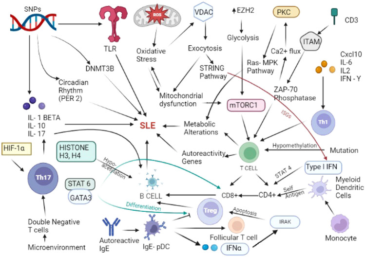
Fig. 3.
Recently elucidated mechanisms and their interactions involved in systemic lupus erythematosus (SLE) modification. Regulation of innate response and enhanced pro-inflammatory cytokines secretion through polymorphism have been recently identified in SLE. Role of mitochondrial dysfunction and associated oxidative stress in metabolic alterations and their impact on immune cell activity and differentiation have been majorly focused in recent studies on SLE. Novel signaling mechanisms have also been elaborated related to metabolic fitness of T cell and their importance in SLE immunobiology. Implications of inflammatory cytokines rich microenvironment on immune cell differentiation specifically, on T cells has been recently focused in SLE related studies. SNP Single nucleotide polymorphism, TLR toll- like receptor, HIF-1α Hypoxia inducing factor 1α, PER2 Period circadian protein homolog 2, DNMT3B DNA-methyltransferase 3 beta, VDAC voltage- dependent anion channel, EZH2 Enhancer of zeste homolog 2, PKC protein kinase C, STAT6 signal transducer and activator of transcription 6, GATA3 GATA binding protein 3, pDC plasmacytoid dendritic cells, ITAM Immunoreceptor tyrosine-based activation motif, ZAP-70 Zeta chain associated protein kinase 70 mTORC1 mammalian target of rapamycin complex 1, IRAK interleukin-1 receptor-associated kinases, IFNα interferon α, Ras-MPK Ras-mitogen-activated protein kinase, Cxcl10 C-X-C motif chemokine ligand 10, ISGs interferon stimulated genes, CD3 cluster of differentiation 3
Table 1.
Function of immune cells in lupus pathogenesis
| Immune cell | Receptor/therapeutic target | Function in lupus pathogenesis | References |
|---|---|---|---|
| Innate immunity | |||
| Neutrophils | IL-18R | Enhance Netosis | [6] |
| TLR | Secretion of IFN gamma | [8] | |
| Plasmacytoid Dendritic cells | TLR-7, 9 | Mediate IFN alpha production | [10] |
| IgE | Enhances the follicular T cell expansion and reduces the Treg population | [18] | |
| Myeloid Dendritic cells | T cells | Phagocytize nuclear material and present antigens to naive CD4+ T cells | [16] |
| Platelets | TLR-7 | Platelet–leukocyte aggregates possibly involved in priming the immune system | [12] |
| Adaptive immunity | |||
| Treg cells | T and B cells | Over-activation of T and B cells | [21] |
| T cells | T cell receptor | Secretion of IL-17 and proliferation of Th17 | [24] |
| CD4+ follicular T cells | B cell | B cell maturation and autoantibodies generation | [27] |
| B cells | ProBDNF/p75NTR | Autoantibody generation | [39] |
Aberrant signaling in SLE
Cellular hyperactivity and hyperresponsiveness associated with deregulated signaling pathways in T and B lymphocytes of SLE suggest detailed analysis for a better understanding of signaling events to pave the path for better management and prevention of this complex disease. T cell receptor (TCR) is a heterodimer, consisting of the TCRα and TCRβ, which recognize antigenic peptides presented by MHC on antigen-presenting cells. CD3 proteins (δ, ε, γ, and ζ) have also been assembled with TCR. CD3ζ contains three immunoreceptors tyrosine-based activation motif (ITAM) domains, and the phosphorylation of ITAM by Src kinase recruits the spleen tyrosine kinase (Syk) family kinase ζ-associated protein kinase 70 (ZAP-70), resulting in the activation of ZAP-70 (Fig. 3) [9]. This results in altered calcium flux, activation of protein kinase C (PKC), and recruitment of Ras guanine-releasing protein 1 leading to activation of the Ras-mitogen protein kinase pathway. The reduced expression level of CD3ζ protein contributes to the aberrant signaling phenotype of SLE T cells [29]. Dual specific phosphatase, a regulator of mitogen-activated protein kinase (MAPK), was also over-expressed (Fig. 3) in T cells from SLE patients [30]. Protein phosphatase 2A (PP2A) regulatory subunit (PPP2R2A) has been demonstrated to regulate the Th1 and Th17 differentiation, but not of Tregs, and their deficiency may trigger autoimmune-like conditions [31]. STAT6-GATA3 signaling axis (Fig. 3) acts as double edge sword in SLE pathology as it enhances the Treg cell differentiation but also leads to the expansion of CD8+ T cells that can secrete IL-13 and IFN-ϒ [32]. Enhanced type-I interferon has been demonstrated in both lupus models and patients where it increases the STAT4 expression which leads to secretion of follicular CD4+ T cells dependent cytokines and is associated with autoantibody generation [33]. An increased level of the mechanistic target of rapamycin (mTOR) (Fig. 3) has been found to have an important role in memory CD8+ cell regulation and maintenance via glyco-metabolism [34]. B cells from SLE expressed high Ca2+ in response to B cell receptor (BCR) stimulation associated with increased tyrosine phosphorylation. Defective B cell signaling recruits inhibitory phosphatase SH2 domain-containing inositol 5′-phosphatase (SHIP) via the inhibitory FcγRIIb receptor [35]. FOXM1, a transcriptional factor of the cell cycle, was found to be highly increased in plasmablast, naïve, and memory B cells, thus, expanding their number in lupus patients [36]. A decrease in Lyn and Syk and an increase in ZAP-70 expression were found in B cells from active SLE patients [37]. Recently, Leptin has been demonstrated to induce B cell dysfunction via activating the JAK/STAT3/5 and ERK1/2 pathways in patients with systemic lupus erythematosus [38]. In a recent study, the higher expression of brain-derived neurotrophic factor precursor and its high-affinity pan-75 receptor (ProBDNF/p75NTR) has been observed in B cells from lupus patients and was positively correlated with disease severity and autoantibody generation [39]. Therefore, the evaluation of these signaling cascades involved in complex immunobiology provides a molecular mechanism that can be used as a diagnostic biomarker or novel targets for various therapies.
Cytokine network in SLE
Cytokines have an important role in the immunobiology of lupus which connects the innate and adaptive immune systems in many ways. The secretion of cytokines from one cell will further modulate the activation and differentiation of other immune cells resulting in the secretion of subsequent cytokines. Cytokines which are comprised of chemotactic activity, known as chemokines, also play an important role in recruiting various immune cells. Different levels of cytokines may regulate the inflammatory and anti-inflammatory immune response, although, in lupus, cytokines associated with inflammatory response were found to be enhanced which triggered the over-activated immune response. The increase in various pro-inflammatory cytokines such as CXCL10, IL-6, IL-2, and interferon-ϒ production in SLE patients results in reinforcement of Th1 differentiation and naïve T cell proliferation leading to IgG production from B cells [40]. Exacerbated production of CXCL10, IL-6, IL-2, and interferon-ϒ (Fig. 3) was associated with a memory-like phenotype in CD4+ polarized B cells towards pro-inflammatory B cells in SLE patients [41]. High mobility group box 1 protein (HMGB1) and type-I IFN are the key molecules that promote the autoreactivity process in SLE and both can be used as the biomarkers for detection of disease severity [42]. Other important cytokines such as high levels of IL-8, macrophage inflammatory protein (MIP) 1α, and MIP1β were found to be associated with high disease severity in SLE patients [43]. IL-25 mRNA serum level was also found to be elevated in SLE patients and correlated with disease severity [44]. Decreased concentration of IL-35 and IL-35+ Breg cells was observed in SLE patients which describes their protective role in SLE pathogenesis [45]. Higher expression of IL-18 was also observed in SLE patients and can be used as a potential biomarker for disease severity [46]. The serum IL-21 level was found to be positively correlated with lupus nephritis activity and can be used potentially as the biomarker [47]. Another cytokine IL-33, which has been demonstrated as the important regulator of TLR-4 was found to be elevated in lupus patients in comparison to healthy controls [48]. As cytokines have an important role in regulating the innate and adaptive immune response, various targeting strategies have been adopted to improve disease outcomes.
The complex immunobiology of lupus includes the over-immune response triggers due to the overactive immune cells towards self-antigens. Various factors including the pro-inflammatory cytokines, chemokines, apoptosis, over-activated APCs, and failure of phagocytotic activity have been well defined to be involved in lupus etiology. These factors can vary from population to population and besides these factors, other important aspects such as genetics, epigenetics, and environmental factors can also be an important part of lupus pathology and can modulate the immunobiology and disease severity.
Genetic factors
Besides the overstimulation of the immune system towards the self-antigens via innate and adaptive immune networks, various other susceptible factors such as genetics can also regulate immunobiology and etiology of lupus. A strong familial aggregation has been observed in SLE along with higher frequency among first-degree relatives, and a higher chance of developing SLE in siblings of the patient shows the polygenic inheritance of the disease. Polygenic inheritance pattern shows the complexity and heterogeneity nature of the disease, which will require an advance and hassle-free analysis method to elucidate the mechanisms behind disease etiology. Genome-wide association studies (GWAS) have been performed to identify the susceptible loci associated with SLE heterogeneity. A novel gene, ILRUN was recently reported by gene-wide association analysis whose expression was significantly low in SLE patients as compared to healthy control [49]. The concurrence of SLE in identical twins is approximately 25–50% and that in dizygotic twins is around 5% [50]. Population studies reveal the positive haplotype associated with HLA genes in SLE (HLA-DR3; DR9; DR15; DQA1*0101), and with lupus nephritis (LN) (DQA1*0101; DR3; DR15), while the possible protective haplotypes noted are HLA-DR4, DR11, DR14 [51]. These population-based genetics studies can enhance the understanding of personalized approaches to genetic susceptibility in lupus pathology that will further assist in solving the complexity of lupus on an individual basis. The non-HLA genes were also found to be of great importance as the single-nucleotide polymorphism (SNP) in genes such as PTPN2, STAT4, RUNX, and SLC have been positively correlated with SLE susceptibility specifically in Belarusian women [52]. TLRs are overstimulated on innate cells from SLE patients and their activation has been linked with SNP (Fig. 3), where specifically the polymorphism in TLR 5 and 9 were found to be associated with a higher risk of nephritis and SLE in the Egyptian population [53]. Polymorphism in DNMT3B (Fig. 3), an important enzyme for DNA methylation, has been found to have a positive correlation with disease severity and with co-existing periodontitis in the Brazilian population, depicting the interplay between genetics and epigenetic factors and lupus pathology [54]. Polymorphism associated with circadian rhythms can also play an important role in disease, where the SNP in the Period 2 gene (PER2), a circadian clock regulatory gene, was also found to be positively associated with the clinical manifestation of SLE pathogenesis [55]. Furthermore, analysis of SNP in various cytokines including IL-1β, IL-17, and IL-10 (Fig. 3) was found to be related to disease etiology in SLE patients of Indian origin [56, 57].
Monogenic lupus
Beyond the complex genetics’ perturbations, rare mutations in genes can also modulate the complexity of lupus pathology in the Mendelian inheritance pattern termed “monogenic lupus”. Recent examples of monogenic lupus such as the homozygous mutation in the DNASE1L3 gene were found to be associated with urticarial skin lesions, recurrent hemoptysis, and renal involvement in pediatric lupus patients [58]. Deleterious mutations in lipopolysaccharide-responsive beige-like anchor (LRBA) genes lead to its deficiency which was found to be associated with juvenile lupus [59]. Mutation (p.His198Gln) in the C1QTNF4 gene was found to be potentially involved in SLE risk in the Iranian population [60]. Current evidence in the genetic susceptibility of lupus shows that genes can also potentially regulates the dysregulated immunological mechanisms associated with disease manifestation that will be further modified through various epigenetic phenomenon. Therefore, the genetic analysis may lead to the identification of novel susceptible targets which are associated with the complex network of lupus and will be further targeted with potent therapeutics. Besides genetics, epigenetic events have been shown to play an important role in the pathophysiology of SLE.
Epigenetic factors
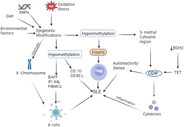
Epigenetics is an important phenomenon that can regulate gene expression in a stable, sometimes heritable fashion. Epigenetics mechanisms such as DNA methylation, post-translational histone modification, and micro RNAs proved to have an important role in SLE (Fig. 4). Alterations such as DNA hypomethylation in both T and B cells were found to be associated with disease pathology [61]. Although major epigenetic investigations have been carried out in T cells, mechanisms such as DNA hypomethylation in T cells were found to be positively correlated with disease activity [62]. Sex-based comparison of methylation pattern in CD4+ T cells shows the dysregulated apoptosis and pro-inflammatory effect in males that were associated with epigenetic over-activation of the Rho family GTPase pathway which enhances the Th17 cell differentiation [63]. Enhancers of zeste homolog 2 (EZH2), a histone methyl transferase and part of polycomb repressive complex 2 (PRC2) which regulates the DNA methyl transferase (DNMT) in several ways were found to be highly expressed in CD4+ T cells from lupus patients [64]. Increased expression of EZH2 can be mediated by higher glycolysis and mechanistic target of rapamycin (mTORC1) activation in lupus T cells leading to metabolic alterations which shows the interplay between immunometabolism and epigenetic modification [65]. Decreased expression of 3-hydroxybutyrate dehydrogenase type 2 (BDH2) has been reported in CD4+ T cells of SLE patients which results in overstimulation of ten-eleven translocation (TET) protein leading to hypomethylation of DNA and activation of autoreactivity related genes [66]. Hypomethylation of the CD-70 promoter region was observed in T cells of juvenile SLE patients and was found to be positively correlated with disease activity [67]. Site-specific hypomethylation of the CD40-ligand gene (CD40L) in CD4+ T cells in SLE patients was also associated with disease activity and severity [68]. Contrary to hypomethylation, the hypermethylated 5-methyl cytosine (m5c) region of the gene has also been observed in CD4+ T cells of SLE patients, which were found to be significantly involved in the modulation of the immune system, cytokines secretion, and various inflammatory mechanisms [69]. Hypermethylation of the FOXP3 gene was also investigated in T cells from children with SLE and demonstrated as one of the causes of increased immune response as FOXP3 acts as an important transcription factor for Treg cell differentiation [70]. Hypermethylation of genes in autoreactive peripheral blood mononuclear cells (PBMCs) from SLE patients can be inhibited via MEK/ERK signaling by co-culture them with mesenchymal stem cells which demonstrated the therapeutic potential of mesenchymal stem in SLE [71]. Methylation in the promoter region of cytotoxic T lymphocytes associated antigen-4 gene (CTLA4) in CD8+ T cells was found to be positively correlated with SLE pathogenesis as CTLA4 has an important role in inhibitory immune response [72]. Oxidative stress in lupus is well determined which can also enhance DNA methylation defects that can result in angiotensin-converting enzyme 2 (ACE2) hypermethylation, which makes lupus patients more prone to COVID-19 infection [73]. Expression of Growth arrest-specific 5 (GAS5), a long non-coding RNA (LncRNAs) that controls cell response and apoptosis, was observed to be low in CD4+ T cells which were further correlated with higher disease activity in lupus patients [74]. N4-acetylcysteine, a novel mRNA modification may have translational efficiency in CD4+ T cells which can be associated with inflammation and critical immune response in SLE patients [75]. Besides the methylation-based mechanisms, micro-RNA such as miR-152-3p was highly expressed in CD4+ T cells and found to be associated with double-stranded DNA and IgG antibodies [76]. miR-183-5p expression levels were found significantly high in patients and were positively correlated with SLEDAI score and anti-dsDNA antibody; thus, this micro-RNA can be a promising biomarker of SLE [77].
Fig. 4.
Epigenetics implications modifies the various mechanisms involved in lupus pathology. Oxidative stress and diet have been recently shown as the important triggers of the epigenetic events demonstrates the importance of environmental factors in disease etiology. Methylation patterns have been found potentially involved in modulation of immunobiology via regulating the T reg and B cell activity. Hypermethylation of the anti-inflammatory sites and hypomethylation of pro-inflammatory genes have been demonstrated in recent studies. Epigenetic events can potentially modulate the lupus outcomes and can be utilized for diagnosis as biomarkers. SNP single-nucleotide polymorphism, BAFF B-cell activating factor, IFI 44L Interferon-inducible 44 like, PBMC Peripheral blood mononuclear cells, FOXP3 forkhead box P3, BDH2 3-hydroxybutyrate dehydrogenase 2, TET ten–eleven translocation, ROS reactive oxygen species
B cells in both pediatric and adult SLE patients have a significant reduction in epigenetic modification on the inactive X chromosome and aberrant X-linked gene expression can underlie the mechanism of female bias of SLE and abnormal autoantibody production [78]. EZH2, a histone methyl transferase, was highly expressed in germinal center (GC) B cells and involved in SLE pathogenesis through enhanced autoantibody generation [79]. MiR-29a, a micro-RNA, can also affect autoantibody secretion in B cells by modulating the Crk-like protein (CKL), thereby contributing to SLE pathogenesis [80]. Inadequate expression of miR-1246 has been found in B cells from lupus patients and was positively correlated with p53 deficiency [81]. DNA methylation patterns may vary during different stages of B cell development. Global DNA methylation in B cells is decreased, but hypermethylation in a few specific genes may be associated with SLE pathogenesis [82]. Hypomethylation of HERS-1 in B cells was found to be associated with SLE disease activity [83]. Hypomethylation of various cytokine genes associated with B cell activation and differentiation, such as interferon-induced protein 44 like (IFI44L) and B cells activation factor (BAFF) in PBMCs was found to be correlated with increased autoantibody secretion from B cells in SLE patients [84]. Global histone modification analysis reveals that H3 and H4 in B cells (Fig. 3) from lupus patients were hypo-acetylated and positively correlated with autoantibody generation and SLEDAI score [85]. Epigenetic alteration in lupus was found to be associated with different cellular mechanisms to guide their nuclear and cytoplasmic factors to regulate the different transcription/translational processes [4]. Epigenetic processes in immune cells regulate their function and bridge the gap between genomics and environmental factors in the etiology of SLE.
Environmental factors
Physical/chemical triggers
Various environmental factors lead to the ensuing of SLE in genetically predisposed individuals towards SLE. These environmental factors include lifestyle factors (cigarette smoking and alcohol drinking) occupational exposure, oral contraceptives, dietary causes, pollution, viral infections especially Epstein–Barr virus, etc. These environmental stimuli may lead to epigenetic changes as they can cause the inhibition of DNA methyl transferases which further leads to hypomethylation of DNA specifically in CD4+ T cells from genetically predisposed individuals. Hypomethylation also leads to oxidative stress, which further activates the signaling pathway mediated by protein kinase C which finally decreases the level of the extracellular signal-regulated kinase (ERK). It is noticeable that the expression of ERK protein was found to be decreased in CD4+ T cells of SLE patients [86]. Exposure to UV radiation is known to worsen pre-existing SLE but its role in the development of SLE is still unclear. However, UV exposure was also known to play an important role in the production of the active form of vitamin D. Vitamin D once converted to 1α,25(OH)2D3 form may prove to be immunosuppressive, hence reducing the risk of SLE [87]. A study on the susceptible subgroup of individuals (relatives of a patient with SLE), suggested an important clue that vitamin D-deficient individuals were more prone to lupus development [88]. Although the cause and consequence relationship between vitamin D and SLE is not specified yet, large-scale studies with controlled experiments are needed to determine the role of UV exposure and SLE incidence.
Occupational exposure to chemical agents is an imperative factor in the development of SLE [89]. Silica exposure and incidence of SLE have been reported in both urban and rural areas. Similarly, silicates such as asbestos have been reported to be associated with the production of anti-nuclear antibodies and proteinuria along with an elevated risk of RA [90]. Lupus-prone NZ-2410 mice exposed to silica have been demonstrated to increase circulating immune complex, autoantibodies, renal deposits of C3, and proteinuria [91].
SLE has been linked to air pollution via a preliminary genome-wide assessment of DNA methylation study which indicates that SLE patients residing near highways have hypomethylated ubiquitin gene (UBE2U gene) encoding enzyme that is involved in ubiquitination and DNA repair [92]. However, more in-depth studies are needed to confirm the above correlation. Epidemiologic studies have established that exposures to petroleum distillates, trichloroethylene, and organochlorines are linked to the intensity of symptoms in lupus patients [93]. Another study of mercury-exposed gold miners showed a higher level of ANA in comparison to miners in diamond and emerald mines with no mercury exposure [94].
Cigarette smoking leads to the inhalation of toxic substances such as tars, nicotine, carbon monoxide, polycyclic aromatic hydrocarbons, and free radicals. Smoking leads to oxidative stress which causes demethylation of DNA and increased expression of inflammatory genes leading to lupus-like manifestations [95]. Various epidemiological studies have confirmed that cigarette smoking is strongly correlated with the risk of SLE incidence [96]. Both cigarette smoking and hypoxia can elevate oxidative stress which has multifactorial effects to induce autoimmunity such as the generation of autoreactive T cells and autoantibodies, inhibition of Treg activity, and enhanced expression of pro-inflammatory mediators [97]. These toxic components bring about damage to biomolecules such as proteins and DNA resulting in genetic aberrations causing gene activation which may be involved in SLE development via an imbalance of antioxidant mechanism and oxidative stress. In a population-based cohort study, it has been demonstrated that nitrogen oxides (NOX) in polluted air and drinking water may lead to significant predilections of SLE, especially for patients with renal involvement [98]. Contrary to the conventional notion, alcohol consumption has been shown to relieve the inflammatory milieu in SLE, diminishing the response to immunogens and decreasing pro-inflammatory cytokines [99].
Infections
SLE patients are generally susceptible to major infections that might be due to profound immunological disturbances [100]. Infections cause changes in normal immune regulation and stimulate immune pathways mediated by molecular mimicry. Adults and children with SLE have higher rates of seropositivity for Epstein–Barr virus (EBV) than other individuals [101]. A possible mechanism involved the complex formation between viral RNA and single-strand binding protein (SSB) which stimulates the TLR3 receptor to induce TNF-α. Furthermore, molecular mimicry between EBV and SLE antigens is another mechanism involved in this process [102]. A study identified a molecular mimicry between SARS-CoV-2 antigen and human molecular chaperons that may induce autoimmunity against the endothelial cells [103]. The mimicry between the symptoms of COVID-19 infections and SLE flares can be of clinical interest to find out the incidence of COVID in SLE patients [104]. Although the incidence of COVID-19 infection in SLE patients was slightly low and the SLE patients with major organ involvement were found to have asymptomatic COVID-19 manifestation that might be due to the high dose of corticosteroids and immunosuppressive treatment regime among SLE patients [105].
Gut microbiota
Recent developments in research have indicated a correlation between gut microbiota and SLE disease activity. In lupus, the ratio of Firmicutes to Bacteroidetes is lower and several other genera are found in abundance [106, 107]. Katz et al. have reported that there is a decrease in the number of Lactobacillaceae and an increase in Lachnospiraceae in patients with SLE [108]. A study in young lupus-prone mice showed clear depletion of lactobacilli and upsurges in Lachnospiraceae in comparison to age-matched healthy controls [109]. However dietary intervention with retinoic acid could restore the number of lactobacilli associated with improvement in symptoms [110]. Furthermore, an increase in Ruminococcus gnavus of the Lachnospiraceae family led to raised serum sCD14 and elevated levels of fecal secretory IgA and calprotectin in female SLE patients [111]. In SLE patients, a leaky gut may elevate the serum endotoxin lipopolysaccharide (LPS) level which is suggestive of chronic microbial translocation that can contribute to the pathogenesis of SLE [112]. Similarly, in lupus-prone NZBxW/F1 mice, complexes of bacterial amyloid and DNA have been shown to stimulate autoimmune responses such as the production of type-I IFN and autoantibodies [113]. Although the definite role of gut symbiotic or pathogenic microbes in SLE is yet to be clarified, the pristane-induced lupus model demonstrated the beneficial role of Lactobacillus probiotics is linked to the reduction of Th1, Th17, and cytotoxic T lymphocytes [114]. The cross-talk between the host and commensal microbiome in addition to infectious bacteria, viruses, and parasites can also impact the presentation of autoimmune diseases. Implications of alteration in the microbiome because of environmental mediators are imperative discoveries and need to be evaluated carefully for their role, especially when establishing a cause-and-effect relationship.
Diet
Intake of excessive carbohydrates has been projected as a risk factor that can worsen the clinical manifestations of autoimmune diseases like rheumatoid arthritis and SLE [115]. Obesity is a well-known mediator of low-grade inflammation mediated by the initiation of several pathways associated with inflammatory cytokine expression such as TNF-α and IL-6 [116, 117]. This favors a continuous inflammatory response, partly contributing to co-morbidities seen in SLE patients [115]. It has also been demonstrated that mice fed with a high-fat diet can have elevated levels of oxidative stress and inflammation leading to autoimmunity [118]. Undoubtedly, SLE patients are at high risk of developing metabolic syndrome, insulin resistance, and type 2 diabetes mellitus leading to a higher risk of cardiovascular co-morbidities and also a major cause of premature death in SLE patients [119]. In SLE, obesity has been linked with higher disease activity and cumulative organ damage [120]. The influence of obesity on gene expression has also been positively correlated with disease severity [121]. Hence, clinical interventions involving meditation and exercise can ease lupus symptoms. Conclusively, SLE patients should maintain a balanced diet avoiding excess calories in addition to following an active lifestyle with daily exercise.
Restriction in protein intake especially in patients with lupus nephropathy has been taken as a beneficial approach to reduce the burden on the kidney. The renal function in SLE patients could be improved by a moderate protein intake of 0.6 g/kg/day [122]. A high salt diet can also accelerate the development of lupus through the regulation of dendritic cell activity via MAPK and STAT1 signaling pathways [123]. Higher dietary sodium and lower dietary potassium intake were significantly correlated with C-reactive protein (CRP), which may lead to an increase in disease severity [124]. A diet rich in eicosapentaenoic acid (EPA) was found to ameliorate the lupus nephritis manifestations including immune complex accumulation and autoantibody generation in the kidney [125].
Vitamins also have an important role to play in the pathogenesis of SLE as vitamins A, B C, D, and E are known to improve clinical manifestations of SLE [126]. Vitamin C has been shown to reduce oxidative stress and inflammation and decrease levels of antibodies against dsDNA, IgG, etc.; and intake of 1 g of vitamin C per day is recommended [122]. A cross-sectional study on 280 patients with SLE demonstrated that the Mediterranean diet can exert a beneficial effect on disease activity and cardiovascular risk [127].
Conclusion
Various complicated and multifactorial aspects have been implicated in SLE pathogenesis. Multiple genes and various epigenetic modifications confer susceptibility to the development of this complex disease. Mitochondrial dysfunction has time and again been demonstrated in SLE, and it is one of the important parameters. The epigenetic modification of various lupus-associated genes has also been implicated in metabolic alterations through direct or indirect mechanisms that can further be modified by environmental factors such as diet. Desirable healthy gut microbiota can be kept in the best form through diet management and can help in regulating homeostasis. These efforts may prevent the adverse effects of various therapies and improve the mental and physical health of SLE patients. Defective immune regulation such as clearance of apoptotic cells and accumulation of antigens, cytokine imbalance, loss of self-tolerance, excess T cells, and immune complex infiltration in various organs are major role players in SLE. Novel approaches towards the signaling defects in T and B cells have been made which reflect the complex nature of the pleiotropic effect in SLE. These signaling abnormalities offer both hope and challenges in the direction of therapeutic interventions. Additionally, analysis of various targets and classification of patients based on microbiota composition may contribute to the development of more personalized strategies in SLE treatment.
Authors contributions
Concept design: AA, RB, and KA. Data collection: AA, AT, and KA. Data analysis and interpretation: AA and RB. Drafting manuscript: AB and AT. Revising manuscript: AA and AB. All authors take full responsibility for the integrity and accuracy of all aspects of the work.
Funding
The authors thank “University Grant Commission, New Delhi” (Grant no. 191620089214) and “Department of Biotechnology (DBT-BUILDER), New Delhi” (Grant no. BT/INF/22/SP41295/2020) for providing fellowship to Mr. Akhil Akhil and Mr. Rohit Bansal, respectively.
Declarations
Conflict of interest
The authors declare no conflict of interest.
Human and animal participants
Not required for this study.
Footnotes
Publisher's Note
Springer Nature remains neutral with regard to jurisdictional claims in published maps and institutional affiliations.
References
- 1.Alaanzy MT, Alsaffar JM, Bari AA. Evaluate the correlation between antioxidant capacity and interferon Γ level with the disease activity of Sle patients in Iraqi Woman. Indian J Public Health Res Dev. 2020;11(1):1278–1282. doi: 10.37506/v11/i1/2020/ijphrd/194018. [DOI] [Google Scholar]
- 2.Terahara N. Flavonoids in foods: a review. Nat Prod Commun. 2015;10(3):19345781501000334. [PubMed] [Google Scholar]
- 3.Mok C, Lau C. Pathogenesis of systemic lupus erythematosus. J Clin Pathol. 2003;56(7):481–490. doi: 10.1136/jcp.56.7.481. [DOI] [PMC free article] [PubMed] [Google Scholar]
- 4.Adams DE, Shao W-H. Epigenetic alterations in immune cells of systemic Lupus erythematosus and therapeutic implications. Cells. 2022;11(3):506. doi: 10.3390/cells11030506. [DOI] [PMC free article] [PubMed] [Google Scholar]
- 5.Katsuyama T, Tsokos GC, Moulton VR. Aberrant T cell signaling and subsets in systemic lupus erythematosus. Front Immunol. 2018;9:1088. doi: 10.3389/fimmu.2018.01088. [DOI] [PMC free article] [PubMed] [Google Scholar]
- 6.Ma J, et al. Elevated interleukin-18 receptor accessory protein mediates enhancement in reactive oxygen species production in neutrophils of systemic lupus erythematosus patients. Cells. 2021;10(5):964. doi: 10.3390/cells10050964. [DOI] [PMC free article] [PubMed] [Google Scholar]
- 7.Bertelli R, et al. Neutrophil extracellular traps in systemic lupus erythematosus stimulate IgG2 production from B lymphocytes. Front Med. 2021;8:635436. doi: 10.3389/fmed.2021.635436. [DOI] [PMC free article] [PubMed] [Google Scholar]
- 8.Liu Y, Kaplan MJ. Neutrophil dysregulation in the pathogenesis of systemic lupus erythematosus. Rheum Dis Clin. 2021;47(3):317–333. doi: 10.1016/j.rdc.2021.04.002. [DOI] [PubMed] [Google Scholar]
- 9.Pan L, et al. Immunological pathogenesis and treatment of systemic lupus erythematosus. World J Pediatr. 2020;16(1):19–30. doi: 10.1007/s12519-019-00229-3. [DOI] [PMC free article] [PubMed] [Google Scholar]
- 10.Nasser N, Kurban M, Abbas O. Plasmacytoid dendritic cells and type I interferons in flares of systemic lupus erythematosus triggered by COVID-19. Rheumatol Int. 2021;41(5):1019–1020. doi: 10.1007/s00296-021-04825-3. [DOI] [PMC free article] [PubMed] [Google Scholar]
- 11.Fillatreau S, Manfroi B, Dörner T. Toll-like receptor signalling in B cells during systemic lupus erythematosus. Nat Rev Rheumatol. 2021;17(2):98–108. doi: 10.1038/s41584-020-00544-4. [DOI] [PMC free article] [PubMed] [Google Scholar]
- 12.Brilland B, Scherlinger M, Khoryati L, Goret J, Duffau P, Lazaro E, Charrier M, Guillotin V, Richez C, Blanco P. Platelets and IgE: shaping the innate immune response in systemic lupus erythematosus. Clin Rev Allerg Immunol. 2019;58:1–19. doi: 10.1007/s12016-019-08744-x. [DOI] [PubMed] [Google Scholar]
- 13.Marques CP, et al. Expression of Toll-like receptors 2 and 4 in the saliva of patients with systemic lupus erythematosus and chronic periodontitis. Clin Rheumatol. 2021;40(7):2727–2734. doi: 10.1007/s10067-020-05560-z. [DOI] [PubMed] [Google Scholar]
- 14.Liu J, Zhang X, Cao X. Dendritic cells in systemic lupus erythematosus: from pathogenesis to therapeutic applications. J Autoimmun. 2022;132:102856. doi: 10.1016/j.jaut.2022.102856. [DOI] [PubMed] [Google Scholar]
- 15.Charrier M, et al. Systemic Lupus Erythematosus. Amsterdam: Elsevier; 2021. The role of dendritic cells in systemic lupus erythematosus; pp. 143–150. [Google Scholar]
- 16.Sim TM, et al. Type I interferons in systemic lupus erythematosus: a journey from bench to bedside. Int J Mol Sci. 2022;23(5):2505. doi: 10.3390/ijms23052505. [DOI] [PMC free article] [PubMed] [Google Scholar]
- 17.Brilland B, et al. Platelets and IgE: shaping the innate immune response in systemic lupus erythematosus. Clin Rev Allergy Immunol. 2020;58(2):194–212. doi: 10.1007/s12016-019-08744-x. [DOI] [PubMed] [Google Scholar]
- 18.Palomares O, et al. Regulatory T cells and immunoglobulin E: a new therapeutic link for autoimmunity? Allergy. 2022;77:3293–3308. doi: 10.1111/all.15449. [DOI] [PubMed] [Google Scholar]
- 19.Li M, et al. Interferon-α activates interleukin-1 receptor-associated kinase 1 to induce regulatory T-cell apoptosis in patients with systemic lupus erythematosus. J Dermatol. 2021;48(8):1172–1185. doi: 10.1111/1346-8138.15899. [DOI] [PubMed] [Google Scholar]
- 20.Scherlinger M, et al. Selectins impair regulatory T cell function and contribute to systemic lupus erythematosus pathogenesis. Sci Transl Med. 2021;13(600):eabi4994. doi: 10.1126/scitranslmed.abi4994. [DOI] [PubMed] [Google Scholar]
- 21.Kurata I, et al. Impaired function of PD-1+ follicular regulatory T cells in systemic lupus erythematosus. Clin Exp Immunol. 2021;206(1):28–35. doi: 10.1111/cei.13643. [DOI] [PMC free article] [PubMed] [Google Scholar]
- 22.Lima G, et al. Exhausted T cells in systemic lupus erythematosus patients in long-standing remission. Clin Exp Immunol. 2021;204(3):285–295. doi: 10.1111/cei.13577. [DOI] [PMC free article] [PubMed] [Google Scholar]
- 23.Buang N, et al. Type I interferons affect the metabolic fitness of CD8+ T cells from patients with systemic lupus erythematosus. Nat Commun. 2021;12(1):1–15. doi: 10.1038/s41467-021-22312-y. [DOI] [PMC free article] [PubMed] [Google Scholar]
- 24.Li H, et al. Systemic lupus erythematosus favors the generation of IL-17 producing double negative T cells. Nat Commun. 2020;11(1):1–12. doi: 10.1038/s41467-020-16636-4. [DOI] [PMC free article] [PubMed] [Google Scholar]
- 25.Abdirama D, et al. Nuclear antigen–reactive CD4+ T cells expand in active systemic lupus erythematosus, produce effector cytokines, and invade the kidneys. Kidney Int. 2021;99(1):238–246. doi: 10.1016/j.kint.2020.05.051. [DOI] [PubMed] [Google Scholar]
- 26.Liao H-J, et al. Increased HIF-1α expression in T cells and associated with enhanced Th17 pathway in systemic lupus erythematosus. J Formos Med Assoc. 2022;121:2446–2456. doi: 10.1016/j.jfma.2022.05.003. [DOI] [PubMed] [Google Scholar]
- 27.Nakayamada S, Tanaka Y. Clinical relevance of T follicular helper cells in systemic lupus erythematosus. Expert Rev Clin Immunol. 2021;17(10):1143–1150. doi: 10.1080/1744666X.2021.1976146. [DOI] [PubMed] [Google Scholar]
- 28.Yang M, et al. AIM2 deficiency in B cells ameliorates systemic lupus erythematosus by regulating Blimp-1–Bcl-6 axis-mediated B-cell differentiation. Signal Transduct Target Ther. 2021;6(1):1–11. doi: 10.1038/s41392-021-00725-x. [DOI] [PMC free article] [PubMed] [Google Scholar]
- 29.Chen P-M, Tsokos GC. T cell abnormalities in the pathogenesis of systemic lupus erythematosus: an update. Curr Rheumatol Rep. 2021;23(2):1–9. doi: 10.1007/s11926-020-00978-5. [DOI] [PMC free article] [PubMed] [Google Scholar]
- 30.Chuang H-C, Tan T-H. MAP4K family kinases and DUSP family phosphatases in T-cell signaling and systemic lupus erythematosus. Cells. 2019;8(11):1433. doi: 10.3390/cells8111433. [DOI] [PMC free article] [PubMed] [Google Scholar]
- 31.Pan W, et al. The regulatory subunit PPP2R2A of PP2A enhances Th1 and Th17 differentiation through activation of the GEF-H1/RhoA/ROCK signaling pathway. J Immunol. 2021;206(8):1719–1728. doi: 10.4049/jimmunol.2001266. [DOI] [PMC free article] [PubMed] [Google Scholar]
- 32.Kato H, Perl A. Double-edged sword: interleukin-2 promotes T regulatory cell differentiation but also expands interleukin-13-and interferon-γ-producing CD8+ T Cells via STAT6-GATA-3 axis in systemic lupus erythematosus. Front Immunol. 2021;12:635531. doi: 10.3389/fimmu.2021.635531. [DOI] [PMC free article] [PubMed] [Google Scholar]
- 33.Dong X, et al. Type I interferon–activated STAT4 regulation of follicular helper T cell–dependent cytokine and immunoglobulin production in Lupus. Arthritis Rheumatol. 2021;73(3):478–489. doi: 10.1002/art.41532. [DOI] [PMC free article] [PubMed] [Google Scholar]
- 34.Cai X, et al. mTOR participates in the formation, maintenance, and function of memory CD8+ T cells regulated by glycometabolism. Biochem Pharmacol. 2022;204:115197. doi: 10.1016/j.bcp.2022.115197. [DOI] [PubMed] [Google Scholar]
- 35.Yap DY, Chan TM. B cell abnormalities in systemic lupus erythematosus and lupus nephritis—role in pathogenesis and effect of immunosuppressive treatments. Int J Mol Sci. 2019;20(24):6231. doi: 10.3390/ijms20246231. [DOI] [PMC free article] [PubMed] [Google Scholar]
- 36.Akita K, et al. Interferon α enhances B cell activation associated with FOXM1 induction: potential novel therapeutic strategy for targeting the plasmablasts of systemic lupus erythematosus. Front Immunol. 2020;11:498703. doi: 10.3389/fimmu.2020.498703. [DOI] [PMC free article] [PubMed] [Google Scholar]
- 37.Vásquez A, et al. Altered recruitment of Lyn, Syk and ZAP-70 into lipid rafts of activated B cells in systemic lupus erythematosus. Cell Signal. 2019;58:9–19. doi: 10.1016/j.cellsig.2019.03.003. [DOI] [PubMed] [Google Scholar]
- 38.Chen H, et al. Leptin accelerates B cell dysfunctions via activating JAK/STAT3/5 and ERK1/2 pathways in patients with systemic lupus erythematosus. Clin Exp Rheumatol. 2022;40(11):2125–2132. doi: 10.55563/clinexprheumatol/84syjo. [DOI] [PubMed] [Google Scholar]
- 39.Shen W-Y, et al. Up-regulation of proBDNF/p75NTR signaling in antibody-secreting cells drives systemic lupus erythematosus. Sci Adv. 2022;8(3):eabj2797. doi: 10.1126/sciadv.abj2797. [DOI] [PMC free article] [PubMed] [Google Scholar]
- 40.Simon Q et al (2021) A cytokine network profile delineates a common Th1/Be1 pro-inflammatory group of patients in four systemic autoimmune diseases. Arthritis Rheumatol 73. 10.1002/art.41697 [DOI] [PubMed]
- 41.Simon Q, et al. A pathogenic cytokine network is associated with pro-inflammatory B cells in systemic lupus erythematosus patients. SSRN J. 2019 doi: 10.2139/ssrn.3420376. [DOI] [Google Scholar]
- 42.Tanaka A, et al. Serum high-mobility group box 1 is correlated with interferon-α and may predict disease activity in patients with systemic lupus erythematosus. Lupus. 2019;28(9):1120–1127. doi: 10.1177/0961203319862865. [DOI] [PubMed] [Google Scholar]
- 43.Park J, et al. Cytokine clusters as potential diagnostic markers of disease activity and renal involvement in systemic lupus erythematosus. J Int Med Res. 2020;48(6):0300060520926882. doi: 10.1177/0300060520926882. [DOI] [PMC free article] [PubMed] [Google Scholar]
- 44.Li Y, et al. Interleukin-25 is upregulated in patients with systemic lupus erythematosus and ameliorates murine lupus by inhibiting inflammatory cytokine production. Int Immunopharmacol. 2019;74:105680. doi: 10.1016/j.intimp.2019.105680. [DOI] [PubMed] [Google Scholar]
- 45.Ye Z, et al. The plasma interleukin (IL)-35 level and frequency of circulating IL-35+ regulatory B cells are decreased in a cohort of Chinese patients with new-onset systemic lupus erythematosus. Sci Rep. 2019;9(1):1–12. doi: 10.1038/s41598-019-49748-z. [DOI] [PMC free article] [PubMed] [Google Scholar]
- 46.Elshal A, et al. Serum interleukin-18 as a novel biomarker for disease activity of systemic lupus erythematosus patients. J Egypt Soc Parasitol. 2019;49(1):145–152. doi: 10.21608/jesp.2019.68297. [DOI] [Google Scholar]
- 47.Shater H, et al. The potential use of serum interleukin-21 as biomarker for lupus nephritis activity compared to cytokines of the tumor necrosis factor (TNF) family. Lupus. 2022;31(1):55–64. doi: 10.1177/09612033211063794. [DOI] [PubMed] [Google Scholar]
- 48.Li Y, et al. Potential role of interleukin-33 in systemic lupus erythematosus by regulating toll like receptor 4. Eur J Inflamm. 2022;20:1721727X221094455. doi: 10.1177/1721727X221094455. [DOI] [Google Scholar]
- 49.Ding X, et al. Gene-based association analysis identified a novel gene associated with systemic lupus erythematosus. Ann Hum Genet. 2021;85(6):213–220. doi: 10.1111/ahg.12439. [DOI] [PubMed] [Google Scholar]
- 50.Putra Gofur NR, Putri Gofur AR, Soesilaningtyas RPGR, Kahdina M. Systemic lupus erythematsous: a review article. Int J Surg Case Rep Images. 2021;2(1):1. [Google Scholar]
- 51.Lever E, Alves MR, Isenberg DA. Towards precision medicine in systemic lupus erythematosus. Pharmacogen Personal Med. 2020;13:39. doi: 10.2147/PGPM.S205079. [DOI] [PMC free article] [PubMed] [Google Scholar]
- 52.Dostanko N, et al. ab0010 association of some non-hla gene polymorphisms with susceptibility to systemic lupus erythematosus in women in belarusian population. London: BMJ Publishing Group Ltd.; 2022. pp. 1140–1141. [Google Scholar]
- 53.Gharbia OM, et al. Toll-like receptor 5 and Toll-like receptor 9 single nucleotide polymorphisms and risk of systemic lupus erythematosus and nephritis in Egyptian patients. Egypt Rheumatol Rehabilit. 2021;48(1):1–10. [Google Scholar]
- 54.Dias LNDS, et al. DNMT3B (rs2424913) polymorphism is associated with systemic lupus erythematosus alone and with co-existing periodontitis in a Brazilian population. J Appl Oral Sci. 2022 doi: 10.1590/1678-7757-2021-0567. [DOI] [PMC free article] [PubMed] [Google Scholar]
- 55.Dan Y-L, et al. Association of PER2 gene single nucleotide polymorphisms with genetic susceptibility to systemic lupus erythematosus. Lupus. 2021;30(5):734–740. doi: 10.1177/0961203321989794. [DOI] [PubMed] [Google Scholar]
- 56.Umare V, et al. Cytokine genes multi-locus analysis reveals synergistic influence on genetic susceptibility in Indian SLE–A multifactor-dimensionality reduction approach. Cytokine. 2020;135:155240. doi: 10.1016/j.cyto.2020.155240. [DOI] [PubMed] [Google Scholar]
- 57.Padhi S, et al. Interleukin 17A rs2275913 polymorphism is associated with susceptibility to systemic lupus erythematosus: a meta and trial sequential analysis. Lupus. 2022;31(6):674–683. doi: 10.1177/09612033221090172. [DOI] [PubMed] [Google Scholar]
- 58.Ekinci RMK, et al. Monogenic lupus due to DNASE1L3 deficiency in a pediatric patient with urticarial rash, hypocomplementemia, pulmonary hemorrhage, and immune-complex glomerulonephritis. Eur J Med Genet. 2021;64(9):104262. doi: 10.1016/j.ejmg.2021.104262. [DOI] [PubMed] [Google Scholar]
- 59.Liphaus BL, et al. LRBA deficiency: a new genetic cause of monogenic lupus. Ann Rheum Dis. 2020;79(3):427–428. doi: 10.1136/annrheumdis-2019-216410. [DOI] [PubMed] [Google Scholar]
- 60.Pakzad B, et al. C1QTNF4 gene p His198Gln mutation is correlated with early-onset systemic lupus erythematosus in Iranian patients. Int J Rheum Dis. 2020;23(11):1594–1598. doi: 10.1111/1756-185X.13981. [DOI] [PubMed] [Google Scholar]
- 61.Hedrich CM. Systemic lupus erythematosus. Amsterdam: Elsevier; 2021. Epigenetics; pp. 277–292. [Google Scholar]
- 62.Zhang Y, et al. Impaired DNA methylation and its mechanisms in CD4+ T cells of systemic lupus erythematosus. J Autoimmun. 2013;41:92–99. doi: 10.1016/j.jaut.2013.01.005. [DOI] [PubMed] [Google Scholar]
- 63.He Z, et al. Comprehensive analysis of epigenetic modifications and immune-cell infiltration in tissues from patients with systemic lupus erythematosus. Epigenomics. 2022;14(2):81–100. doi: 10.2217/epi-2021-0318. [DOI] [PubMed] [Google Scholar]
- 64.Tsou PS, et al. EZH2 modulates the DNA methylome and controls T cell adhesion through junctional adhesion molecule A in lupus patients. Arthritis Rheumatol. 2018;70(1):98–108. doi: 10.1002/art.40338. [DOI] [PMC free article] [PubMed] [Google Scholar]
- 65.Zheng X, Tsou P-S, Sawalha AH. Increased expression of EZH2 is mediated by higher glycolysis and mTORC1 activation in lupus CD4+ T cells. Immunometabolism. 2020 doi: 10.20900/immunometab20200013. [DOI] [PMC free article] [PubMed] [Google Scholar]
- 66.Zhao M, et al. Downregulation of BDH2 modulates iron homeostasis and promotes DNA demethylation in CD4+ T cells of systemic lupus erythematosus. Clin Immunol. 2018;187:113–121. doi: 10.1016/j.clim.2017.11.002. [DOI] [PubMed] [Google Scholar]
- 67.Keshavarz-Fathi M, et al. DNA methylation of CD70 promoter in juvenile systemic lupus erythematosus. Fetal Pediatr Pathol. 2022;41(1):58–67. doi: 10.1080/15513815.2020.1764681. [DOI] [PubMed] [Google Scholar]
- 68.Vordenbäumen S, et al. Associations of site-specific CD4+-T-cell hypomethylation within CD40-ligand promotor and enhancer regions with disease activity of women with systemic lupus erythematosus. Lupus. 2021;30(1):45–51. doi: 10.1177/0961203320965690. [DOI] [PubMed] [Google Scholar]
- 69.Guo G, et al. Disease activity-associated alteration of mRNA m5 C methylation in CD4+ T cells of systemic lupus erythematosus. Front Cell Dev Biol. 2020;8:430. doi: 10.3389/fcell.2020.00430. [DOI] [PMC free article] [PubMed] [Google Scholar]
- 70.Hanaei S, et al. The status of FOXP3 gene methylation in pediatric systemic lupus erythematosus. Allergol Immunopathol. 2020;48(4):332–338. doi: 10.1016/j.aller.2020.03.014. [DOI] [PubMed] [Google Scholar]
- 71.Xiong H, et al. Mesenchymal stem cells activate the MEK/ERK signaling pathway and enhance DNA methylation via DNMT1 in PBMC from systemic lupus erythematosus. BioMed Res Int. 2020;2020:1. doi: 10.1155/2020/4174082. [DOI] [PMC free article] [PubMed] [Google Scholar]
- 72.NosratZehi S, et al. Promoter methylation and expression status of cytotoxic T-lymphocyte-associated antigen-4 gene in patients with lupus. J Epigenetics. 2020;2(1):33–39. [Google Scholar]
- 73.Sawalha AH, et al. Epigenetic dysregulation of ACE2 and interferon-regulated genes might suggest increased COVID-19 susceptibility and severity in lupus patients. Clin Immunol. 2020;215:108410. doi: 10.1016/j.clim.2020.108410. [DOI] [PMC free article] [PubMed] [Google Scholar]
- 74.Liu Q, et al. LncRNA GAS5 suppresses CD4+ T cell activation by upregulating E4BP4 via inhibiting miR-92a-3p in systemic lupus erythematosus. Immunol Lett. 2020;2021:41–47. doi: 10.1016/j.imlet.2020.08.001. [DOI] [PubMed] [Google Scholar]
- 75.Guo G, et al. Epitranscriptomic N4-acetylcytidine profiling in CD4+ T cells of systemic lupus erythematosus. Front Cell Dev Biol. 2020;8:842. doi: 10.3389/fcell.2020.00842. [DOI] [PMC free article] [PubMed] [Google Scholar]
- 76.Tao B, et al. Regulation of Toll-like receptor-mediated inflammatory response by microRNA-152-3p-mediated demethylation of MyD88 in systemic lupus erythematosus. Inflamm Res. 2021;70(3):285–296. doi: 10.1007/s00011-020-01433-y. [DOI] [PubMed] [Google Scholar]
- 77.Zhou S, Zhang J, Luan P, Ma Z, Dang J, Zhu H, Huo Z. miR-183–5p is a potential molecular marker of systemic lupus erythematosus. J Immunol Res. 2021;2021:1–11. doi: 10.1155/2021/5547635. [DOI] [PMC free article] [PubMed] [Google Scholar]
- 78.Pyfrom S, et al. The dynamic epigenetic regulation of the inactive X chromosome in healthy human B cells is dysregulated in lupus patients. Proc Natl Acad Sci. 2021 doi: 10.1073/pnas.2024624118. [DOI] [PMC free article] [PubMed] [Google Scholar]
- 79.Zhen Y, et al. Ezh2-mediated epigenetic modification is required for allogeneic T cell-induced lupus disease. Arthritis Res Ther. 2020;22:1–10. doi: 10.1186/s13075-020-02225-9. [DOI] [PMC free article] [PubMed] [Google Scholar]
- 80.Shi X, et al. Downregulated miR-29a promotes B cell overactivation by upregulating Crk-like protein in systemic lupus erythematosus. Mol Med Rep. 2020;22(2):841–849. doi: 10.3892/mmr.2020.11166. [DOI] [PMC free article] [PubMed] [Google Scholar]
- 81.Zhang Q, et al. Deficiency of p53 causes the inadequate expression of miR-1246 in B cells of systemic lupus erythematosus. J Immunol. 2022;209(8):1492–1498. doi: 10.4049/jimmunol.2200307. [DOI] [PMC free article] [PubMed] [Google Scholar]
- 82.Scharer CD, et al. Epigenetic programming underpins B cell dysfunction in human SLE. Nat Immunol. 2019;20(8):1071–1082. doi: 10.1038/s41590-019-0419-9. [DOI] [PMC free article] [PubMed] [Google Scholar]
- 83.Fali T, et al. DNA methylation modulates HRES1/p28 expression in B cells from patients with Lupus. Autoimmunity. 2014;47(4):265–271. doi: 10.3109/08916934.2013.826207. [DOI] [PMC free article] [PubMed] [Google Scholar]
- 84.Fan H, et al. Gender differences of B cell signature related to estrogen-induced IFI44L/BAFF in systemic lupus erythematosus. Immunol Lett. 2017;181:71–78. doi: 10.1016/j.imlet.2016.12.002. [DOI] [PubMed] [Google Scholar]
- 85.Gautam P, Sharma A, Bhatnagar A. Global histone modification analysis reveals hypoacetylated H3 and H4 histones in B Cells from systemic lupus erythematosus patients. Immunol Lett. 2021;240:41–45. doi: 10.1016/j.imlet.2021.09.007. [DOI] [PubMed] [Google Scholar]
- 86.Luo M, et al. The regulatory effect of ERK1 pathway in the DNA hypomethylation of MRL/lpr Mice. Int J Immunol. 2021;9(3):41. doi: 10.11648/j.iji.20210903.11. [DOI] [Google Scholar]
- 87.Ao T, Kikuta J, Ishii M. The effects of vitamin D on immune system and inflammatory diseases. Biomolecules. 2021;11(11):1624. doi: 10.3390/biom11111624. [DOI] [PMC free article] [PubMed] [Google Scholar]
- 88.Hayashi K, et al. Real-world data on vitamin D supplementation and its impacts in systemic lupus erythematosus: cross-sectional analysis of a lupus registry of nationwide institutions (LUNA) PLoS One. 2022;17(6):e0270569. doi: 10.1371/journal.pone.0270569. [DOI] [PMC free article] [PubMed] [Google Scholar]
- 89.Ehrlich R. Silica—a multisystem hazard. Int J Epidemiol. 2021;50(4):1226–1228. doi: 10.1093/ije/dyab020. [DOI] [PubMed] [Google Scholar]
- 90.Boudigaard SH, et al. Occupational exposure to respirable crystalline silica and risk of autoimmune rheumatic diseases: a nationwide cohort study. Int J Epidemiol. 2021;50(4):1213–1226. doi: 10.1093/ije/dyaa287. [DOI] [PMC free article] [PubMed] [Google Scholar]
- 91.Brown J, et al. Silica accelerated systemic autoimmune disease in lupus-prone New Zealand mixed mice. Clin Exp Immunol. 2003;131(3):415–421. doi: 10.1046/j.1365-2249.2003.02094.x. [DOI] [PMC free article] [PubMed] [Google Scholar]
- 92.Gilcrease GW, et al. Is air pollution affecting the disease activity in patients with systemic lupus erythematosus? State of the art and a systematic literature review. Eur J Rheumatol. 2020;7(1):31. doi: 10.5152/eurjrheum.2019.19141. [DOI] [PMC free article] [PubMed] [Google Scholar]
- 93.Kilburn KH, Warshaw RH. Prevalence of symptoms of systemic lupus erythematosus (SLE) and of fluorescent antinuclear antibodies associated with chronic exposure to trichloroethylene and other chemicals in well water. Environ Res. 1992;57(1):1–9. doi: 10.1016/S0013-9351(05)80014-3. [DOI] [PubMed] [Google Scholar]
- 94.Cooper GS, et al. Occupational and environmental exposures and risk of systemic lupus erythematosus: silica, sunlight, solvents. Rheumatology. 2010;49(11):2172–2180. doi: 10.1093/rheumatology/keq214. [DOI] [PMC free article] [PubMed] [Google Scholar]
- 95.Perricone C, et al. Smoke and autoimmunity: the fire behind the disease. Autoimmun Rev. 2016;15(4):354–374. doi: 10.1016/j.autrev.2016.01.001. [DOI] [PubMed] [Google Scholar]
- 96.Speyer CB, Costenbader KH. Cigarette smoking and the pathogenesis of systemic lupus erythematosus. Expert Rev Clin Immunol. 2018;14(6):481–487. doi: 10.1080/1744666X.2018.1473035. [DOI] [PMC free article] [PubMed] [Google Scholar]
- 97.Hussain MS, Tripathi V. Smoking under hypoxic conditions: a potent environmental risk factor for inflammatory and autoimmune diseases. Mil Med Res. 2018;5(1):1–14. doi: 10.1186/s40779-018-0158-5. [DOI] [PMC free article] [PubMed] [Google Scholar]
- 98.Chen J, et al. The relationship of polluted air and drinking water sources with the prevalence of systemic lupus erythematosus: a provincial population-based study. Sci Rep. 2021;11(1):1–11. doi: 10.1038/s41598-021-98111-8. [DOI] [PMC free article] [PubMed] [Google Scholar]
- 99.Wang J, et al. Association between alcohol intake and the risk of systemic lupus erythematosus: a systematic review and meta-analysis. Lupus. 2021;30(5):725–733. doi: 10.1177/0961203321991918. [DOI] [PubMed] [Google Scholar]
- 100.Wang H, et al. Major infections in newly diagnosed systemic lupus erythematosus: an inception cohort study. Lupus Sci Med. 2022;9(1):e000725. doi: 10.1136/lupus-2022-000725. [DOI] [PMC free article] [PubMed] [Google Scholar]
- 101.James JA, et al. Systemic lupus erythematosus in adults is associated with previous Epstein-Barr virus exposure. Arthritis Rheum. 2001;44(5):1122–1126. doi: 10.1002/1529-0131(200105)44:5<1122::AID-ANR193>3.0.CO;2-D. [DOI] [PubMed] [Google Scholar]
- 102.Poole BD, et al. Epstein-Barr virus and molecular mimicry in systemic lupus erythematosus. Autoimmunity. 2006;39(1):63–70. doi: 10.1080/08916930500484849. [DOI] [PubMed] [Google Scholar]
- 103.Anand P, et al. SARS-CoV-2 strategically mimics proteolytic activation of human ENaC. Elife. 2020;9:e58603. doi: 10.7554/eLife.58603. [DOI] [PMC free article] [PubMed] [Google Scholar]
- 104.Fu X-L, et al. COVID-19 in patients with systemic lupus erythematosus: a systematic review. Lupus. 2022;31(6):684–696. doi: 10.1177/09612033221093502. [DOI] [PMC free article] [PubMed] [Google Scholar]
- 105.Schioppo T, et al. Clinical and peculiar immunological manifestations of SARS-CoV-2 infection in systemic lupus erythematosus patients. Rheumatology. 2022;61(5):1928–1935. doi: 10.1093/rheumatology/keab611. [DOI] [PMC free article] [PubMed] [Google Scholar]
- 106.de Oliveira GL. Probiotics-current knowledge and future prospects. Houston: InTech; 2018. Probiotic applications in autoimmune diseases; pp. 69–89. [Google Scholar]
- 107.Luo XM, et al. Gut microbiota in human systemic lupus erythematosus and a mouse model of lupus. Appl Environ Microbiol. 2018;84(4):e02288–e2317. doi: 10.1128/AEM.02288-17. [DOI] [PMC free article] [PubMed] [Google Scholar]
- 108.Neuman H, Koren O. The gut microbiota: a possible factor influencing systemic lupus erythematosus. Curr Opin Rheumatol. 2017;29(4):374–377. doi: 10.1097/BOR.0000000000000395. [DOI] [PubMed] [Google Scholar]
- 109.Zhang H, et al. Dynamics of gut microbiota in autoimmune lupus. Appl Environ Microbiol. 2014;80(24):7551–7560. doi: 10.1128/AEM.02676-14. [DOI] [PMC free article] [PubMed] [Google Scholar]
- 110.Abdelhamid L, Luo XM. Retinoic acid, leaky gut, and autoimmune diseases. Nutrients. 2018;10(8):1016. doi: 10.3390/nu10081016. [DOI] [PMC free article] [PubMed] [Google Scholar]
- 111.Azzouz D, et al. Lupus nephritis is linked to disease-activity associated expansions and immunity to a gut commensal. Ann Rheum Dis. 2019;78(7):947–956. doi: 10.1136/annrheumdis-2018-214856. [DOI] [PMC free article] [PubMed] [Google Scholar]
- 112.Ogunrinde E, et al. A link between plasma microbial translocation, microbiome, and autoantibody development in first-degree relatives of systemic lupus erythematosus patients. Arthritis Rheumatol. 2019;71(11):1858–1868. doi: 10.1002/art.40935. [DOI] [PMC free article] [PubMed] [Google Scholar]
- 113.Tursi SA, et al. Bacterial amyloid curli acts as a carrier for DNA to elicit an autoimmune response via TLR2 and TLR9. PLoS Pathog. 2017;13(4):e1006315. doi: 10.1371/journal.ppat.1006315. [DOI] [PMC free article] [PubMed] [Google Scholar]
- 114.Mardani F, et al. In vivo study: Th1–Th17 reduction in pristane-induced systemic lupus erythematosus mice after treatment with tolerogenic Lactobacillus probiotics. J Cell Physiol. 2019;234(1):642–649. doi: 10.1002/jcp.26819. [DOI] [PubMed] [Google Scholar]
- 115.dos Santos FDMM, et al. Excess weight and associated risk factors in patients with systemic lupus erythematosus. Rheumatol Int. 2013;33(3):681–688. doi: 10.1007/s00296-012-2402-8. [DOI] [PubMed] [Google Scholar]
- 116.Kono M, et al. The impact of obesity and a high-fat diet on clinical and immunological features in systemic lupus erythematosus. Nutrients. 2021;13(2):504. doi: 10.3390/nu13020504. [DOI] [PMC free article] [PubMed] [Google Scholar]
- 117.Kono M, et al. The impact of obesity and a high-fat diet on clinical and immunological features in systemic lupus erythematosus. Nutrients. 2021;13:504. doi: 10.3390/nu13020504. [DOI] [PMC free article] [PubMed] [Google Scholar]
- 118.Yang Y, et al. Spirulina lipids alleviate oxidative stress and inflammation in mice fed a high-fat and high-sucrose diet. Mar Drugs. 2020;18(3):148. doi: 10.3390/md18030148. [DOI] [PMC free article] [PubMed] [Google Scholar]
- 119.Liu Y, Kaplan MJ. Cardiovascular disease in systemic lupus erythematosus: an update. Curr Opin Rheumatol. 2018;30(5):441–448. doi: 10.1097/BOR.0000000000000528. [DOI] [PubMed] [Google Scholar]
- 120.Kang J-H, et al. Obesity increases the incidence of new-onset lupus nephritis and organ damage during follow-up in patients with systemic lupus erythematosus. Lupus. 2020;29(6):578–586. doi: 10.1177/0961203320913616. [DOI] [PubMed] [Google Scholar]
- 121.La Cava A. The influence of diet and obesity on gene expression in SLE. Genes. 2019;10(5):405. doi: 10.3390/genes10050405. [DOI] [PMC free article] [PubMed] [Google Scholar]
- 122.Constantin MM, et al. Significance and impact of dietary factors on systemic lupus erythematosus pathogenesis. Exp Ther Med. 2019;17(2):1085–1090. doi: 10.3892/etm.2018.6986. [DOI] [PMC free article] [PubMed] [Google Scholar]
- 123.Xiao ZX, et al. High salt diet accelerates the progression of murine lupus through dendritic cells via the p38 MAPK and STAT1 signaling pathways. Signal Transduct Target Ther. 2020;5(1):1–13. doi: 10.1038/s41392-020-0139-5. [DOI] [PMC free article] [PubMed] [Google Scholar]
- 124.Correa-Rodríguez M, et al. Dietary sodium, potassium, and sodium to potassium ratio in patients with systemic lupus erythematosus. Biol Res Nurs. 2022;24:10998004211065491. doi: 10.1177/10998004211065491. [DOI] [PubMed] [Google Scholar]
- 125.Kobayashi A, et al. Dietary supplementation with eicosapentaenoic acid inhibits plasma cell differentiation and attenuates lupus autoimmunity. Front Immunol. 2021 doi: 10.3389/fimmu.2021.650856. [DOI] [PMC free article] [PubMed] [Google Scholar]
- 126.Islam MA, et al. Immunomodulatory effects of diet and nutrients in systemic lupus erythematosus (SLE): a systematic review. Front Immunol. 2020;11:1477. doi: 10.3389/fimmu.2020.01477. [DOI] [PMC free article] [PubMed] [Google Scholar]
- 127.Pocovi-Gerardino G, et al. Beneficial effect of Mediterranean diet on disease activity and cardiovascular risk in systemic lupus erythematosus patients: a cross-sectional study. Rheumatology. 2021;60(1):160–169. doi: 10.1093/rheumatology/keaa210. [DOI] [PubMed] [Google Scholar]


