Abstract
Objectives
This study aims to investigate the inflammatory effect of the microRNA (miRNA) miR-30e-5p on rheumatoid arthritis (RA) development in RA mice and fibroblast-like synoviocytes (FLS).
Materials and methods
MiR-30e-5p and atlastin GTPase 2 (Atl2) expression in RA tissues and RA-FLS was evaluated using real-time quantitative polymerase chain reaction. The function of miR-30e-5p in inflammation of RA mice and RA-FLS was analyzed by enzyme-linked immunosorbent assay (ELISA) and Western blotting. 5-ethynyl-2ˊ-deoxyuridine (EdU) assay was used to detect RA-FLS proliferation. Luciferase reporter assay was to confirm the interaction between miR-30e-5p and Atl2.
Results
MiR-30e-5p expression was upregulated in the tissues from RA mice. Silencing miR-30e-5p alleviated inflammation in RA mice and RA-FLS. MiR-30e-5p negatively modulated Atl2 expression. Atl2 knockdown exerted a proinflammatory effect on RA-FLS. Atl2 knockdown rescued the inhibitory effect of miR-30e-5p knockdown on proliferation and inflammatory response of RA-FLS.
Conclusion
MiR-30e-5p knockdown inhibited the inflammatory response in RA mice and RA-FLS through Atl2.
Keywords: Atl2, fibroblast-like synoviocytes, inflammatory response, MiR-30e-5p, rheumatoid arthritis.
Introduction
As a commonly diagnosed systemic autoimmune disease, rheumatoid arthritis (RA) causes chronic inflammation of the synovial joint, cartilage damage, as well as bone erosion,[1] with an incidence of about 0.3 to 1.1% of the population worldwide.[2] Patients with RA often experience stiffness, fatigue, work disorders, or even disability.[3] Notably, the inflammatory response has been confirmed to be a main cause of RA, and the expression of inflammatory factors such as tumor necrosis factor (TNF), interleukin (IL)-1, IL-17, and IL-6 are upregulated in RA, and effective small molecule kinase inhibitors targeting inflammatory cytokines have greatly improved the clinical efficacy of RA.[4] Fibroblast-like synoviocytes (FLS) are in the intima of the synovium membrane and participate in the proinflammatory cytokine network and play a key role in RA development.[5-8] However, RA is still currently incurable, thus, to explore the role of possible molecule in inflammatory response of RA-FLS or RA mice is important for further RA study.
Micro ribonucleic acids (miRNAs) can regulate gene expression by binding in the 3’-UTR of target messenger RNAs (mRNAs).[9] Many researchers have investigated the role of several miRNAs in RA-FLS or in animal models. To illustrate, miR20a regulates FLS proliferation and apoptosis in RA.[10] The miR-21 relieves RA in rats by downregulating the Wnt signaling pathway.[11] The miR-124a exerts an inhibitory effect on RA-FLS proliferation and inflammation via PIK3/NF-κB signaling pathway.[12] According to the results of GSE115885, miR-30e-5p has been confirmed as one of the upregulated miRNAs in the blood samples from early RA patients, compared to the blood samples from healthy controls. Moreover, miR-30e-5p is also involved in the prediction of coronary events in RA.[13] However, the potential role of miR-30e-5p in inflammatory response of RA-FLS and RA mice is still undocumented.
As a member of the Atlastin GTPase (Atl2) group, Atl2 has been reported as one of the direct targets of miR-30b-5p in the mammary epithelial cells.[14] The Atl2 expression was also regulated by miR-214-5p in gastric cancer cells.[15] However, the mechanism of miR-30e-5p/Atl2 in RA have not been discussed, yet.
In the present study, we aimed to detect the impact of miR-30b-5p targeting Atl2 on inflammatory response of in RA mice and RA-FLS, which identified a promising new research strategy for RA development.
Patients and Methods
Establishment of a mouse model with R
Forty-eight male DBA/1 mice (23-25 g, 9 to 10-week-old) were obtained from Shanghai SLAC Laboratory Animal Co. Ltd. and housed in quiet and clean cages with normal circadian rhythm of water and food intake for one week with the temperature of 25±2°C. Then, all mice were divided into four groups as follows: sham (n=12); RA (n=12); RA+ Adeno-associated virus (AAV)-NC (n=12); and RA+AAV-miR-30e-5p inhibitor (n=12). Thirty-six mice were used to induce RA according to the previous study.[16] Specifically, mice were treated with bovine-II collagen (200 mg) via intradermal injection. Twenty-one days later, mice were treated with bovine-II collagen agonist via intradermal injection again. The sham operation group was given the same amount of phosphate-buffered saline (PBS) as control. Adeno-associated virus vectors carrying miR-30e-5p inhibitor, or a negative control were generated by the GeneChem Technology Company (Shanghai, China). The AAV-miR-30e-5p inhibitor or AAV-NC were injected intradermally in PBS (50 μL) per RA mouse. After three weeks, when the RA mice showed adequate transfection, mouse foot swelling was assessed with a vernier caliper. At the end, all mice were euthanized with pentobarbital sodium (100 mg/kg), synovial tissues were collected, and the hind paws were prepared for the following experiments.
RA-FLS culture and transfection
The isolation and culture of FLS were performed as previously described.[17] Synovial tissues from the RA mice model were digested and then cultured in prescribed Dulbecco's modified eagle medium (DMEM; Sigma-Aldrich, St. Louis, Missouri, USA) for seven days at 5% carbon dioxide (CO2) and 37°C. Cells were, then, emerged and subcultured in complete DMEM containing 10% FCS. The FLSs at passages 3 to 4 were used for the present study. For subsequent experiments, cells (1X10⁴) were treated with lentiviruses encoding miR-30e-5p inhibitor, sh-Atl2 and their controls (Sangon Biotechnology Co., Ltd., Shanghai, China) using Lipofectamine™ 2000 (Invitrogen, Carlsbad, CA, USA) for 48 h.
Reverse transcription quantitative polymerase chain reaction (RT-qPCR)
The total RNA was extracted from synovial tissues and FLSs using a TRIzol kit (Invitrogen, Carlsbad, CA, USA) and was reverse transcribed to complementary deoxyribonucleic acid DNA (cDNA). Then, the relative expression levels were evaluated using an ABI real-time qPCR System (Thermo Fisher Scientific, Massachusetts, USA). The GAPDH and U6 were used as internal parameters. The data were analyzed by 2-ΔΔCt method. Sequences of primers are provided in Table 1.
Table 1. Primers used for quantitative RT-PCR.
| Name | Sequence (5'-3') forward | Sequence (5'-3') reverse |
| miR-30e-5p | TGTAAACATCCTTGACTGGAAGG | CCAGTGCGAATACCTCGGAC |
| Atl2 | ATGGAACAGGTATGTGGAGG | CACTTCCTTGAGATCCAAGTG |
| GAPDH | TCAAGATCATCAGCAATGCC | CGATACCAAAGTTGTCATGGA |
| U6 | GGTCGGGCAGGAAAGAGGGC | GCTAATCTTCTCTGTATCGTTCC |
| RT-PCR: Reverse transcription-polymerase chain reaction. | ||
Enzyme-linked immunosorbent assay (ELISA)
The synovial tissues of the mice and supernatant of RA-FLS were collected, and the corresponding ELISA kits (Abcam Co., Ltd., NY, USA) were used to detect the protein levels of TNF-alpha (TNF-α), IL-1β, and IL-6.
Western blotting analysis
Total protein from synovial tissues and RA-FLS was extracted and separated by SDS-PAGE (10%). Subsequently, the equal amount of protein was transferred onto polyvinylidene fluoride (PVDF) membrane, which was then blocked for 2 h using 5% skim milk. The membranes were, then, incubated with primary antibody against IL-1β (ab254360; 1:1000; Abcam Co., Ltd., NY, USA), IL-6 (ab259341; 1:1000; Abcam Co., Ltd., NY, USA), TNF-α (ab183218; 1:1000; Abcam Co., Ltd., NY, USA), Atl2 (ab224825; 1:1000; Abcam Co., Ltd., NY, USA) and GAPDH (ab8245; 1:500; Abcam Co., Ltd., NY, USA) at 4°C for 24 h. After washing, the membranes were incubated with horseradish peroxidase (HPR)-conjugated secondary at room temperature for 2 h. The ImageJ software (NIH, Bethesda, MD, USA) was used to scan blots and evaluate the optical density (OD).
5-ethynyl-2ˊ-deoxyuridine (EdU) labeling assay
The RA-FLSs (5X10³ cells/well) were seeded into each well of 96-well plates and exposed to EdU solution (100 μL) for 4 h at 37°C. Subsequently, cells were fixed in formaldehyde (4%) for half an hour at room temperature and permeabilized in Triton X-100 (0.5%) for 10 min. Next, the cells were washed with PBS and added with Apollo staining reaction solution in the dark. Nuclei were labeled using 4ˊ,6-diamidino2-phenylindole 2hci (DAPI) for 30 min, and the images were captured using a fluorescence microscope (Olympus, Corporation, Tokyo, Japan).
Luciferase activity assay
The TargetScan (https://www.targetscan.org/ vert_71/) forecasted the binding site between miR-30e-5p and Atl2 3ˊUTR. Wild sequences and mutant sequences of the binding site were designed and then cloned into luciferase reporter vector pmirGLO (Promega, Madison, WI, USA) to obtain Atl2 3ˊUTR wild type plasmid (Atl2-WT) and Atl2 3ˊUTR mutant type plasmid (Atl2-MUT). Cells were, then, seeded in 96-well plates and co-transfected with the luciferase reporter plasmid and LV-miR-30e-5p inhibitor or LV-NC using Lipofectamine™ 2000 (Invitrogen, Carlsbad, CA, USA) for 48 h, and then the luciferase activity was tested with luciferase detection kit (BioVision, San Francisco, CA, USA).
Statistical analysis
Statistical analysis was performed using the IBM SPSS version 21.0 software (IBM Corp., Armonk, NY, USA). Descriptive data were expressed in mean ± standard deviation (SD). Comparisons between two groups and among multiple groups were assessed by t-test and one-way analysis of variance (ANOVA), followed by Tukey’s post-hoc analysis. A p value of <0.05 was considered statistically significant.
Results
Silencing of miR-30e-5p alleviates inflammation in RA mice
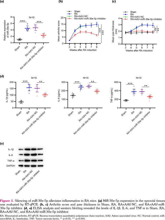
MiR-30e-5p expression was higher in the synovial tissues of RA mice than that in control group, as shown by RT-qPCR. While miR30e-5p knockdown significantly downregulated miR-30e-5p expression, compared to that in RA+AAV-NC group (Figure 1a). In addition, the RA scores and swelling in mice was also reduced by miR-30e-5p knockdown (Figure 1b, c). The ELISA and Western blotting indicated that the levels of IL-1β, IL-6, and TNF-α were significantly elevated in the synovial tissues of the RA mice compared to that in control group. However, miR-30e-5p knockdown markedly decreased the levels of IL-1β, IL-6, and TNF-α in RA mice (Figure 1d, e). Thus, miR-30e-5p knockdown mitigated the inflammatory response in RA mice.
Figure 1. Silencing of miR-30e-5p alleviates inflammation in RA mice. (a) MiR-30e-5p expression in the synovial tissues was evaluated by RT-qPCR. (b, c) Arthritis score and paw thickness in Sham, RA, RA+AAV-NC, and RA+AAV-miR30e-5p inhibitor. (d, e) ELISA analysis and western blotting revealed the levels of IL-1b, IL-6, and TNF-a in Sham, RA, RA+AAV-NC, and RA+AAV-miR-30e-5p inhibitor. RA: Rheumatoid arthritis; RT-qPCR: Reverse transcription quantitative polymerase chain reaction; AAV: Adeno-associated virus: NC: Normal control; miR: microRNA; IL: Interleukin; TNF: Tumor necrosis factor; ** p<0.01; *** p<0.001.
MiR-30e-5p knockdown exerts an anti-inflammatory effect on RA-FLS
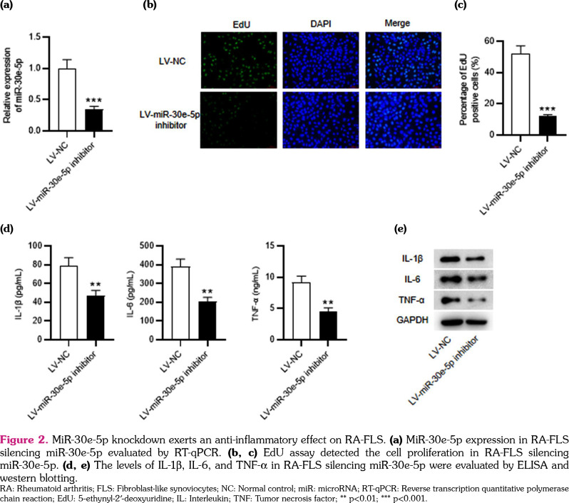
We, then, extracted RA-FLS from the synovial tissues to conduct the subsequent experiments. The miR-30e-5p expression was downregulated in RA-FLS silencing miR-30e-5p (Figure 2a). The number of EdU-positive cells was obviously reduced by miR-30e-5p knockdown relative to the control group (Figure 2b, c). According to the results of ELISA, the levels of IL-1β, IL-6, and TNF-α in the cells were decreased after miR-30e-5p knockdown, and the similar alterations were observed in Western blotting (Figure 2d, e). Collectively, miR-30e-5p knockdown inhibited the inflammatory response in RA-FLS.
Figure 2. MiR-30e-5p knockdown exerts an anti-inflammatory effect on RA-FLS. (a) MiR-30e-5p expression in RA-FLS silencing miR-30e-5p evaluated by RT-qPCR. (b, c) EdU assay detected the cell proliferation in RA-FLS silencing miR-30e-5p. (d, e) The levels of IL-1b, IL-6, and TNF-a in RA-FLS silencing miR-30e-5p were evaluated by ELISA and western blotting. RA: Rheumatoid arthritis; FLS: Fibroblast-like synoviocytes; NC: Normal control; miR: microRNA; RT-qPCR: Reverse transcription quantitative polymerase chain reaction; EdU: 5-ethynyl-2ˊ-deoxyuridine; IL: Interleukin; TNF: Tumor necrosis factor; ** p<0.01; *** p<0.001.
MiR-30e-5p negatively modulates Atl2 expression
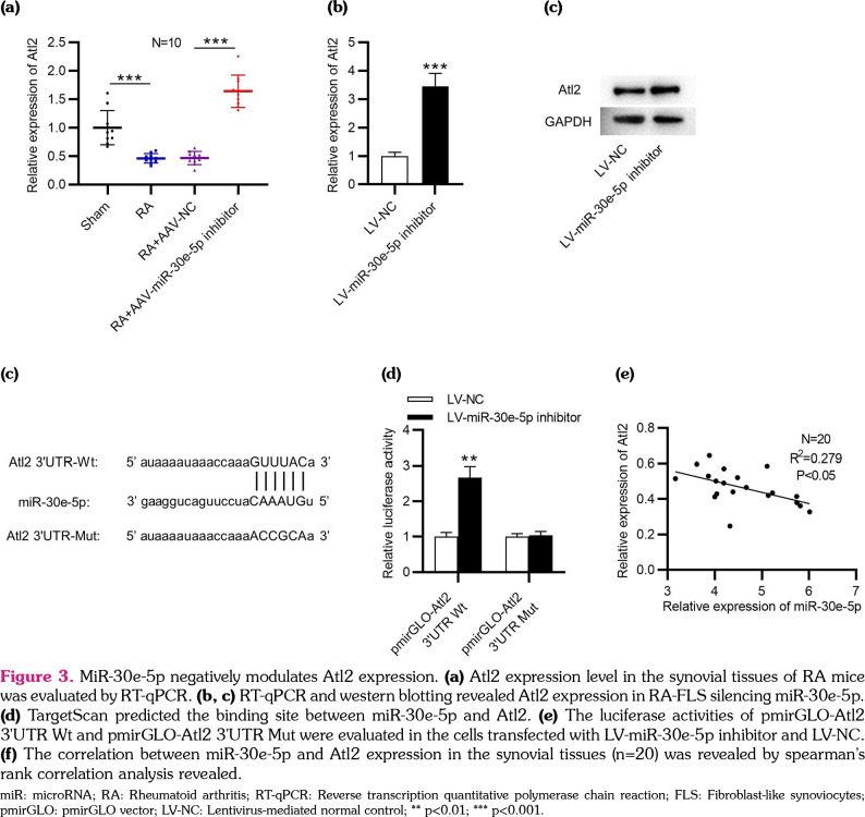
Atl2 was predicted as a potential target of miR-30e-5p by the starBase with the screening condition of CLIP data ≥5 and Degradome data ≥2. A lower Atl2 expression was observed in RA mice, which was, then, upregulated by miR-30e-5p knockdown (Figure 3a). The RT-qPCR and Western blotting revealed that silencing of miR-30e-5p increased Atl2 expression in RA-FLS (Figure 3b, c). The TargetScan predicted the binding site between miR-30e-5p and Atl2 (Figure 3d). The luciferase reporter activity of pmirGLO-Atl2 3'UTR Wt was significantly increased by LV-miR-30e-5p inhibitor, compared to that in the LV-NC group. While The luciferase reporter activity of pmirGLO-Atl2 3'UTR Mut had no significant change in the cells transfected with LV-miR-30e-5p inhibitor and LV-NC (Figure 3e). Furthermore, Atl2 expression was negatively correlated with miR-30e-5p expression in the synovial tissues (n=20), according to the results of Spearman rank correlation analysis (Figure 3f). Thus, Atl2 was a target of miR-30e-5p.
Figure 3. MiR-30e-5p negatively modulates Atl2 expression. (a) Atl2 expression level in the synovial tissues of RA mice was evaluated by RT-qPCR. (b, c) RT-qPCR and western blotting revealed Atl2 expression in RA-FLS silencing miR-30e-5p. (d) TargetScan predicted the binding site between miR-30e-5p and Atl2. (e) The luciferase activities of pmirGLO-Atl2 3'UTR Wt and pmirGLO-Atl2 3'UTR Mut were evaluated in the cells transfected with LV-miR-30e-5p inhibitor and LV-NC. (f) The correlation between miR-30e-5p and Atl2 expression in the synovial tissues (n=20) was revealed by spearman’s rank correlation analysis revealed. miR: microRNA; RA: Rheumatoid arthritis; RT-qPCR: Reverse transcription quantitative polymerase chain reaction; FLS: Fibroblast-like synoviocytes; pmirGLO: pmirGLO vector; LV-NC: Lentivirus-mediated normal control; ** p<0.01; *** p<0.001.
Atl2 knockdown exerts a proinflammatory effect on RA-FLS
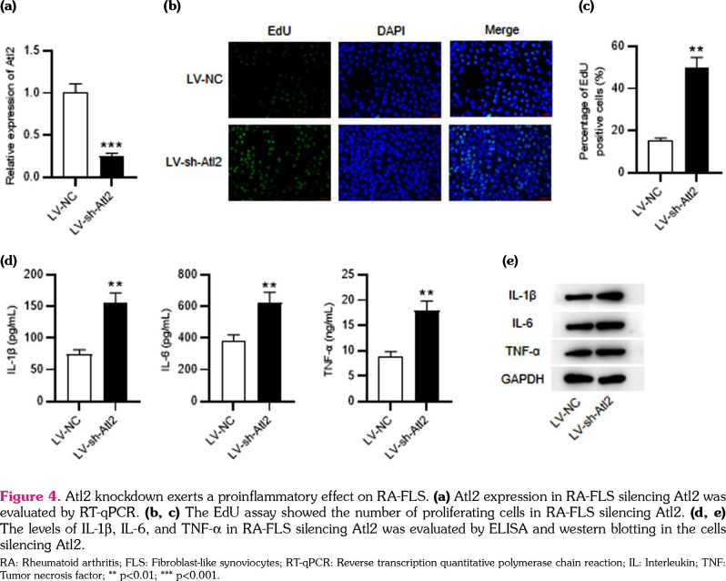
Subsequently, we transfected RA-FLS with LV-sh-Atl2 and LV-NC to explore the effect of Atl2 knockdown on inflammation. The Atl2 expression was significantly decreased in RA-FLS silencing Atl2 (Figure 4a). The EdU assay showed the number of proliferating cells was obviously increased by Atl2 knockdown (Figure 4b, c). The Atl2 knockdown aggravated the inflammation with increased levels of IL-1β, IL-6, and TNF-α expression, as shown in ELISA and Western blotting (Figure 4d, e). It was inferred that Atl2 knockdown exerted a proinflammatory effect on RA-FLS.
Figure 4. Atl2 knockdown exerts a proinflammatory effect on RA-FLS. (a) Atl2 expression in RA-FLS silencing Atl2 was evaluated by RT-qPCR. (b, c) The EdU assay showed the number of proliferating cells in RA-FLS silencing Atl2. (d, e) The levels of IL-1b, IL-6, and TNF-a in RA-FLS silencing Atl2 was evaluated by ELISA and western blotting in the cells silencing Atl2. RA: Rheumatoid arthritis; FLS: Fibroblast-like synoviocytes; RT-qPCR: Reverse transcription quantitative polymerase chain reaction; IL: Interleukin; TNF: Tumor necrosis factor; ** p<0.01; *** p<0.001.
Atl2 knockdown rescues the effect of miR-30e-5p knockdown on inflammatory response of RA-FLS
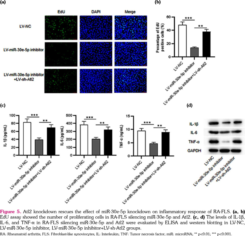
We, then, transfected sh-Atl2 into miR30e-5p silencing RA-FLS. As shown in EdU assay, the proliferation of RA-FLS was inhibited by miR-30e-5p knockdown. However, Atl2 knockdown partially recovered the results (Figure 5a, b). The levels of IL-1β, IL-6, and TNF-α was downregulated in RA-FLS silencing miR-30e-5p, compared to those in the LV-NC group. While Atl2 knockdown significantly upregulated the levels of IL-1β, IL-6, and TNF-α relative to those in LV-miR-30e-5p group (Figure 5c, d). Collectively, Atl2 knockdown reversed the inhibitory effect of miR-30e-5p knockdown on proliferation and inflammatory response of RA-FLS.
Figure 5. Atl2 knockdown rescues the effect of miR-30e-5p knockdown on inflammatory response of RA-FLS. (a, b) EdU assay showed the number of proliferating cells in RA-FLS silencing miR-30e-5p and Atl2. (c, d) The levels of IL-1b, IL-6, and TNF-a in RA-FLS silencing miR-30e-5p and Atl2 were evaluated by ELISA and western blotting in LV-NC, LV-miR-30e-5p inhibitor, LV-miR-30e-5p inhibitor+LV-sh-Atl2 groups. RA: Rheumatoid arthritis; FLS: Fibroblast-like synoviocytes; IL: Interleukin; TNF: Tumor necrosis factor; miR: microRNA; ** p<0.01; *** p<0.001.
Discussion
The inflammation, proliferation, and invasion of the synovial layer are the main features of RA.[18] It has been confirmed that etiology of RA is related to the induction of inflammatory cytokines and chemokines.[19] Of these inflammatory factors, IL-1β, IL-6, and TNF-α have been emphasized, as high levels of IL-1β, IL-6, and TNF-α are detected in the synovial fluid and the plasma for RA patients.[20,21] In addition, as a fundamental regulator of joint impairment, RA-FLS can invade articular cartilage.[22] Importantly, increasing evidence have confirmed the significant role of gene therapy in RA treatment.[23] Thus, in our study, we identified a novel biomarker of RA, and explored its role and mechanism in inflammation of RA mice and RA-FLS.
Many miRNAs including miR-146a,[24] miR-22[25] and miR-130a[26] have been found to participate in the regulation of RA development.[27] Notably, GSE115885 data suggested that miR-30e-5p expression was upregulated in the blood samples from early RA patients, compared to the blood samples from healthy controls. In the current study, miR-30e-5p expression was higher in the synovial tissues of RA mice. More importantly, miR-30e-5p knockdown significantly reduced the arthritis score and paw thickness, as well as the levels of IL-1β, IL-6, and TNF-α both in RA mice and RA-FLS.
In general, miRNAs mediated the biological processes through regulating the expression of its downstream targeting mRNA.[28-30] We bioinformatically predicted and confirmed that Atl2 was a target gene of miR-30e-5p. However, the role and mechanism of Atl2 were merely reported in any diseases. We, for the first time, revealed that miR-30e-5p expression was negatively correlated with Atl2 expression. After transfecting RA-FLS with LV-sh-Atl2, we surprisingly found that Atl2 knockdown increased the number of proliferating cells and the levels of IL-1β, IL-6, and TNF-α. In rescue assay, Atl2 knockdown also reversed the inhibitory effect of miR-30e-5p knockdown and promoted proliferation and inflammatory response of RA-FLS. However, we still wonder whether certain signaling pathway are activated to contribute to this mechanism, which would be further verified in the future.
In conclusion, we found that miR-30e-5p knockdown repressed the inflammatory response in RA mice and RA-FLS by regulating Atl2 expression, providing a new research strategy for further studying the development of RA.
Footnotes
Conflict of Interest: The authors declared no conflicts of interest with respect to the authorship and/or publication of this article.
Author Contributions: Idea/concept: S.L.; Design, critical review: S.L.; K.W.; Control/supervision, data collection and/or processing: J.L.; Analysis and/or interpretation: Y.L.; Literature review, writing the article: Z.Z., D.M.; References and fundings, materials: J.L., Y.L.
Financial Disclosure: The authors received no financial support for the research and/or authorship of this article.
References
- 1.Zerbini CAF, Clark P, Mendez-Sanchez L, Pereira RMR, Messina OD, Uña CR, et al. Biologic therapies and bone loss in rheumatoid arthritis. Osteoporos Int. 2017;28:429–446. doi: 10.1007/s00198-016-3769-2. [DOI] [PubMed] [Google Scholar]
- 2.Deane KD, Holers VM. Rheumatoid arthritis pathogenesis, prediction, and prevention: An emerging paradigm shift. Arthritis Rheumatol. 2021;73:181–193. doi: 10.1002/art.41417. [DOI] [PMC free article] [PubMed] [Google Scholar]
- 3.Davis JM 3rd. Rheumatoid arthritis: A severe disease that preventive approaches would greatly benefit. Clin Ther. 2019;41:1240–1245. doi: 10.1016/j.clinthera.2019.04.026. [DOI] [PubMed] [Google Scholar]
- 4.Rivellese F, Lobasso A, Barbieri L, Liccardo B, de Paulis A, Rossi FW. Novel therapeutic approaches in rheumatoid arthritis: Role of janus kinases inhibitors. Curr Med Chem. 2019;26:2823–2843. doi: 10.2174/0929867325666180209145243. [DOI] [PubMed] [Google Scholar]
- 5.Bottini N, Firestein GS. Duality of fibroblast-like synoviocytes in RA: Passive responders and imprinted aggressors. Nat Rev Rheumatol. 2013;9:24–33. doi: 10.1038/nrrheum.2012.190. [DOI] [PMC free article] [PubMed] [Google Scholar]
- 6.You S, Koh JH, Leng L, Kim WU, Bucala R. The tumor-like phenotype of rheumatoid synovium: Molecular profiling and prospects for precision medicine. Arthritis Rheumatol. 2018;70:637–652. doi: 10.1002/art.40406. [DOI] [PMC free article] [PubMed] [Google Scholar]
- 7.Korb-Pap A, Bertrand J, Sherwood J, Pap T. Stable activation of fibroblasts in rheumatic arthritiscauses and consequences. ii64-ii67Rheumatology (Oxford) 2016;55(suppl 2) doi: 10.1093/rheumatology/kew347. [DOI] [PubMed] [Google Scholar]
- 8.Falconer J, Murphy AN, Young SP, Clark AR, Tiziani S, Guma M, et al. Review: Synovial cell metabolism and chronic inflammation in rheumatoid arthritis. Arthritis Rheumatol. 2018;70:984–999. doi: 10.1002/art.40504. [DOI] [PMC free article] [PubMed] [Google Scholar]
- 9.Davidson-Moncada J, Papavasiliou FN, Tam W. MicroRNAs of the immune system: Roles in inflammation and cancer. Ann N Y Acad Sci. 2010;1183:183–194. doi: 10.1111/j.1749-6632.2009.05121.x. [DOI] [PMC free article] [PubMed] [Google Scholar]
- 10.Wei XJ, Li XW, Lu JL, Long ZX, Liang JQ, Wei SB, et al. MiR-20a regulates fibroblast-like synoviocyte proliferation and apoptosis in rheumatoid arthritis. Eur Rev Med Pharmacol Sci. 2020;24:7578–7578. doi: 10.26355/eurrev_202007_22253. [DOI] [PubMed] [Google Scholar]
- 11.Liu XG, Zhang Y, Ju WF, Li CY, Mu YC. MiR-21 relieves rheumatoid arthritis in rats via targeting Wnt signaling pathway. Eur Rev Med Pharmacol Sci. 2019;23(3 Suppl):96–103. doi: 10.26355/eurrev_201908_18635. [DOI] [PubMed] [Google Scholar]
- 12.Yang B, Ge Y, Zhou Y, Wang J, Xie X, Li S, et al. miR-124a inhibits the proliferation and inflammation in rheumatoid arthritis fibroblast-like synoviocytes via targeting PIK3/NF-kB pathway. Cell Biochem Funct. 2019;37:208–215. doi: 10.1002/cbf.3386. [DOI] [PubMed] [Google Scholar]
- 13.Ormseth MJ, Solus JF, Sheng Q, Chen SC, Ye F, Wu Q, et al. Plasma miRNAs improve the prediction of coronary atherosclerosis in patients with rheumatoid arthritis. Clin Rheumatol. 2021;40:2211–2219. doi: 10.1007/s10067-020-05573-8. [DOI] [PMC free article] [PubMed] [Google Scholar]
- 14.Le Guillou S, Laubier J, Péchoux C, Aujean E, Castille J, Leroux C, et al. Defects of the endoplasmic reticulum and changes to lipid droplet size in mammary epithelial cells due to miR-30b-5p overexpression are correlated to a reduction in Atlastin 2 expression. Biochem Biophys Res Commun. 2019;512:283–288. doi: 10.1016/j.bbrc.2019.03.022. [DOI] [PubMed] [Google Scholar]
- 15.Zhong S, Wang J, Hou J, Zhang Q, Xu H, Hu J, et al. Circular RNA hsa_circ_0000993 inhibits metastasis of gastric cancer cells. Epigenomics. 2018;10:1301–1313. doi: 10.2217/epi-2017-0173. [DOI] [PubMed] [Google Scholar]
- 16.Zhao F, Dong J, Guo J, Bi L. Inhibiting role of long non-coding RNA LINC01197 in inflammation in rheumatoid arthritis through the microRNA-150/ THBS2 axis. Exp Cell Res. 2020;394:112136–112136. doi: 10.1016/j.yexcr.2020.112136. [DOI] [PubMed] [Google Scholar]
- 17.Hückel M, Schurigt U, Wagner AH, Stöckigt R, Petrow PK, Thoss K, et al. Attenuation of murine antigen-induced arthritis by treatment with a decoy oligodeoxynucleotide inhibiting signal transducer and activator of transcription-1 (STAT-1) R17Arthritis Res Ther. 2006;8 doi: 10.1186/ar1869. [DOI] [PMC free article] [PubMed] [Google Scholar]
- 18.Firestein GS. Evolving concepts of rheumatoid arthritis. Nature. 2003;423:356–361. doi: 10.1038/nature01661. [DOI] [PubMed] [Google Scholar]
- 19.Shrivastava AK, Pandey A. Inflammation and rheumatoid arthritis. J Physiol Biochem. 2013;69:335–347. doi: 10.1007/s13105-012-0216-5. [DOI] [PubMed] [Google Scholar]
- 20.Ahsan H, Irfan HM, Alamgeer , Jahan S, Shahzad M, Asim MH. Potential of ephedrine to suppress the gene expression of TNF-a, IL-1b, IL-6 and PGE2: A novel approach towards management of rheumatoid arthritis. Life Sci. 2021;282:119825–119825. doi: 10.1016/j.lfs.2021.119825. [DOI] [PubMed] [Google Scholar]
- 21.Kaczyński T, Wroński J, Głuszko P, Kryczka T, Miskiewicz A, Górski B, et al. Salivary interleukin 6, interleukin 8, interleukin 17A, and tumour necrosis factor a levels in patients with periodontitis and rheumatoid arthritis. Cent Eur J Immunol. 2019;44:269–276. doi: 10.5114/ceji.2019.89601. [DOI] [PMC free article] [PubMed] [Google Scholar]
- 22.Asif Amin M, Fox DA, Ruth JH. Synovial cellular and molecular markers in rheumatoid arthritis. Semin Immunopathol. 2017;39:385–393. doi: 10.1007/s00281-017-0631-3. [DOI] [PMC free article] [PubMed] [Google Scholar]
- 23.Deviatkin AA, Vakulenko YA, Akhmadishina LV, Tarasov VV, Beloukhova MI, Zamyatnin AA Jr, et al. Emerging concepts and challenges in rheumatoid arthritis gene therapy. Biomedicines. 2020;8:9–9. doi: 10.3390/biomedicines8010009. [DOI] [PMC free article] [PubMed] [Google Scholar]
- 24.Bae SC, Lee YH. MiR-146a levels in rheumatoid arthritis and their correlation with disease activity: A meta-analysis. Int J Rheum Dis. 2018;21:1335–1342. doi: 10.1111/1756-185X.13338. [DOI] [PubMed] [Google Scholar]
- 25.Yang QY, Yang KP, Li ZZ. MiR-22 restrains proliferation of rheumatoid arthritis by targeting IL6R and may be concerned with the suppression of NF-kB pathway. Kaohsiung J Med Sci. 2020;36:20–26. doi: 10.1002/kjm2.12124. [DOI] [PMC free article] [PubMed] [Google Scholar]
- 26.Su X, Zhang H, Wang H, Sun P. MiR-130a/ Ndrg2 axis inhibits the proliferation of fibroblast-like synoviocytes in rheumatoid arthritis. Inflammation. 2020;43:2048–2060. doi: 10.1007/s10753-019-01118-7. [DOI] [PubMed] [Google Scholar]
- 27.Filková M, Jüngel A, Gay RE, Gay S. MicroRNAs in rheumatoid arthritis: Potential role in diagnosis and therapy. BioDrugs. 2012;26:131–141. doi: 10.2165/11631480-000000000-00000. [DOI] [PubMed] [Google Scholar]
- 28.Ritchie W, Rasko JE, Flamant S. MicroRNA target prediction and validation. Adv Exp Med Biol. 2013;774:39–53. doi: 10.1007/978-94-007-5590-1_3. [DOI] [PubMed] [Google Scholar]
- 29.Witkos TM, Koscianska E, Krzyzosiak WJ. Practical aspects of microRNA target prediction. Curr Mol Med. 2011;11:93–109. doi: 10.2174/156652411794859250. [DOI] [PMC free article] [PubMed] [Google Scholar]
- 30.Zheng L, Zhang Y, Liu Y, Zhou M, Lu Y, Yuan L, et al. MiR-106b induces cell radioresistance via the PTEN/ PI3K/AKT pathways and p21 in colorectal cancer. J Transl Med. 2015;13:252–252. doi: 10.1186/s12967-015-0592-z. [DOI] [PMC free article] [PubMed] [Google Scholar]


